

Retracted Article: Aclarubicin regulates glioma cell growth and DNA damage through the SIRT1/PI3K/AKT signaling pathway
- PMID: 35529648
- PMCID: PMC9071234
- DOI: 10.1039/c9ra05572j
Retracted Article: Aclarubicin regulates glioma cell growth and DNA damage through the SIRT1/PI3K/AKT signaling pathway
Retraction in
-
Retraction: Aclarubicin regulates glioma cell growth and DNA damage through the SIRT1/PI3K/AKT signaling pathway.RSC Adv. 2021 Jan 26;11(9):5022. doi: 10.1039/d1ra90038b. eCollection 2021 Jan 25. RSC Adv. 2021. PMID: 35427023 Free PMC article.
Abstract
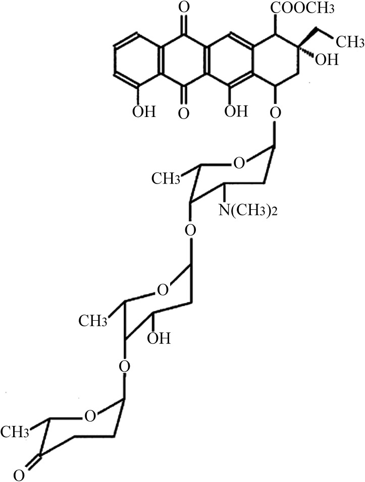
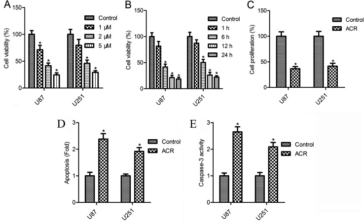
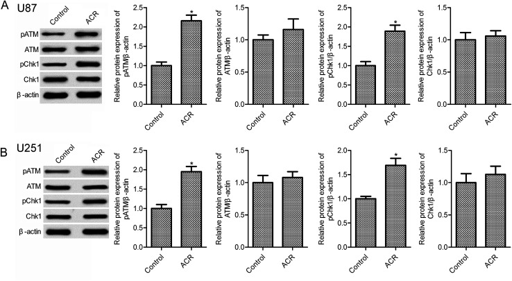
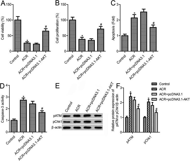
Aclarubicin (ACR), an anthracycline anti-tumor agent, is known to play important roles in cancer. Evidence has suggested that ACR has therapeutic effects on rats intracranially implanted with C6 glioma cells. However, the function and mechanism of ACR in glioma cells remain elusive. In this study, we examined the effects of ACR on glioma cell growth, apoptosis, and DNA damage. Our results showed that treatment with different concentrations of ACR (1, 2, and 5 μM) markedly impeded glioma cell survival, significantly decreased cell proliferation, and increased cell apoptosis and caspase-3 activity. Furthermore, ACR treatment promoted DNA damage through phosphorylation of ATM and CHK1 in U87 and U251 cells. Treatment with ACR also increased sirtuin 1 (SIRT1) expression and inhibited phosphatidylinositol 3'-kinase (PI3K)/AKT pathway activation. Interestingly, we found that AKT overexpression reversed the effects of ACR on glioma cell survival, proliferation, apoptosis, and DNA damage. Thus, our data suggest that ACR induces apoptosis and DNA damage in U87 and U251 cells through the SIRT1/PI3K/AKT signaling pathway.
This journal is © The Royal Society of Chemistry.
Conflict of interest statement
There is no conflict of interest to be declared by the authors.
Figures


Similar articles
-
Tenascin C Promotes Glioma Cell Malignant Behavior and Inhibits Chemosensitivity to Paclitaxel via Activation of the PI3K/AKT Signaling Pathway.J Mol Neurosci. 2021 Aug;71(8):1636-1647. doi: 10.1007/s12031-021-01832-8. Epub 2021 Apr 19. J Mol Neurosci. 2021. PMID: 33876384 Free PMC article.
-
Effects of Long Form of CAPON Overexpression on Glioma Cell Proliferation are Dependent on AKT/mTOR/P53 Signaling.Int J Med Sci. 2019 Apr 25;16(4):614-622. doi: 10.7150/ijms.31579. eCollection 2019. Int J Med Sci. 2019. PMID: 31171914 Free PMC article.
-
miR-489 inhibits proliferation, cell cycle progression and induces apoptosis of glioma cells via targeting SPIN1-mediated PI3K/AKT pathway.Biomed Pharmacother. 2017 Sep;93:435-443. doi: 10.1016/j.biopha.2017.06.058. Epub 2017 Jun 27. Biomed Pharmacother. 2017. PMID: 28666210
-
Neuroglobin promotes the proliferation and suppresses the apoptosis of glioma cells by activating the PI3K/AKT pathway.Mol Med Rep. 2018 Feb;17(2):2757-2763. doi: 10.3892/mmr.2017.8132. Epub 2017 Nov 22. Mol Med Rep. 2018. PMID: 29207186
-
Induction of Apoptosis in Human Glioma Cells by Fucoxanthin via Triggering of ROS-Mediated Oxidative Damage and Regulation of MAPKs and PI3K-AKT Pathways.J Agric Food Chem. 2019 Feb 27;67(8):2212-2219. doi: 10.1021/acs.jafc.8b07126. Epub 2019 Feb 13. J Agric Food Chem. 2019. PMID: 30688446
References
-
- Stupp R. Hegi M. E. Mason W. P. van den Bent M. J. Taphoorn M. J. Janzer R. C. Ludwin S. K. Allgeier A. Fisher B. Belanger K. Hau P. Brandes A. A. Gijtenbeek J. Marosi C. Vecht C. J. Mokhtari K. Wesseling P. Villa S. Eisenhauer E. Gorlia T. Weller M. Lacombe D. Cairncross J. G. Mirimanoff R. O. Lancet Oncol. 2009;10:459–466. doi: 10.1016/S1470-2045(09)70025-7. - DOI - PubMed
Publication types
LinkOut - more resources
Full Text Sources
Research Materials
Miscellaneous