

Sandwiching analytes with structurally diverse plasmonic nanoparticles on paper substrates for surface enhanced Raman spectroscopy
- PMID: 35529713
- PMCID: PMC9073094
- DOI: 10.1039/c9ra05399a
Sandwiching analytes with structurally diverse plasmonic nanoparticles on paper substrates for surface enhanced Raman spectroscopy
Abstract
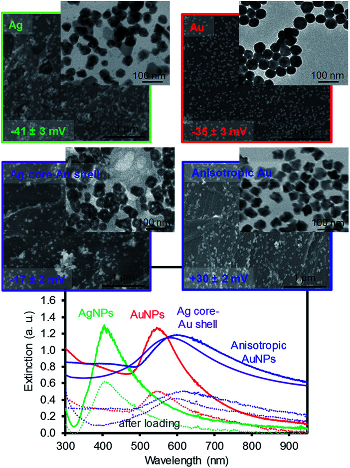
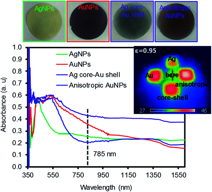
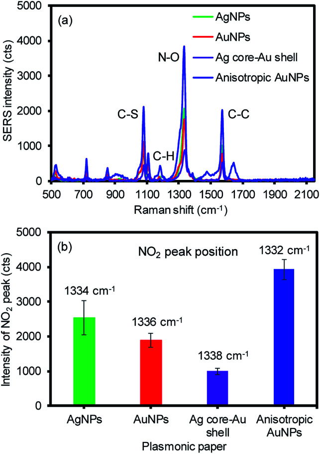
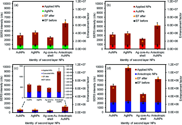
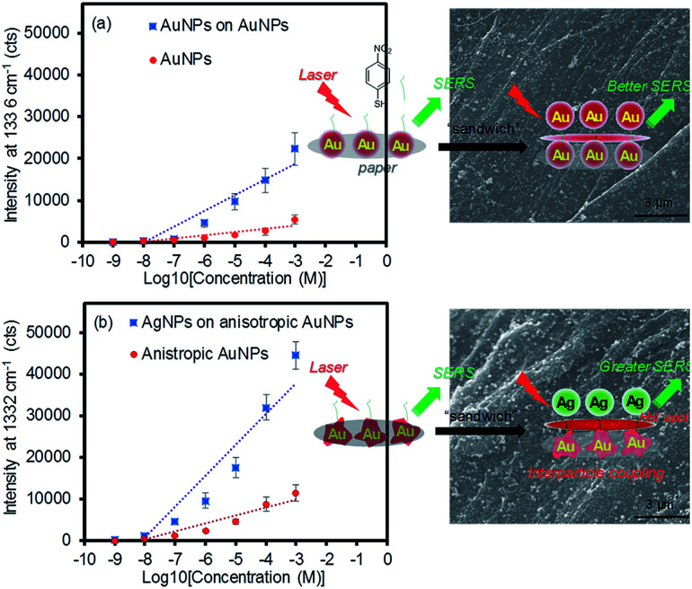
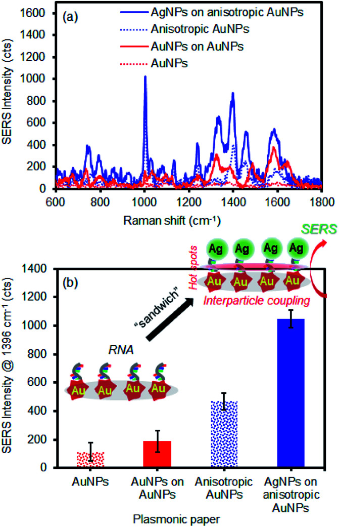
This report describes the systematic combination of structurally diverse plasmonic metal nanoparticles (AgNPs, AuNPs, Ag core-Au shell NPs, and anisotropic AuNPs) on flexible paper-based materials to induce signal-enhancing environments for surface enhanced Raman spectroscopy (SERS) applications. The anisotropic AuNP-modified paper exhibits the highest SERS response due to the surface area and the nature of the broad surface plasmon resonance (SPR) neighboring the Raman excitation wavelength. The subsequent addition of a second layer with these four NPs (e.g., sandwich arrangement) leads to the notable increase of the SERS signals by inducing a high probability of electromagnetic field environments associated with the interparticle SPR coupling and hot spots. After examining sixteen total combinations, the highest SERS response is obtained from the second layer with AgNPs on the anisotropic AuNP paper substrate, which allows for a higher calibration sensitivity and wider dynamic range than those of typical AuNP-AuNP arrangement. The variation of the SERS signals is also found to be below 20% based on multiple measurements (both intra-sample and inter-sample). Furthermore, the degree of SERS signal reductions for the sandwiched analytes is notably slow, indicating their increased long-term stability. The optimized combination is then employed in the detection of let-7f microRNA to demonstrate their practicability as SERS substrates. Precisely introducing interparticle coupling and hot spots with readily available plasmonic NPs still allows for the design of inexpensive and practical signal enhancing substrates that are capable of increasing the calibration sensitivity, extending the dynamic range, and lowering the detection limit of various organic and biological molecules.
This journal is © The Royal Society of Chemistry.
Conflict of interest statement
There are no conflicts to declare.
Figures
Similar articles
-
Growth of Spherical Gold Satellites on the Surface of Au@Ag@SiO2 Core-Shell Nanostructures Used for an Ultrasensitive SERS Immunoassay of Alpha-Fetoprotein.ACS Appl Mater Interfaces. 2019 Jan 23;11(3):3617-3626. doi: 10.1021/acsami.8b21238. Epub 2019 Jan 11. ACS Appl Mater Interfaces. 2019. PMID: 30608142
-
The Effect of Nanoparticle Composition on the Surface-Enhanced Raman Scattering Performance of Plasmonic DNA Origami Nanoantennas.ACS Nano. 2023 Nov 14;17(21):21227-21239. doi: 10.1021/acsnano.3c05464. Epub 2023 Oct 17. ACS Nano. 2023. PMID: 37847540
-
Three-Dimensional SERS Substrates Formed with Plasmonic Core-Satellite Nanostructures.Sci Rep. 2017 Oct 12;7(1):13066. doi: 10.1038/s41598-017-13577-9. Sci Rep. 2017. PMID: 29026173 Free PMC article.
-
SPR/SERS dual-mode plasmonic biosensor via catalytic hairpin assembly-induced AuNP network.Biosens Bioelectron. 2021 Oct 15;190:113376. doi: 10.1016/j.bios.2021.113376. Epub 2021 May 29. Biosens Bioelectron. 2021. PMID: 34098358
-
Dimensional Design for Surface-Enhanced Raman Spectroscopy.ACS Mater Au. 2022 May 10;2(5):552-575. doi: 10.1021/acsmaterialsau.2c00005. eCollection 2022 Sep 14. ACS Mater Au. 2022. PMID: 36855623 Free PMC article. Review.
Cited by
-
Silver Microparticle-Enhanced Laser-Induced Breakdown Spectroscopy.Appl Spectrosc. 2022 Aug;76(8):905-916. doi: 10.1177/00037028221096483. Epub 2022 May 28. Appl Spectrosc. 2022. PMID: 35634979 Free PMC article.
References
LinkOut - more resources
Full Text Sources
Other Literature Sources
Miscellaneous