. 2022 Apr;11(4):572-587.
doi: 10.21037/tlcr-21-729.

Plasma extracellular vesicle long RNA profiling identifies a diagnostic signature for stage I lung adenocarcinoma

Affiliations

Plasma extracellular vesicle long RNA profiling identifies a diagnostic signature for stage I lung adenocarcinoma

Wei Guo et al. Transl Lung Cancer Res. 2022 Apr.

Abstract

Background: The early diagnosis of lung adenocarcinoma (LUAD) is particularly challenging. Recent studies have reported that extracellular vesicles (EVs) include both small and long RNA. However, the profile and diagnosis-related significance of EV long RNA (exLR) profiles for early LUAD remain unclear.

Methods: A case-control analysis was carried out involving 110 participants, including 64 stage I LUAD cases, 24 benign pulmonary nodule (BPN) cases, and 22 healthy controls (HCs). The analysis was performed on the plasma samples' exLR profile based on exLR sequencing. The d-signature was identified using the least absolute shrinkage and selection operator (LASSO) method and a training set (n=48), and validation was completed through use of an internal validation set (n=32) and an external validation set (n=30).

Results: A diagnostic signature (d-signature) encompassing 8 exLR markers (NFKBIA, NDUFB10, SLC7A7, ARPC5, SEPTIN9, HMGN1, H4C2, and lnc-PLA2G1B-2:3) was identified for the detection of LUAD. This d-signature exhibited a high level of accuracy, with an area under the receiver operating characteristic (ROC) curve (AUC) of 0.991 in the training group, 0.921 in the internal validation group, and 0.9 in the external validation group. Moreover, the d-signature could distinguish adenocarcinomas in situ (AIS) and minimally invasive adenocarcinomas (MIA) from the noncancerous controls (NCs), with AUCs of 0.934 and 0.909, respectively, in the combined cohorts.

Conclusions: This study initially characterized the plasma exLR profile of early LUAD and reported on an exLR-based diagnostic signature for the detection of LUAD. This d-signature could be a promising noninvasive biomarker for the early detection and routine screening of LUAD.

Keywords: Extracellular vesicles (EVs); biomarker; diagnostic signature; long RNAs; lung adenocarcinoma (LUAD).


Conflict of interest statement

Conflicts of Interest: All authors have completed the ICMJE uniform disclosure form (available at https://tlcr.amegroups.com/article/view/10.21037/tlcr-21-729/coif). XL is an employee of Echo Biotech Co., Ltd. The other authors have no conflicts of interest to declare.

Figures

Figure 1
Figure 1
Plasma exLR-seq results. (A) Electron microscopy image of isolated vesicles. (B) Size distribution measurements of isolated EVs. (C) Western blot analysis of unenriched and exosomes-enriched proteins in isolated EVs. (D) Distribution of total mapped reads to the annotated genes with high confidence detection (mRNA and lncRNA). (E) Pie chart showing the reads distribution of these exLRs. (F) Boxplot lines represent the medians and interquartile ranges of the exLRs (mRNA and lncRNA). (G) The distribution of different types of RNA (mRNA and lncRNA) on chromosomes. (H) The basic the coverage of exLRs. The green part is mRNA, and the gray part is lncRNA. (I) The comparison of the mitochondrial gene expression ratio in our samples with the exoRbase. exLR, EV long RNA; EV, extracellular vesicle.
Figure 2
Figure 2
Comparison of exLR between the LUAD, the BPN patients and the HCs. (A) sEVs enriched fraction RNA concentration among LUAD patients, BPN patients and HCs; (B) Distribution of exLRs per sample among LUAD patients, BPN patients and HCs. (C) T-SNE analysis for the differential exLR profiles of LUAD patients from NCs. (D) PCA analysis for the differential exLR profiles of LUAD patients from NCs. (E) Heatmap of unsupervised hierarchical clustering of the exLRs differentially expressed between LUAD patients and NCs. (F) KEGG pathway analysis of the exLRs differentially expressed between LUAD patients and NCs. *, P<0.05; **, P<0.01; ***, P<0.001. exLR, EV long RNA; sEV, small extracellular vesicle; LUAD, lung adenocarcinoma; BPN, benign pulmonary nodule; HC, healthy control; T-SNE, T-distributed stochastic neighbor embedding; NC, non-cancerous control; PCA, principal components analysis; KEGG, Kyoto Encyclopedia of Genes and Genomes; ns, not significant.
Figure 3
Figure 3
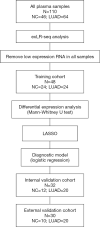
Workflow of data generation and analysis. Diagnostic marker selection: LASSO was applied to a training cohort of 24 LUAD patients and 11 BPN patients, and 13 HCs, leading to a final selection of eight markers. These eight markers were applied to a validation cohort of 24 LUAD patients and 8 BPN patients, and 8 HCs. LASSO, least absolute shrinkage and selection operator; NC, non-cancerous control; LUAD, lung adenocarcinoma; BPN, benign pulmonary nodule; HC, healthy control.
Figure 4
Figure 4
Establishment and validation of the exLR d-signature for LUAD. (A) Receiver operating characteristic curve and confusion table for performance of the exLR d-signature in the training cohort, internal validation cohort and external validation cohort. (B) Confusion table for performance of the exLR d-signature in the training cohort, internal validation cohort and external validation cohort. exLR, EV long RNA; NC, non-cancerous control; LUAD, lung adenocarcinoma.
Figure 5
Figure 5
The exLR d-signature for diagnosis of early stage LUAD. (A) ExLR d-signature score in AIS (n=22), MIA patients (n=17) and IAC patients (n=25). (B) Receiver operating characteristic (ROC) for the performance of the exLR d-signature in LUAD in the combined cohorts. (C) ExLR d-signature score in HCs (n=24), BPN patients (n=22) and LUAD patients (n=64). (D) ROC for the performance of the exLR d-signature for AIS, MIA and IAC in the combined cohorts. ****, P<0.0001. exLR, EV long RNA; LUAD, lung adenocarcinoma; HC, healthy control; BPN, benign pulmonary nodule; ROC, receiver operating characteristic; IAC, invasive adenocarcinoma; AIS, adenocarcinoma in situ; MIA, minimal invasive adenocarcinoma; ns, not significant.
Figure 6
Figure 6
The expression levels of the 8 EV long RNA markers between lung adenocarcinoma cases and noncancerous controls.
Figure 7
Figure 7
sEVs-associated characteristic evaluation of the 8 exLR markers. (A) Coomassie blue staining showed the total protein level in EV enriched fractions when treated by proteinase K and RNase A. (B) Representative Agilent 2100 Bioanalyzer results of sEVs enriched fractions derived RNA with and without the pretreatment of Proteinase K and RNase A before RNA extraction procedure. (C) The levels of eight genes in the exLR d-signature detected from sEVs enriched fraction samples with and without the pretreatment of Proteinase K and RNase A. exLR, EV long RNA; sEV, small extracellular vesicle; FU, fluorescence unit; nt, nucleotide.
Figure 8
Figure 8
Biological process network of 8 exLR markers. Red nodes are 8 exLR markers. Green nodes are target genes of lnc-PLA2G1B-2:3. Yellow nodes are important pathway/function. Grey nodes are other pathways that we are not concerned. exLR, EV long RNA; EV, extracellular vesicle.

References

    1. Siegel RL, Miller KD, Jemal A. Cancer statistics, 2019. CA Cancer J Clin 2019;69:7-34. 10.3322/caac.21551 - DOI - PubMed
    1. Chen W, Zheng R, Baade PD, et al. Cancer statistics in China, 2015. CA Cancer J Clin 2016;66:115-32. 10.3322/caac.21338 - DOI - PubMed
    1. Goldstraw P, Chansky K, Crowley J, et al. The IASLC Lung Cancer Staging Project: Proposals for Revision of the TNM Stage Groupings in the Forthcoming (Eighth) Edition of the TNM Classification for Lung Cancer. J Thorac Oncol 2016;11:39-51. 10.1016/j.jtho.2015.09.009 - DOI - PubMed
    1. Maeshima AM, Tochigi N, Yoshida A, et al. Histological scoring for small lung adenocarcinomas 2 cm or less in diameter: a reliable prognostic indicator. J Thorac Oncol 2010;5:333-9. 10.1097/JTO.0b013e3181c8cb95 - DOI - PubMed
    1. Yim J, Zhu LC, Chiriboga L, et al. Histologic features are important prognostic indicators in early stages lung adenocarcinomas. Mod Pathol 2007;20:233-41. 10.1038/modpathol.3800734 - DOI - PubMed