

Micro- and nanotechnology in biomedical engineering for cartilage tissue regeneration in osteoarthritis
- PMID: 35529803
- PMCID: PMC9039523
- DOI: 10.3762/bjnano.13.31
Micro- and nanotechnology in biomedical engineering for cartilage tissue regeneration in osteoarthritis
Abstract
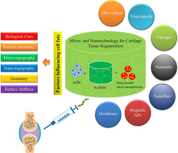
Osteoarthritis, which typically arises from aging, traumatic injury, or obesity, is the most common form of arthritis, which usually leads to malfunction of the joints and requires medical interventions due to the poor self-healing capacity of articular cartilage. However, currently used medical treatment modalities have reported, at least in part, disappointing and frustrating results for patients with osteoarthritis. Recent progress in the design and fabrication of tissue-engineered microscale/nanoscale platforms, which arises from the convergence of stem cell research and nanotechnology methods, has shown promising results in the administration of new and efficient options for treating osteochondral lesions. This paper presents an overview of the recent advances in osteochondral tissue engineering resulting from the application of micro- and nanotechnology approaches in the structure of biomaterials, including biological and microscale/nanoscale topographical cues, microspheres, nanoparticles, nanofibers, and nanotubes.
Keywords: biological cues; cartilage regeneration; micro/nanotopographical cues; nanotechnology; osteoarthritis; regenerative medicine.
Copyright © 2022, Nabizadeh et al.
Figures
Similar articles
-
Advances of nanotechnology in osteochondral regeneration.Wiley Interdiscip Rev Nanomed Nanobiotechnol. 2019 Nov;11(6):e1576. doi: 10.1002/wnan.1576. Epub 2019 Jul 22. Wiley Interdiscip Rev Nanomed Nanobiotechnol. 2019. PMID: 31329375 Review.
-
Treatment of osteochondral defects in the rabbit's knee joint by implantation of allogeneic mesenchymal stem cells in fibrin clots.J Vis Exp. 2013 May 21;(75):e4423. doi: 10.3791/4423. J Vis Exp. 2013. PMID: 23728213 Free PMC article.
-
Functional Biomolecule Delivery Systems and Bioengineering in Cartilage Regeneration.Curr Pharm Biotechnol. 2019;20(1):32-46. doi: 10.2174/1389201020666190206202048. Curr Pharm Biotechnol. 2019. PMID: 30727886 Review.
-
Nanomaterials/Nanocomposites for Osteochondral Tissue.Adv Exp Med Biol. 2018;1058:79-95. doi: 10.1007/978-3-319-76711-6_4. Adv Exp Med Biol. 2018. PMID: 29691818 Review.
-
Progress in Osteochondral Regeneration with Engineering Strategies.Ann Biomed Eng. 2022 Oct;50(10):1232-1242. doi: 10.1007/s10439-022-03060-6. Epub 2022 Aug 22. Ann Biomed Eng. 2022. PMID: 35994165 Review.
Cited by
-
Combinatorial Effect of Mesenchymal Stem Cells and Extracellular Vesicles in a Hydrogel on Cartilage Regeneration.Tissue Eng Regen Med. 2023 Feb;20(1):143-154. doi: 10.1007/s13770-022-00509-6. Epub 2022 Dec 8. Tissue Eng Regen Med. 2023. PMID: 36482140 Free PMC article.
-
Current Utilization of Gel-Based Scaffolds and Templates in Foot and Ankle Surgery-A Review.Gels. 2025 Apr 24;11(5):316. doi: 10.3390/gels11050316. Gels. 2025. PMID: 40422336 Free PMC article. Review.
-
Biophysical control of plasticity and patterning in regeneration and cancer.Cell Mol Life Sci. 2023 Dec 15;81(1):9. doi: 10.1007/s00018-023-05054-6. Cell Mol Life Sci. 2023. PMID: 38099951 Free PMC article. Review.
-
Benefits of Applying Nanotechnologies to Hydrogels in Efficacy Tests in Osteoarthritis Models-A Systematic Review of Preclinical Studies.Int J Mol Sci. 2022 Jul 26;23(15):8236. doi: 10.3390/ijms23158236. Int J Mol Sci. 2022. PMID: 35897805 Free PMC article.
-
Preparation and in vitro evaluation of biological agents based on Zinc-laponite- curcumin incorporated in alginate hydrogel.J Biol Eng. 2023 Nov 24;17(1):73. doi: 10.1186/s13036-023-00391-3. J Biol Eng. 2023. PMID: 38001515 Free PMC article.
References
Publication types
LinkOut - more resources
Full Text Sources