

Inversed Ratio of CD39/CD73 Expression on γδ T Cells in HIV Versus Healthy Controls Correlates With Immune Activation and Disease Progression
- PMID: 35529864
- PMCID: PMC9074873
- DOI: 10.3389/fimmu.2022.867167
Inversed Ratio of CD39/CD73 Expression on γδ T Cells in HIV Versus Healthy Controls Correlates With Immune Activation and Disease Progression
Abstract
Background: γδ T cells are unconventional T cells that have been demonstrated to be crucial for the pathogenesis and potentially for the cure of HIV-1 infection. The ectonucleotidase CD39 is part of the purinergic pathway that regulates immune responses by degradation of pro-inflammatory ATP in concert with CD73. Few studies on the expression of the ectoenzymes CD73 and CD39 on human γδ T cells in HIV have been performed to date.
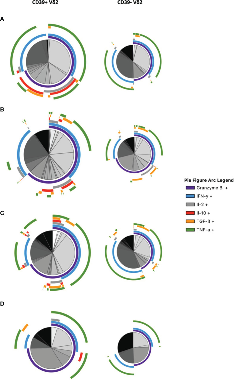
Methods: PBMC of n=86 HIV-1-infected patients were compared to PBMC of n=26 healthy individuals using 16-color flow cytometry determining the surface expression of CD39 and CD73 on Vδ1 and Vδ2 T cells in association with differentiation (CD45RA, CD28, CD27), activation and exhaustion (TIGIT, PD-1, CD38, and HLA-DR), and assessing the intracellular production of pro- and anti-inflammatory cytokines (IL-2, TGF-ß, TNF-α, Granzyme B, IL-10, IFN-γ) after in vitro stimulation with PMA/ionomycin.
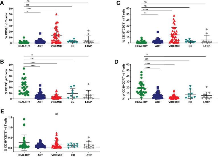
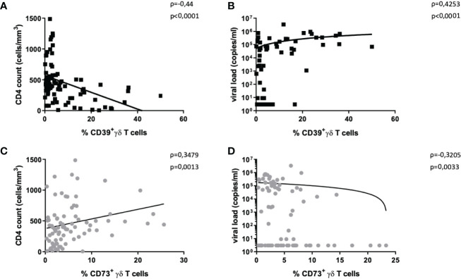
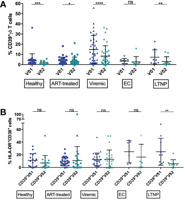
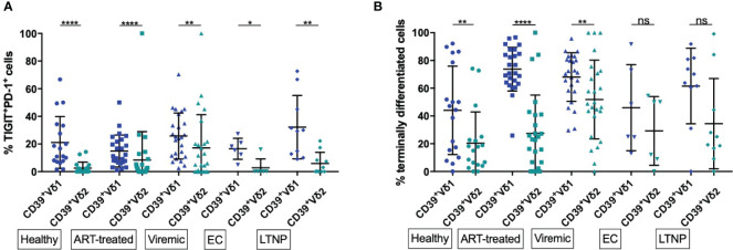
Results: CD39 and CD73 expression on γδ T cells were inversed in HIV infection which correlated with HIV disease progression and immune activation. CD39, but not CD73 expression on γδ T cells of ART-treated patients returned to levels comparable with those of healthy individuals. Only a small subset (<1%) of γδ T cells co-expressed CD39 and CD73 in healthy or HIV-infected individuals. There were significantly more exhausted and terminally differentiated CD39+ Vδ1 T cells regardless of the disease status. Functionally, IL-10 was only detectable in CD39+ γδ T cells after in vitro stimulation in all groups studied. Viremic HIV-infected patients showed the highest levels of IL-10 production. The highest percentage of IL-10+ cells was found in the small CD39/CD73 co-expressing γδ T-cell population, both in healthy and HIV-infected individuals. Also, CD39+ Vδ2 T cells produced IL-10 more frequently than their CD39+ Vδ1 counterparts in all individuals regardless of the HIV status.
Conclusions: Our results point towards a potential immunomodulatory role of CD39+ and CD73+ γδ T cells in the pathogenesis of chronic HIV infection that needs further investigation.
Keywords: CD39; CD73; HIV-1; IL-10; T cell; Vδ2; elite controllers; γδ T cells.
Copyright © 2022 Kolbe, Wittner, Hartjen, Hüfner, Degen, Ackermann, Cords, Stellbrink, Haag and Schulze zur Wiesch.
Conflict of interest statement
The authors declare that the research was conducted in the absence of any commercial or financial relationships that could be construed as a potential conflict of interest.
Figures

Similar articles
-
Bone Marrow-Resident Vδ1 T Cells Co-express TIGIT With PD-1, TIM-3 or CD39 in AML and Myeloma.Front Med (Lausanne). 2021 Nov 8;8:763773. doi: 10.3389/fmed.2021.763773. eCollection 2021. Front Med (Lausanne). 2021. PMID: 34820398 Free PMC article.
-
Tissue-Specific Expression of TIGIT, PD-1, TIM-3, and CD39 by γδ T Cells in Ovarian Cancer.Cells. 2022 Mar 11;11(6):964. doi: 10.3390/cells11060964. Cells. 2022. PMID: 35326415 Free PMC article.
-
Vδ1 Effector and Vδ2 γδ T-Cell Subsets Shift in Frequency and Are Linked to Plasma Inflammatory Markers During Antiretroviral Therapy-Suppressed HIV Infection.J Infect Dis. 2024 May 15;229(5):1317-1327. doi: 10.1093/infdis/jiae091. J Infect Dis. 2024. PMID: 38390982 Free PMC article.
-
Decreased Frequency of Intestinal CD39+ γδ+ T Cells With Tissue-Resident Memory Phenotype in Inflammatory Bowel Disease.Front Immunol. 2020 Sep 24;11:567472. doi: 10.3389/fimmu.2020.567472. eCollection 2020. Front Immunol. 2020. PMID: 33072107 Free PMC article.
-
The Role of Cluster of Differentiation 39 (CD39) and Purinergic Signaling Pathway in Viral Infections.Pathogens. 2023 Feb 8;12(2):279. doi: 10.3390/pathogens12020279. Pathogens. 2023. PMID: 36839551 Free PMC article. Review.
Cited by
-
Pannexin-1 channels, extracellular ATP, and purinergic receptors are essential for CCR5/CXCR4 clustering and HIV entry.NeuroImmune Pharm Ther. 2025 May 23;4(2):217-236. doi: 10.1515/nipt-2025-0005. eCollection 2025 Jun. NeuroImmune Pharm Ther. 2025. PMID: 40740680 Free PMC article.
-
CD73: a new immune checkpoint for leukemia treatment.Front Immunol. 2025 Mar 6;16:1486868. doi: 10.3389/fimmu.2025.1486868. eCollection 2025. Front Immunol. 2025. PMID: 40114928 Free PMC article. Review.
-
High and Sustained Ex Vivo Frequency but Altered Phenotype of SARS-CoV-2-Specific CD4+ T-Cells in an Anti-CD20-Treated Patient with Prolonged COVID-19.Viruses. 2022 Jun 10;14(6):1265. doi: 10.3390/v14061265. Viruses. 2022. PMID: 35746736 Free PMC article.
-
Single-cell analyses identify monocyte gene expression profiles that influence HIV-1 reservoir size in acutely treated cohorts.Nat Commun. 2025 May 29;16(1):4975. doi: 10.1038/s41467-025-59833-9. Nat Commun. 2025. PMID: 40442100 Free PMC article.
-
Research progress on V delta 1+ T cells and their effect on pathogen infection.PeerJ. 2024 Oct 30;12:e18313. doi: 10.7717/peerj.18313. eCollection 2024. PeerJ. 2024. PMID: 39494290 Free PMC article. Review.
References
Publication types
MeSH terms
Substances
LinkOut - more resources
Full Text Sources
Medical
Research Materials