. 2022 Apr 22:13:852830.
doi: 10.3389/fimmu.2022.852830. eCollection 2022.

Lentiviral Mediated ADA2 Gene Transfer Corrects the Defects Associated With Deficiency of Adenosine Deaminase Type 2

Affiliations

Lentiviral Mediated ADA2 Gene Transfer Corrects the Defects Associated With Deficiency of Adenosine Deaminase Type 2

Ying Hong et al. Front Immunol. .

Abstract

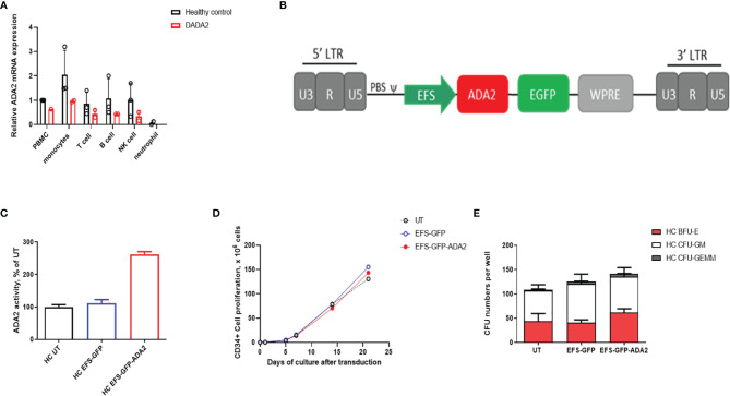
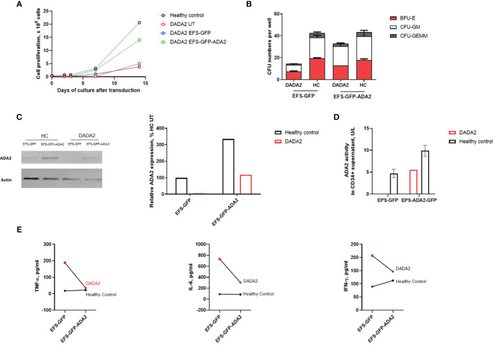
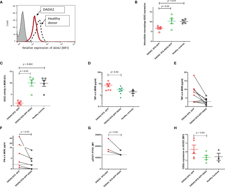
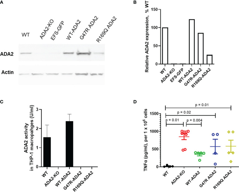
Deficiency of adenosine deaminase type 2 (DADA2) is an autosomal recessive disease caused by bi-allelic loss-of-function mutations in ADA2. Treatment with anti-TNF is effective for the autoinflammatory and vasculitic components of the disease but does not correct marrow failure or immunodeficiency; and anti-drug antibodies cause loss of efficacy over time. Allogeneic haematopoietic stem cell transplantation may be curative, but graft versus host disease remains a significant concern. Autologous gene therapy would therefore be an attractive longer-term therapeutic option. We investigated whether lentiviral vector (LV)-mediated ADA2 gene correction could rescue the immunophenotype of DADA2 in primary immune cells derived from patients and in cell line models. Lentiviral transduction led to: i) restoration of ADA2 protein expression and enzymatic activity; (ii) amelioration of M1 macrophage cytokine production, IFN-γ and phosphorylated STAT1 expression in patient-derived macrophages; and (iii) amelioration of macrophage-mediated endothelial activation that drives the vasculitis of DADA2. We also successfully transduced human CD34+ haematopoietic stem progenitor cells (HSPC) derived from a DADA2 patient with pure red cell aplasia and observed restoration of ADA2 expression and enzymatic activity in CD34+HSPC, alongside recovery of stem-cell proliferative and colony forming unit capacity. These preclinical data now expand the evidence for the efficacy of gene transfer strategies in DADA2, and strongly support clinical translation of a lentivirus-mediated gene therapy approach to treat DADA2.

Keywords: DADA2; anti-TNF; gene therapy; macrophages; stem cells.


Conflict of interest statement

PB has received institutional grants from: Novartis, SOBI, Roche, Chemocentryx, and Novimmune; consultancy fees from Roche, Novartis and SOBI; and speaker fees from UCB. DE received institutional grants from Lilly, Sobi, Roche and Pfizer. All research at Great Ormond Street Hospital NHS Foundation Trust and UCL Great Ormond Street Institute of Child Health is made possible by the NIHR Great Ormond Street Hospital Biomedical Research Centre. The views expressed are those of the author(s) and not necessarily those of the NHS, the NIHR or the Department of Health. The remaining authors declare that the research was conducted in the absence of any commercial or financial relationships that could be construed as a potential conflict of interest.

Figures

Figure 1
Figure 1
Ex vivo lentiviral mediated transduction delivers ADA2 protein expression and enzyme activity with no detrimental effect to the proliferative potential and colony forming capacity of CD34+haematopoietic stem progenitor cells (HSPC). (A) Relative ADA2 mRNA expression was examined in FACS sorted lymphocytes, monocytes, NK cells and neutrophils derived from both healthy controls (n = 2) and patients with DADA2 (n = 2). Both monocytes and lymphocytes were confirmed to express ADA2. (B) Lentivirus construct design containing codon optimised ADA2 c-DNA employing an EFS promoter and tagged to GFP. (C) ADA2 enzymatic activity was increased in macrophages derived from EFS-ADA2-GFP transduced healthy control CD34+HSPC compared to EFS-GFP alone treated cells (n = 3). (D, E) EFS-ADA2-GFP transduction of healthy control CD34+HSPC had no impact on cell proliferation and colony forming capacity across all myeloid and erythroid lineages (n=3). DADA2, deficiency of adenosine deaminase type 2; HSPC, haematopoietic stem cells; EFS, elongation factor 1α short; GFP, green fluorescent protein; ADA2, adenosine deaminase 2; GEMM, granulocyte, erythroid, macrophage, megakaryocyte; GM, granulocyte–macrophage; BFU-E, burst-forming units erythroid; NK, natural killer; UT, untransduced. WPRE, Woodchuck Hepatitis Virus Posttranscriptional Regulatory Element.
Figure 2
Figure 2
Ex vivo lentiviral mediated gene transfer restores ADA2 protein expression, enzyme activity and ameliorated the proliferative potential and colony forming capacity of CD34+cells from a patient with DADA2. (A, B). EFS-ADA2-GFP transduction of CD34+HSPC derived from a patient with DADA2 with bone marrow aplasia (genotype ADA2 p.G47W/p.G47W) improved the ability of these cells to proliferate and their colony forming capacity across all myeloid and erythroid lineages. (C, D). In addition, there was recovery of cellular ADA2 protein expression and enzyme activity in macrophages derived from EFS-ADA2-GFP transduced patient CD34+HSPC compared to EFS-GFP alone treated cells. (E) There was also reduction in the levels of proinflammatory cytokines (TNF-α, IL-6 and IFN-γ) released by macrophages derived from EFS-GFP-ADA2 transduced patient CD34+HSPC compared to EFS-GFP alone treated cells. Blots were performed in cell lysates. DADA2, deficiency of adenosine deaminase type2; HSPC, haematopoietic stem cells; EFS, elongation factor 1α short; GFP, green fluorescent protein; ADA2, adenosine deaminase 2; GEMM, granulocyte, erythroid, macrophage, megakaryocyte; GM, granulocyte–macrophage; BFU-E, burst-forming units erythroid; UT, untransduced; TNF, tumour necrosis factor; IL, interleukin; IFN, interferon.
Figure 3
Figure 3
Lentivirus mediated ADA2 gene transfer restores ADA2 protein expression, enzyme activity and rescues immunophenotype in monocyte derived macrophages (MDM) from DADA2 patients (A). Representative flow cytometric histogram showing ADA2 expression (MFI, median fluorescence intensity) in MDM obtained from patients with DADA2 and healthy controls. (B) EFS-GFP-ADA2 transduction of macrophage cells derived from patients with DADA2 resulted in restoration of ADA2 protein expression assessed by flow cytometry compared to no change in protein expression in EFS-GFP alone treated cells. (C) ADA2 enzyme activity assessed by an automated spectrophotometric assay also recovered in the culture supernatant of EFS-GFP-ADA2 transduced MDM from DADA2 patients compared to supernatants derived from EFS-GFP transduced cells. (D, E) EFS-GFP-ADA2 transduction of MDM derived from patients also led to a reduction in levels of TNF-α cytokine production in culture supernatants compared to EFS-GFP alone treated cells. Cumulative results and individual changes for each sample assessed are shown. (F, G) There was also suppression of IFN-γ release expression in culture supernatants and downregulation of p-STAT1 expression in EFS-GFP-ADA2 transduced MDM compared to EFS-GFP treated cells. (H) There was significant improvement in CD62E expression (MFI) on HUVEC incubated with culture supernatants from EFS-GFP-ADA2 transduced MDM from patients with DADA2 compared to co incubation with supernatants from EFS-GFP treated MDM. Dotted line represents levels observed in healthy control samples. MDM, monocyte derived macrophages; MFI, median fluorescence intensity; TNFα, tumour necrosis factor-α; IL, interleukin; EFS, elongation factor 1α short; IFN, interferon; GFP, green fluorescent protein; HUVEC, human umbilical vein endothelial cells; INF, interferon; UT, untransduced: ADA2, adenosine deaminase 2.
Figure 4
Figure 4
ADA2 protein expression, enzyme activity and macrophage responses in cell line models of DADA2. (A, B) ADA2-KO THP-1 cells were transduced with a lentivirus encoding wild-type ADA2 (WT-ADA2), or with ADA2 p.G47R or p.R169Q mutants and protein expression assessed; results of blot testing shown in 4A and cumulative protein quantification in 4B. This resulted in detectable ADA2 protein expression for EFS-WT ADA2 and EFS-G47R ADA2, EFS-R169Q ADA2 vectors. (C) There was restoration of ADA2 enzyme activity in the supernatants of cells transduced with EFS-WT ADA2, compared to no recovery of ADA2 activity when cells were treated with EFS-GFP; or when the EFS-G47R ADA2, EFS-R169Q ADA2 vectors were used. (D) EFS-ADA2-GFP transduction of these cells to express wild type ADA2 also reduced the levels of TNFα released in culture supernatants compared to no change observed in EFS-GFP or EFS-G47R ADA2, EFS-R169Q ADA2 treated cells. Blots were performed in cell lysates. TNFα, tumour necrosis factor-α; IL, interleukin; EFS, the elongation factor 1α short; GFP, green fluorescent protein; HUVEC, human umbilical vein endothelial cells; UT, untransduced: ADA2, adenosine deaminase 2; KO, knock out; WT, wild type.

References

    1. Zhou Q, Yang D, Ombrello AK, Zavialov AV, Toro C, Zavialov AV, et al. Early-Onset Stroke and Vasculopathy Associated With Mutations in ADA2. N Engl J Med (2014) 370(10):911–20. doi: 10.1056/NEJMoa1307361 - DOI - PMC - PubMed
    1. Navon Elkan P, Pierce SB, Segel R, Walsh T, Barash J, Padeh S, et al. Mutant Adenosine Deaminase 2 in a Polyarteritis Nodosa Vasculopathy. N Engl J Med (2014) 370(10):921–31. doi: 10.1056/NEJMoa1307362 - DOI - PubMed
    1. Nanthapisal S, Murphy C, Omoyinmi E, Hong Y, Standing A, Berg S, et al. Deficiency of Adenosine Deaminase Type 2: A Description of Phenotype and Genotype in Fifteen Cases: ADA2 Deficiency. Arthritis Rheumatol (2016) 68(9):2314–22. doi: 10.1002/art.39699 - DOI - PubMed
    1. Moens L, Hershfield M, Arts K, Aksentijevich I, Meyts I. Human Adenosine Deaminase 2 Deficiency: A Multi-Faceted Inborn Error of Immunity. Immunol Rev (2019) 287(1):62–72. doi: 10.1111/imr.12722 - DOI - PubMed
    1. Ombrello AK, Qin J, Hoffmann PM, Kumar P, Stone D, Jones A, et al. Treatment Strategies for Deficiency of Adenosine Deaminase 2. N Engl J Med (2019) 380(16):1582–4. doi: 10.1056/NEJMc1801927 - DOI - PMC - PubMed

Publication types

MeSH terms

Substances

Supplementary concepts