. 2019 Oct 25;9(59):34451-34456.
doi: 10.1039/c9ra06114b. eCollection 2019 Oct 23.

Normal vibrations of ternary DOPC/DPPC/cholesterol lipid bilayers by low-frequency Raman spectroscopy

Affiliations

Normal vibrations of ternary DOPC/DPPC/cholesterol lipid bilayers by low-frequency Raman spectroscopy

Dmitry V Leonov et al. RSC Adv. .

Abstract

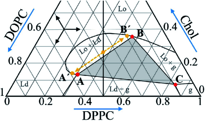
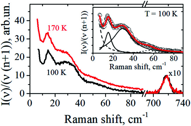
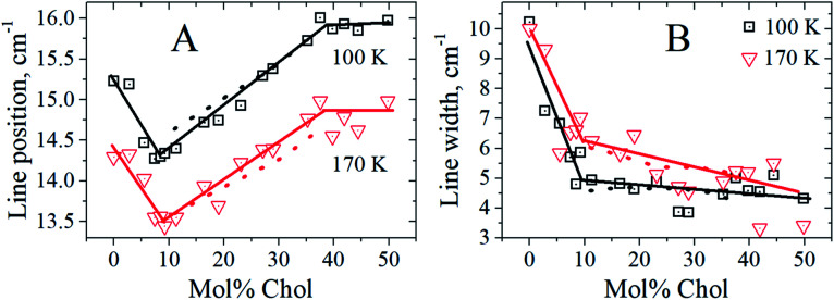
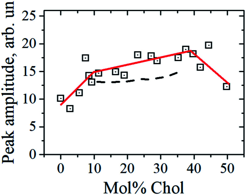
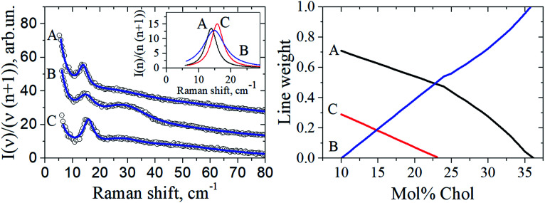
A lipid bilayer containing a ternary mixture of low- and high-melting lipids and cholesterol (Chol) can give rise to domain formation, referred to as lipid rafts. Low-frequency Raman spectroscopy at reduced temperatures allows detection of normal membrane mechanical vibrations. In this work, Raman spectra were obtained in the spectral range between 5 and 90 cm-1 for bilayers prepared from dioleoyl-glycero-phosphocholine (DOPC), dipalmitoyl-glycero-phosphocholine (DPPC) and Chol. A narrow peak detected between 13 and 16 cm-1 was attributed to the vibrational eigenmode of a lipid monolayer (a leaflet). For the equimolar DOPC/DPPC ratio, the Chol concentration dependence for the peak position, width and amplitude may be divided into three distinct ranges: below 9 mol%, the intermediate range between 9 mol% and 38 mol%, and above 38 mol%. In the intermediate range the peak position attains its minimum, and the peak width drops approximately by a factor of two as compared with the Chol-free bilayers. Meanwhile, this range is known for raft formation in a fluid state. The obtained results may be interpreted as evidence that bilayer structures in the raft-containing fluid state may be frozen at low temperatures. The drop of peak width indicates that at the spatial scale of the experiment (∼2.5 nm) the intermolecular bilayer structure with raft formation becomes more homogeneous and more cohesive.


Conflict of interest statement

There are no conflicts to declare.

Figures

Fig. 1
Fig. 1. Phase diagram of the DOPC/DPPC/Chol ternary system at 18 °C retrieved from literature data. The vertical line corresponds to the compositions studied in this work. The grey triangle with vertexes A, B and C corresponds to the compositions where the Ld, Lo and g phases are present; see text for meaning of the A′ and B′ points.
Fig. 2
Fig. 2. Low-frequency Raman spectra of cholesterol-free DOPC/DPPC bilayers obtained at 100 K and 170 K. In the inset the experimental spectrum at T = 100 K (circles) is approximated by four Lorentzians shown by dashed, dotted and two solid lines, with their sum presented by the solid line drawn through the circles.
Fig. 3
Fig. 3. The peak position (A) and the peak width (B) versus Chol concentration at 100 K (black squares) and 170 K (red triangles). The solid straight-line segments are drawn to guide the eye, and the dotted lines are predictions of eqn (3) (see text).
Fig. 4
Fig. 4. The peak amplitude versus Chol concentration at 100 K. Three solid straight-line segments are drawn to guide the eye, and the dashed lines are predictions of eqn (3) (see text).
Fig. 5
Fig. 5. Left: The circles show experimental Raman spectra for the DOPC/DPPC/Chol bilayers taken at 100 K for the compositions A, B, and C of the triangle vertexes shown in Fig. 1. Data are shifted upwards for convenience. The solid lines drawn through the experimental points are fits by a sum of Loretzians (cf.Fig. 2). The insert shows the Lorentzians describing the peak between 13 and 16 cm−1, for the A, B and C compositions. Right: the peak weights a, b and c found from eqn (4), as a function of Chol concentration.

Similar articles

Cited by

References

    1. Marsh D. Cholesterol-induced fluid membrane domains: a compendium of lipid-raft ternary phase diagrams. Biochim. Biophys. Acta. 2009;1788:2114–2123. doi: 10.1016/j.bbamem.2009.08.004. - DOI - PubMed
    1. Davis J. H. Clair J. J. Juhasz J. Phase equilibria in DOPC/DPPC-d62/cholesterol mixtures. Biophys. J. 2009;96:521–539. doi: 10.1016/j.bpj.2008.09.042. - DOI - PMC - PubMed
    1. Juhasz J. Sharom F. J. Davis J. H. Quantitative characterization of coexisting phases in DOPC/DPPC/cholesterol mixtures: comparing confocal fluorescence microscopy and deuterium nuclear magnetic resonance. Biochim. Biophys. Acta. 2009;1788:2541–2552. doi: 10.1016/j.bbamem.2009.10.006. - DOI - PubMed
    1. Heftberger P. Kollmitzer B. Rieder A. A. Amenitsch H. Pabst J. In situ determination of structure and fluctuations of coexisting fluid membrane domains. Biophys. J. 2015;108:854–862. doi: 10.1016/j.bpj.2014.11.3488. - DOI - PMC - PubMed
    1. Fischer T. Vink R. L. C. Domain formation in membranes with quenched protein obstacles: Lateral heterogeneity and the connection to universality classes. J. Chem. Phys. 2011;134:055106. doi: 10.1063/1.3530587. - DOI - PubMed