

Novel vinyl-modified RGD conjugated silica nanoparticles based on photo click chemistry for in vivo prostate cancer targeted fluorescence imaging
- PMID: 35530054
- PMCID: PMC9070015
- DOI: 10.1039/c9ra04513a
Novel vinyl-modified RGD conjugated silica nanoparticles based on photo click chemistry for in vivo prostate cancer targeted fluorescence imaging
Abstract
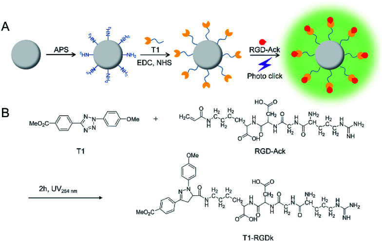
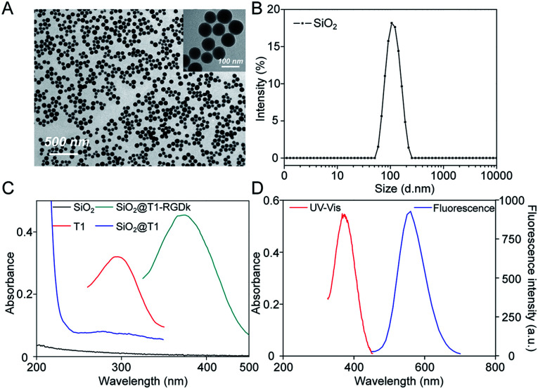
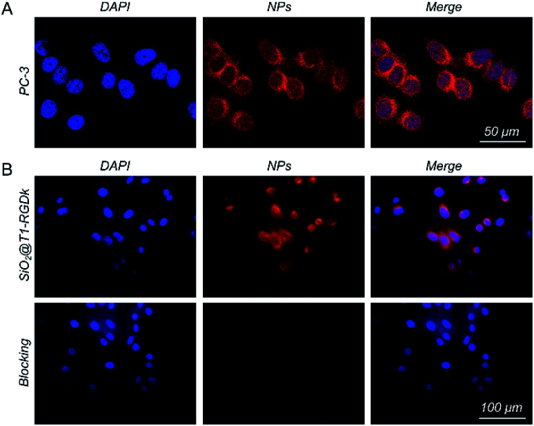
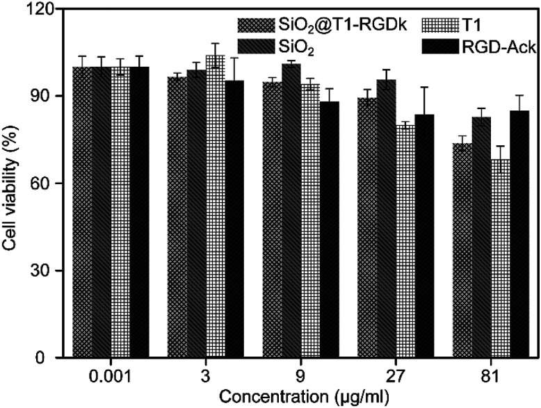
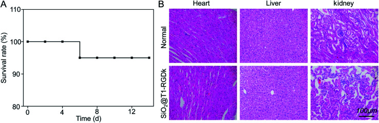
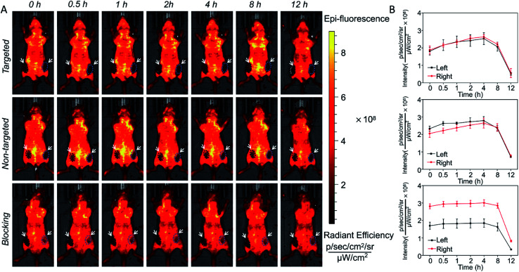
Molecular imaging is a powerful tool for non-invasive visualization of tumors that plays an important role in their diagnosis and treatment. The specificity of molecular imaging probes for cancer cells is important for accurate tumor visualization, with antibody and polypeptide nanoprobe conjugates having often been used as targeting agents for tumor detection. However, many traditional chemical conjugation methods employ complex conjugation reactions that result in poor efficiency and poor bioactivity. Herein, we describe the use of photo click methodology for the rapid synthesis of nanoprobes comprised of silica nanoparticles functionalized with RGD targeting units (SiO2@T1-RGDk NPs) (∼80 nm) for in vivo prostate cancer fluorescent imaging applications. These SiO2@T1-RGDk NPs exhibit a maximum absorption wavelength of 380 nm in their UV absorption spectra with a maximum fluorescence emission wavelength of 550 nm. Confocal immunofluorescent imaging reveal that SiO2@T1-RGDk NPs exhibit excellent targeting ability for visualizing cancer cells, with in vivo fluorescence imaging intensity in a subcutaneous tumor model of prostate cancer reaching a maxima after 4 h. Biosafety assessments showed that SiO2@T1-RGDk NPs demonstrate no obvious toxicity in mice, thus demonstrated that these novel NPs may prove to be promising fluorescent imaging agents for the accurate detection and treatment of tumors.
This journal is © The Royal Society of Chemistry.
Conflict of interest statement
There are no conflicts to declare.
Figures

Similar articles
-
PEG-coated and Gd-loaded fluorescent silica nanoparticles for targeted prostate cancer magnetic resonance imaging and fluorescence imaging.Int J Nanomedicine. 2019 Jul 23;14:5611-5622. doi: 10.2147/IJN.S207098. eCollection 2019. Int J Nanomedicine. 2019. PMID: 31413566 Free PMC article.
-
A photo-triggered conjugation approach for attaching RGD ligands to biodegradable mesoporous silica nanoparticles for the tumor fluorescent imaging.Nanomedicine. 2019 Jul;19:136-144. doi: 10.1016/j.nano.2019.04.005. Epub 2019 Apr 29. Nanomedicine. 2019. PMID: 31048083
-
In vivo near infrared fluorescence imaging and dynamic quantification of pancreatic metastatic tumors using folic acid conjugated biodegradable mesoporous silica nanoparticles.Nanomedicine. 2018 Aug;14(6):1867-1877. doi: 10.1016/j.nano.2018.04.018. Epub 2018 May 5. Nanomedicine. 2018. PMID: 29733890
-
Nanomedicine and Early Cancer Diagnosis: Molecular Imaging using Fluorescence Nanoparticles.Curr Top Med Chem. 2020;20(30):2737-2761. doi: 10.2174/1568026620666200922112640. Curr Top Med Chem. 2020. PMID: 32962614 Review.
-
Molecular imaging based on metabolic glycoengineering and bioorthogonal click chemistry.Biomaterials. 2017 Jul;132:28-36. doi: 10.1016/j.biomaterials.2017.04.003. Epub 2017 Apr 4. Biomaterials. 2017. PMID: 28399460 Review.
Cited by
-
New opportunities for RGD-engineered metal nanoparticles in cancer.Mol Cancer. 2023 May 25;22(1):87. doi: 10.1186/s12943-023-01784-0. Mol Cancer. 2023. PMID: 37226188 Free PMC article. Review.
-
Influence of the Surface Functionalization on the Fate and Performance of Mesoporous Silica Nanoparticles.Nanomaterials (Basel). 2020 May 9;10(5):916. doi: 10.3390/nano10050916. Nanomaterials (Basel). 2020. PMID: 32397449 Free PMC article. Review.
-
The Advancing Role of Nanocomposites in Cancer Diagnosis and Treatment.Int J Nanomedicine. 2024 Jun 19;19:6099-6126. doi: 10.2147/IJN.S471360. eCollection 2024. Int J Nanomedicine. 2024. PMID: 38911500 Free PMC article. Review.
References
LinkOut - more resources
Full Text Sources