Review
. 2019 Aug 13;9(44):25285-25302.
doi: 10.1039/c9ra04508b.

Recent progress in the imaging detection of enzyme activities in vivo

Affiliations
Review

Recent progress in the imaging detection of enzyme activities in vivo

Chunjie Yang et al. RSC Adv. .

Abstract

Enzymatic activities are important for normal physiological processes and are also critical regulatory mechanisms for many pathologies. Identifying the enzyme activities in vivo has considerable importance in disease diagnoses and monitoring of the physiological metabolism. In the past few years, great strides have been made towards the imaging detection of enzyme activity in vivo based on optical modality, MRI modality, nuclear modality, photoacoustic modality and multifunctional modality. This review summarizes the latest advances in the imaging detection of enzyme activities in vivo reported within the past years, mainly concentrating on the probe design, imaging strategies and demonstration of enzyme activities in vivo. This review also highlights the potential challenges and the further directions of this field.


Conflict of interest statement

There are no conflicts to declare.

Figures

Fig. 1
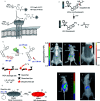
Fig. 1. Fig. 1 (A) Structure of AR-2. (B) Schematic representation of the NIRF imaging of a mouse bearing an MDA-MB-231 tumor (arrow) after being injected with AR-2. (C) There is a significant negative relativity between the tumor fluorescence and ATX. (D) Preparation of enzyme-responsive micellar nanoparticles. (E) Schematic representation of the relative retention levels of enzyme-responsive nanoparticles vs. control particles with HT-1080 tumors. (E1) Nanoparticle M injected. (E2) Nanoparticle MD injected. (A–C) Reproduced from ref. 56. Copy right 2013 Madan et al. D and E) Reproduced from ref. 37. Copy right 2013 American Chemical Society.
Fig. 2
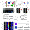
Fig. 2. (A) Schematic representation of the mechanism of in vivo imaging by caspase-3/7 activity. Reproduced from ref. 58. Copyright 2014 Springer Nature. (B) Schematic representation of the mechanism of in vivo imaging by legumain. Reproduced from ref. 66. Copyright 2018 American Chemical Society.
Fig. 3
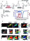
Fig. 3. (A) Conceptual scheme of β-Gal enzymatic activation of DCM-βgal. (B) Three-dimensional in vivo imaging of DCM-βgal. (C) Conceptual scheme of DCDHF-βgal when encountering β-Gal and the following fluorescence change. (D) In vivo fluorescence imaging of DCDHF-βgal. (A and B) Reproduced from ref. 54. Copyright 2016 American Chemical Society. (C and D) Reproduced from ref. 53. Copyright 2017 Elsevier.
Fig. 4
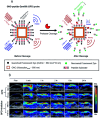
Fig. 4. (a–e) Recognition mechanism of the probes. (A–E) Real-time activity measurement in deep tissues of a mouse. (a and A) Reproduced from ref. 61. Copyright 2012 American Chemical Society. (b and B) Reproduced from ref. 60. Copyright 2018 American Chemical Society. (c and C) Reproduced from ref. 64. Copyright 2011 National Academy of Sciences. (d and D) Reproduced from ref. 63. Copyright 2016 Elsevier. (e and E) Reproduced from ref. 52. Copyright 2017 Royal Society of Chemistry.
Fig. 5
Fig. 5. (A) Schematic of bioluminogenic probes for detecting APN activity. (B) Bioluminescence imaging of APN activity in the implanted ES-2-luc cells for probe 1(left), probe 2(right). (C) Schematic of caspase-3/7 activity imaging with DEVD-(d-Cys) peptide and NH2-CBT. (D) Representative image of mice treated with LPS and D-GalN or vehicle, injection of DEVD-aminoluciferin or a combination of DEVD-(d-Cys) and NH2-CBT reagents. (A and B): Reproduced from ref. 90. Copyright 2014 American Chemical Society. (C and D) Reproduced from ref. 95. Copyright 2013 American Chemical Society.
Fig. 6
Fig. 6. (A) Schematic illustration of the self-assembly followed by disassembly of 1-NPs, and the self-assembly of 2-NPs. (B) Representative image of tumour-bearing or healthy zebrafish after being injected with the probes. White arrows indicate the tumour sites, and black arrows indicate the injection sites. White circle indicates the tumour location. (C) Schematic of FLAME-DEVD X (X = 1, 2), enzyme-responsive 19F MRI nanoprobes for detecting caspase-3/7 activity. (D) 19F MRI of caspase-3/7 activity in a mouse. FLAME-DEVD was intravenously injected into a mouse (before). Then, clodronate liposome was intravenously injected (after). L: liver. S: spleen. (E) Schematic of MRI contrast for detecting β-gal activity. (F) F1: imaging β-gal activity in vivo by 1H MRI. Green arrow indicates anomalous injection outside the tumour. White arrows indicate injection inside th tumour. Pink arrows indicate the change in T-weighted images and T2 values was obvious in the lacZ-transfected tumours post-injection of the agent and FAC. F2: Detection of β-gal activity in vivo by 19F NMR. NaTFA served as a chemical shift reference at 0 ppm and remained quite constant. (A and B) Reproduced from ref. 103. Copyright 2015 American Chemical Society. (C and D) Reproduced from ref. 107. Copyright 2018 American Chemical Society. (E and F) Reproduced from ref. 109. Copyright 2012 American Chemical Society.
Fig. 7
Fig. 7. (A) The CEST-FISP MRI protocol of the agent. (B) CatalyCEST MRI of GGT activity in vivo. Reproduced from ref. 118. Copyright 2017 International Society for Magnetic Resonance in Medicine.
Fig. 8
Fig. 8. (A) Schematic representation of the MMP activity-based PET strategy. (B) Micro-PET imaging of MMP activity in vivo. Mice bearing established HT1080 (left hind legs) and MCF-7 (right hind legs) tumours were injected with PEG-peptide-18F-TMR. The red arrows indicate HT1080 human fibrosarcomas in left hind legs. The green arrows indicate MCF-7 human breast adenocarcinomas in right legs. (C) Chemical structures of fluor-18 and indium-111 labelled [111In]MICA-401. (D) Representative SPECT images at 95 h post injection of the uPA activity-based probe [111In]MICA-401 in the three models. (a) Healthy control mouse; (b) HT-29 tumour-bearing mouse; (c) MDA-MB-231 tumour-bearing mouse. The white arrows indicate tumours and yellow arrows indicate liver. (A and B) Reproduced from ref. 126. Copyright 2012 American Association for Cancer Research. (C and D) Reproduced from ref. 129. Copyright 2016 John Wiley & Sons, Ltd.
Fig. 9
Fig. 9. (A) Schematic design of the activatable probe GPD. The probe is expected to produce a strong PA signal from the GNCs and Dye680, conjugated by a cleavable peptide substrate. (B) PAI after intratumoral injections of GPD probe ± inhibitor. Reproduced from ref. 142. Copyright 2018 World Molecular Imaging Society.
Fig. 10
Fig. 10. (A) Structures of the activity based probe BMV101. (B and C) Application of the 64Cu-BMV101 as a dual optical (B)/PET/CT (C) imaging probe. (D) First-in-human application of imaging probe 68Ga-BMV101. Representative scans of patients with idiopathic pulmonary fibrosis (IPF) and unclassifiable pulmonary fibrosis (fibrosis) compared to healthy controls. Reproduced from ref. 48. Copyright 2016 Springer Nature.
None
Chunjie Yang
None
Qian Wang
None
Wu Ding

Similar articles

Cited by

References

    1. Palamakumbura A. H. Trackman P. C. Anal. Biochem. 2002;300:245–251. doi: 10.1006/abio.2001.5464. - DOI - PubMed
    1. Wijdeven R. H. Neefjes J. Ovaa H. Trends Cell Biol. 2014;24:751–760. doi: 10.1016/j.tcb.2014.07.002. - DOI - PubMed
    1. Youdim M. B. Edmondson D. Tipton K. F. Nat. Rev. Neurosci. 2006;7:295–309. doi: 10.1038/nrn1883. - DOI - PubMed
    1. Yuan J. Xu Y. Q. Zhou N. N. Wang R. Qian X. H. Xu Y. F. RSC Adv. 2014;4:56207–56210. doi: 10.1039/C4RA10044A. - DOI
    1. Leto G. Gebbia N. Rausa L. Tumminello F. M. Anticancer Res. 1992;12:235–240. - PubMed