. 2019 Aug 19;9(44):25836-25846.
doi: 10.1039/c9ra03903a. eCollection 2019 Aug 13.

Novel cationic tannin/glycosaminoglycan-based polyelectrolyte multilayers promote stem cells adhesion and proliferation

Affiliations

Novel cationic tannin/glycosaminoglycan-based polyelectrolyte multilayers promote stem cells adhesion and proliferation

Paulo C F da Câmara et al. RSC Adv. .

Abstract

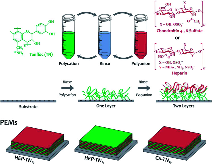
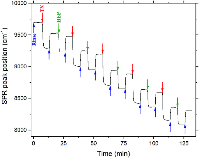
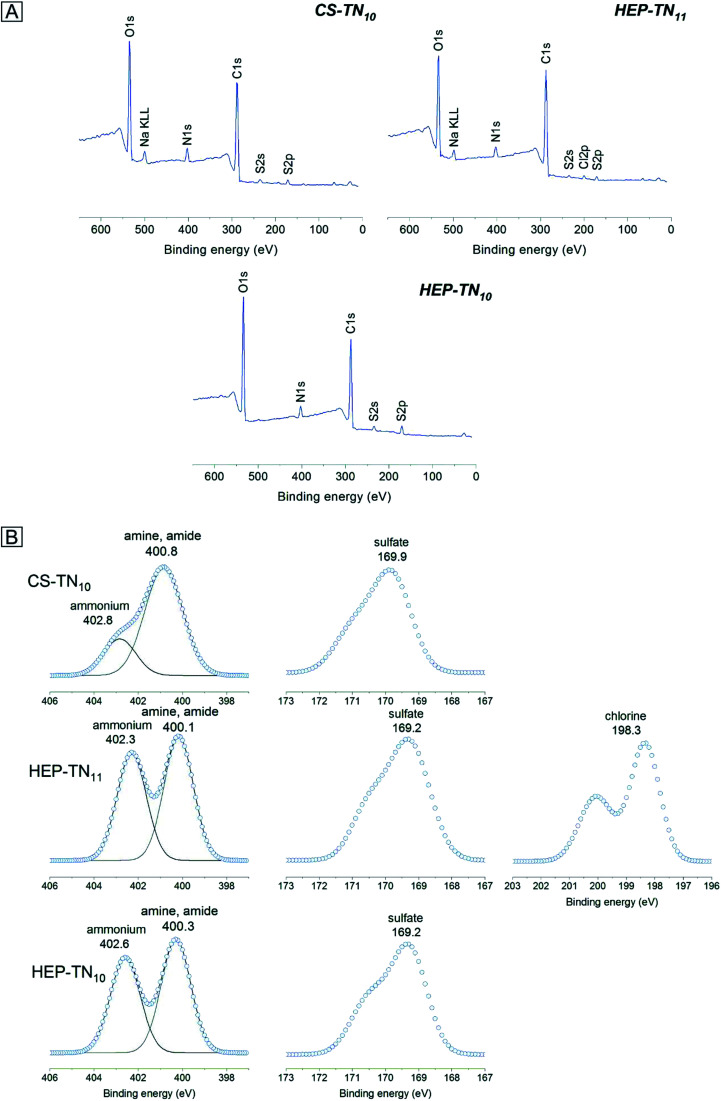
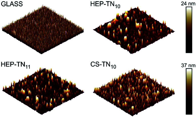
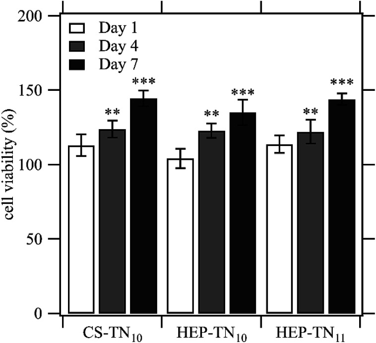
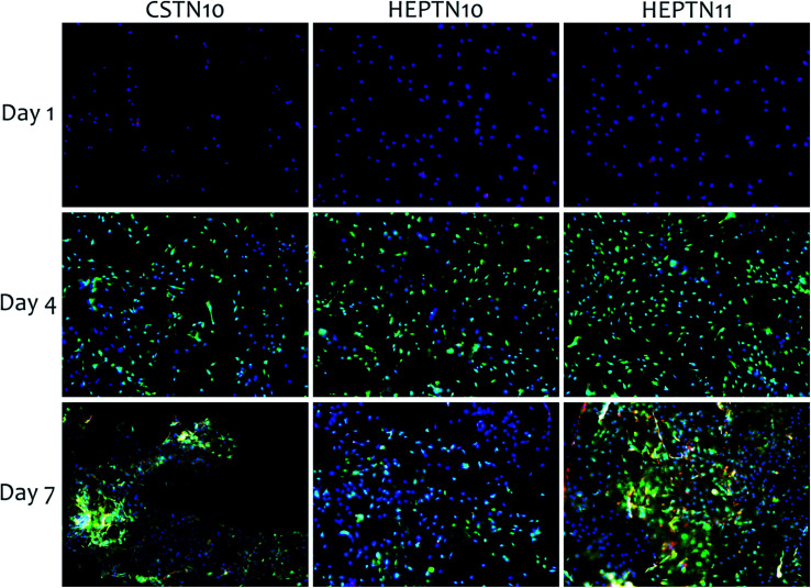
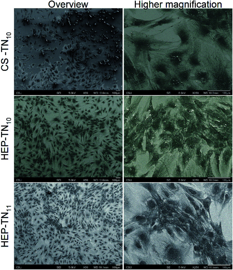
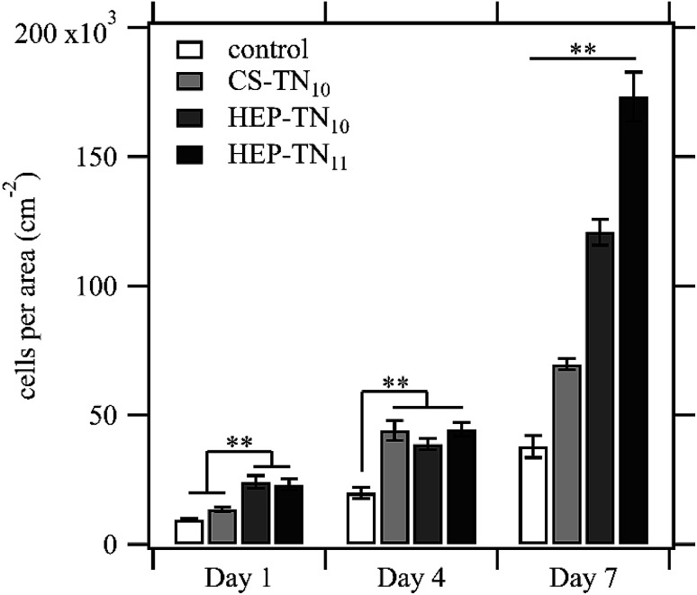
Modifying materials with biocompatible surface coatings is an important method for controlling cell responses to biomaterials. In this work, tanfloc (TN), a cationic tannin-derivative polymer was assembled with heparin (HEP) and chondroitin sulfate (CS), using the layer-by-layer (LbL) approach, to build polyelectrolyte multilayers (PEMs) and to design cytocompatible coatings. LbL deposition was monitored through Fourier-transform surface plasmon resonance, and characterized by X-ray photoelectron spectroscopy, atomic force microscopy, and contact angle measurements. The response of human adipose-derived stem cells (ADSCs) was evaluated in vitro. All of the TN-containing PEMs exhibit cytocompatibility and support adhesion, proliferation, and the spreading of ADSCs after 7 days of culture. HEP-TN PEM assembly with 11 layers (HEP-TN11) supports the greatest rate of cell proliferation. When TN is the terminal layer of the PEM, the surfaces promote the spreading of ADSCs, indicating that the surface charge and PEM terminal layer are key determinants of the microenvironmental niche that control cellular response. The promotion of stem cell attachment and proliferation makes these surface coatings potentially useful for biomedical implants and regenerative medicine.


Conflict of interest statement

There are no conflicts to declare.

Figures

Fig. 1
Fig. 1. Schematic representation for the PEM construction and chemical structures of the polyelectrolytes.
Fig. 2
Fig. 2. PEM assembly monitored by in situ FT-SPR. Arrows represent the beginning of acidified water rinse (blue), TN deposition layers (red), and HEP deposition layers (green).
Fig. 3
Fig. 3. Survey (A) and high-resolution X-ray spectra (B) of the N 1s, S 2p and Cl 2s envelopes determined on PEM surfaces.
Fig. 4
Fig. 4. Representative 10 μm × 10 μm AFM topographic images of the glass (non-modified), CS–TN10, HEP–TN10 and HEP–TN11 surfaces.
Fig. 5
Fig. 5. Effect of the contact with CS–TN10, HEP–TN10 and HEP–TN11 on ADSC activity normalized to the control (cell culture with standard supplemented culture medium on TCPS). Cell viability at day 4 and day 7 were statistically different from the control on days 4 and 7 (n = 5; ** indicates p < 0.01 compared to the control; *** indicates p < 0.0001 compared to the control).
Fig. 6
Fig. 6. Fluorescence microscopy images of ADSC cells to visualize cytoplasm (green), cytoskeleton (red), and nucleus (blue) on CS–TN10, HEP–TN10 and HEP–TN11 PEMs. The figure shows day 1, 4 and 7 of cell culture.
Fig. 7
Fig. 7. SEM overview and higher magnification micrographs of ADSCs seeded on CS–TN10, HEP–TN10 and HEP–TN11 PEMs, after 7 days of cell culture.
Fig. 8
Fig. 8. Quantification of ADSC cells per area on control (unmodified glass), CS–TN10, HEP–TN10 and HEP–TN11, after 1, 4 and 7 days of cell culture. Significant differences between different surface types on the same day are indicted (n = 5; ** indicates p < 0.01).

Similar articles

Cited by

References

    1. Martins J. G. Camargo S. E. A. Bishop T. T. Popat K. C. Kipper M. J. Martins A. F. Carbohydr. Polym. 2018;197:47–56. doi: 10.1016/j.carbpol.2018.05.062. - DOI - PubMed
    1. Silva J. M. Reis R. L. Mano J. F. Small. 2016;12:4308–4342. doi: 10.1002/smll.201601355. - DOI - PubMed
    1. Martins A. F. Facchi S. P. da Câmara P. C. F. Camargo S. E. A. Camargo C. H. R. Popat K. C. Kipper M. J. J. Colloid Interface Sci. 2018;525:21–30. doi: 10.1016/j.jcis.2018.04.060. - DOI - PubMed
    1. Simon-Walker R. Romero R. Staver J. M. Zang Y. Reynolds M. M. Popat K. C. Kipper M. J. ACS Biomater. Sci. Eng. 2017;3:68–77. doi: 10.1021/acsbiomaterials.6b00572. - DOI - PubMed
    1. Wang Y. Hong Q. Chen Y. Lian X. Xiong Y. Colloids Surf., B. 2012;100:77–83. doi: 10.1016/j.colsurfb.2012.05.030. - DOI - PubMed