Review
. 2022 Mar;12(3):1041-1053.
doi: 10.1016/j.apsb.2021.09.010. Epub 2021 Sep 16.

The generation of PD-L1 and PD-L2 in cancer cells: From nuclear chromatin reorganization to extracellular presentation

Affiliations
Review

The generation of PD-L1 and PD-L2 in cancer cells: From nuclear chromatin reorganization to extracellular presentation

Zhiwei Fan et al. Acta Pharm Sin B. 2022 Mar.

Abstract

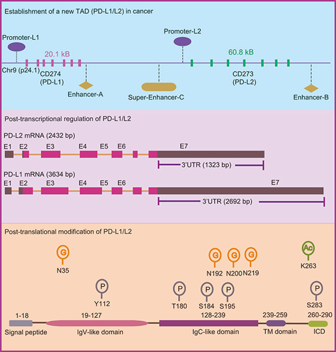
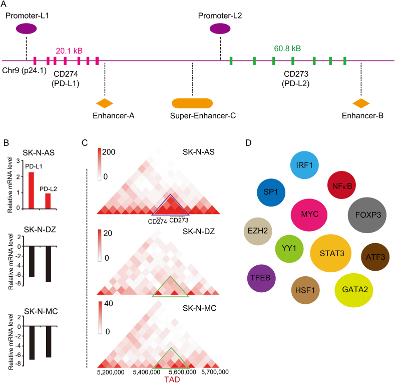
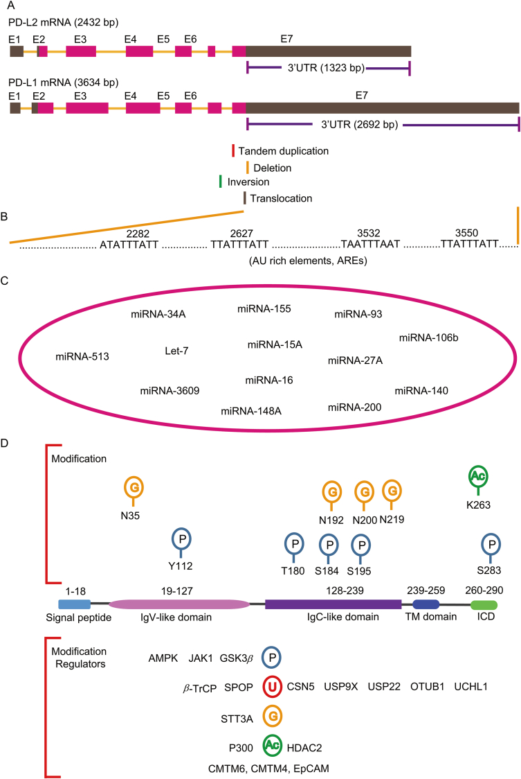
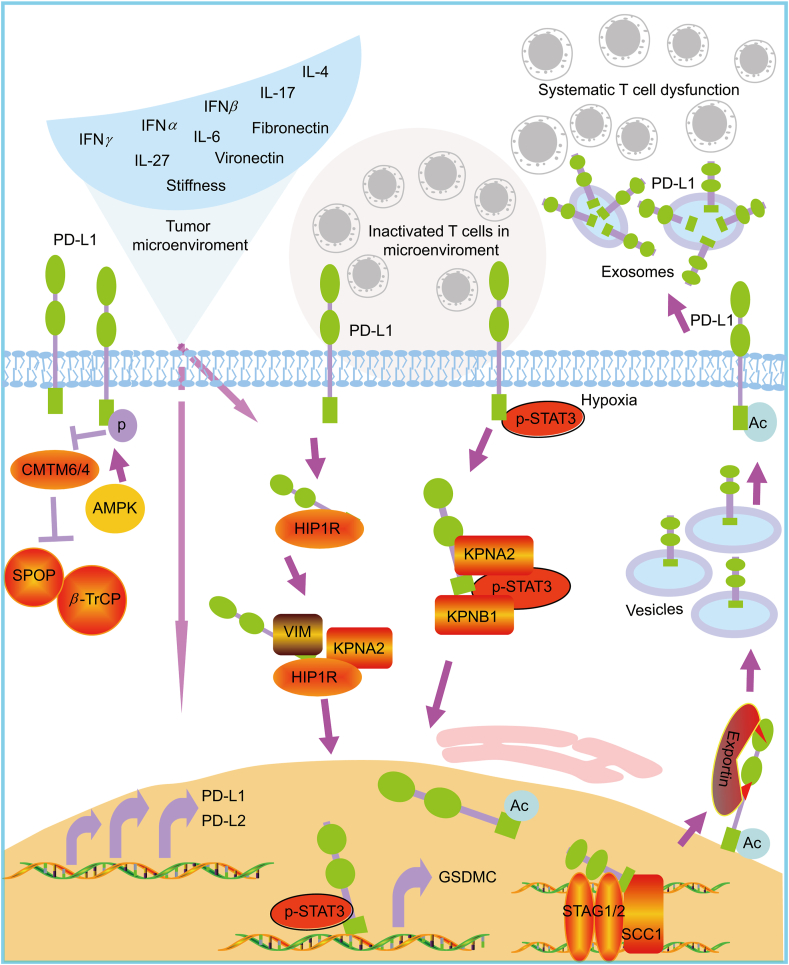
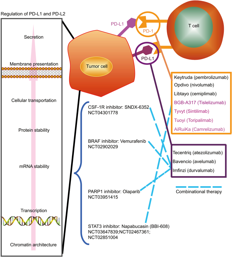
The immune checkpoint blockade (ICB) targeting on PD-1/PD-L1 has shown remarkable promise in treating cancers. However, the low response rate and frequently observed severe side effects limit its broad benefits. It is partially due to less understanding of the biological regulation of PD-L1. Here, we systematically and comprehensively summarized the regulation of PD-L1 from nuclear chromatin reorganization to extracellular presentation. In PD-L1 and PD-L2 highly expressed cancer cells, a new TAD (topologically associating domain) (chr9: 5,400,000-5,600,000) around CD274 and CD273 was discovered, which includes a reported super-enhancer to drive synchronous transcription of PD-L1 and PD-L2. The re-shaped TAD allows transcription factors such as STAT3 and IRF1 recruit to PD-L1 locus in order to guide the expression of PD-L1. After transcription, the PD-L1 is tightly regulated by miRNAs and RNA-binding proteins via the long 3'UTR. At translational level, PD-L1 protein and its membrane presentation are tightly regulated by post-translational modification such as glycosylation and ubiquitination. In addition, PD-L1 can be secreted via exosome to systematically inhibit immune response. Therefore, fully dissecting the regulation of PD-L1/PD-L2 and thoroughly detecting PD-L1/PD-L2 as well as their regulatory networks will bring more insights in ICB and ICB-based combinational therapy.

Keywords: 3′-UTR, 3′-untranslated region; ADAM17, a disintegrin and metalloprotease 17; APCs, antigen-presenting cells; AREs, adenylate and uridylate (AU)-rich elements; ATF3, activating transcription factor 3; CD273/274, cluster of differentiation 273/274; CDK4, cyclin-dependent kinase 4; CMTM6, CKLF like MARVEL transmembrane domain containing 6; CSN5, COP9 signalosome subunit 5; CTLs, cytotoxic T lymphocytes; EMT, epithelial to mesenchymal transition; EpCAM, epithelial cell adhesion molecule; Exosome; FACS, fluorescence-activated cell sorting; GSDMC, Gasdermin C; GSK3β, glycogen synthase kinase 3 beta; HSF1, heat shock transcription factor 1; Hi-C, high throughput chromosome conformation capture; ICB, immune checkpoint blockade; IFN, interferon; IL-6, interleukin 6; IRF1, interferon regulatory factor 1; Immune checkpoint blockade; JAK, Janus kinase 1; NFκB, nuclear factor kappa B; NSCLC, non-small cell lung cancer; OTUB1, OTU deubiquitinase, ubiquitin aldehyde binding 1; PARP1, poly(ADP-ribose) polymerase 1; PD-1, programmed cell death-1; PD-L1; PD-L1, programmed death-ligand 1; PD-L2; PD-L2, programmed death ligand 2; Post-transcriptional regulation; Post-translational regulation; SP1, specificity protein 1; SPOP, speckle-type POZ protein; STAG2, stromal antigen 2; STAT3, signal transducer and activator of transcription 3; T2D, type 2 diabetes; TADs, topologically associating domains; TFEB, transcription factor EB; TFs, transcription factors; TNFα, tumor necrosis factor-alpha; TTP, tristetraprolin; Topologically associating domain; Transcription; UCHL1, ubiquitin carboxy-terminal hydrolase L1; USP22, ubiquitin specific peptidase 22; dMMR, deficient DNA mismatch repair; irAEs, immune related adverse events.


Figures

Image 1
Graphical abstract
Figure 1
Figure 1
Genomic alternation and the transcriptional regulation of CD274 and CD273. (A) CD274 and CD273 are the encoding genes of PD-L1 and PD-L2 respectively. The genomic location of CD274 and CD273 locus as well as the identified promoters, enhancer-A, enhancer-B and super-enhancer-C are depictured. (B) The expression of PD-L1 and PD-L2 in SK-N-AS, SK-N-DZ and SK-N-MC cells is indicated. The expression of PD-L1 and PD-L2 is higher in SK-N-AS cells than it in SK-N-DZ and SK-N-MC cells. The data was searched from the 3D genome browser. (C) The HiC-data (the genome-wide chromosome conformation capture) around CD274 and CD273 locus was explored. The TAD around CD274 and CD273 (topologically associating domains) in SK-N-AS, SK-N-DZ, SK-N-MC cells is shown in triangle. The data was generated from the 3D genome browser. (D) The reported transcription factors that regulate the expression of PD-L1 are demonstrated. These TFs include IRF1, GATA2, SP1, NFκB, MYC, EZH2, FOXP3, YY1, STAT3, TFEB, HSF1 and ATF3.
Figure 2
Figure 2
Post-transcriptional and post-translational regulation of PD-L1. (A) The diagram showing the PD-L1 encoding gene CD274 and CD273. The exons and the 3′-untranslated regions (3′-UTR) of CD274 and CD273 are listed. The frequently observed breakpoints in CD274 resulting from tandem duplication, deletion, inversion and translocation around 3′UTR region are indicated. (B) Four potential AU rich regions in 3′-UTR of CD274 are shown. (C) The reported miRNAs that target on 3′UTR to regulate PD-L1 are summarized. These miRNAs include but not limited to miRNA-513, miRNA-34A, miRNA-3609, Let-7, miRNA-155, miRNA-148A, miRNA-15A, miRNA-16, miRNA-200, miRNA-93, miRNA-27A, miRNA-106b and miRNA-140. (D) The protein modification of PD-L1 and the associated enzymes are demonstrated. The upper panel shows the protein modification and the modified amino acids including glycosylation at N35, N192, N200, N219 and phosphorylation at Y112, T180, S184, S195 and S283 as well as acetylation at K263. The low panel shows the associated enzymes that regulate protein modification. The PD-L1 associated regulators include but not limited to AMPK, JAK1, GSK3β, β-TrCP, SPOP, CSN5, USP9X, USP22, OTUB1, UCHL1, STT3A, P300, HDAC2, CMTM6, CMTM4 and EpCAM.
Figure 3
Figure 3
The regulation of PD-L1 in tumor microenviroment and the dynamic cellular distribution. In tumor microenviroment, there are lots of reported factors, such as IFNγ, IFNα, IFNβ, IL-17, IL-4, IL-27, IL-6, Fibronectin, Vironectin and the stiffness, that can induce the expression of PD-L1 and PD-L2 via different mechanism at different levels. Expressed PD-L1 and PD-L2 resident on membrane to inactivate T cells locally. The membrane PD-L1 can translocate to nucleus through HIP1R/KPNA2/KPNB1 pathway. Nuclear PD-L1 is acetylated and translocates to membrane via exportin. Intracellular PD-L1 can secrete to tumor microenviroment or even long distance via exosome. Unlike the membrane resident PD-L1 inactivates T cells locally, the exosome-containing PD-L1 can systematically inactivate T cell function. The protein stability of PD-L1 was regulated by CMTM6 or CMTM4 by blocking SPOP- and β-TrCP-mediated degradation. However, in low energy condition, the activated AMPK can phosphorylate PD-L1 and disrupt the interaction between PD-L1 and CMTM4. Thus, the tumor microenviroment can substantially affects the distribution and function of PD-L1 not only locally but also systematically.
Figure 4
Figure 4
The approved PD-1/PD-L1 antibodies and its combinational therapy with inhibitors targeting on the regulatory pathway of PD-L1 and PD-L2. Currently, there are 10 antibodies that have been approved in clinic including 7 anti-PD1 antibodies (pembrolizumab, nivolumab, cemiplimab, tislelizumab, sintilimab, toripalimab, and amrelizumab) and three anti-PD-L1 antibodies (atezolizumab, avelumab, and durvalumab). ICIs-based combinational therapy with other inhibitors is an emerging and promising trend. Several clinic trials have been already ongoing to test the inhibitors targeting the PD-L1/PD-L2 regulators such as STAT3. The number of 6 clinic trials are listed. Results from the clinical trial targeting STAT3 (napabucasin) and PD-1 (pembrolizumab) show anti-tumor activity in metastatic colorectal cancer with microsatellite stable. Although the results did not meet the primary end point, it is still very promising to consider no biomarkers were used in recruiting patients. Thus, ICI-based combinational therapy with inhibitors targeting regulatory factors of PD-L1/PD-L2, especially cancer-specific regulatory pathways, is deserved to be extensively examined by considering patients' recruitment with designed biomarkers.

Similar articles

Cited by

References

    1. Pardoll D.M. The blockade of immune checkpoints in cancer immunotherapy. Nat Rev Cancer. 2012;12:252–264. - PMC - PubMed
    1. Ribas A., Wolchok J.D. Cancer immunotherapy using checkpoint blockade. Science. 2018;359:1350–1355. - PMC - PubMed
    1. Sanmamed M.F., Chen L. A paradigm shift in cancer immunotherapy: from enhancement to normalization. Cell. 2018;175:313–326. - PMC - PubMed
    1. Zaretsky J.M., Garcia-Diaz A., Shin D.S., Escuin-Ordinas H., Hugo W., Hu-Lieskovan S., et al. Mutations associated with acquired resistance to PD-1 blockade in melanoma. N Engl J Med. 2016;375:819–829. - PMC - PubMed
    1. Postow M.A., Sidlow R., Hellmann M.D. Immune-related adverse events associated with immune checkpoint blockade. N Engl J Med. 2018;378:158–168. - PubMed