. 2022 Mar;12(3):1288-1304.
doi: 10.1016/j.apsb.2021.08.015. Epub 2021 Aug 14.

The FAP α -activated prodrug Z-GP-DAVLBH inhibits the growth and pulmonary metastasis of osteosarcoma cells by suppressing the AXL pathway

Affiliations

The FAP α -activated prodrug Z-GP-DAVLBH inhibits the growth and pulmonary metastasis of osteosarcoma cells by suppressing the AXL pathway

Geni Ye et al. Acta Pharm Sin B. 2022 Mar.

Erratum in

Abstract

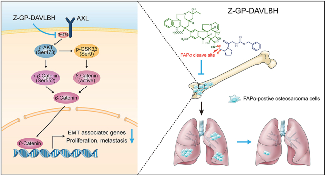
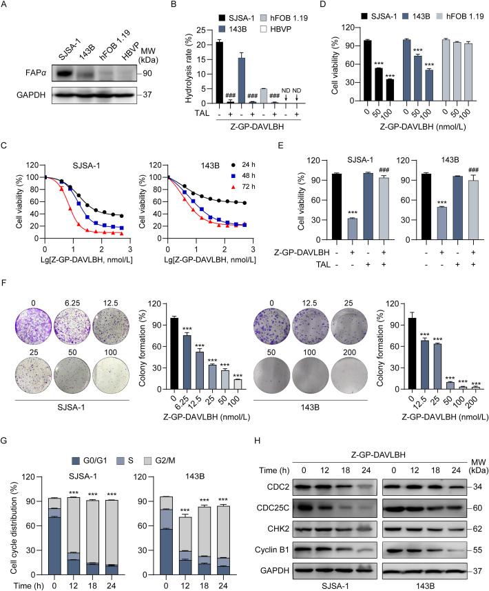
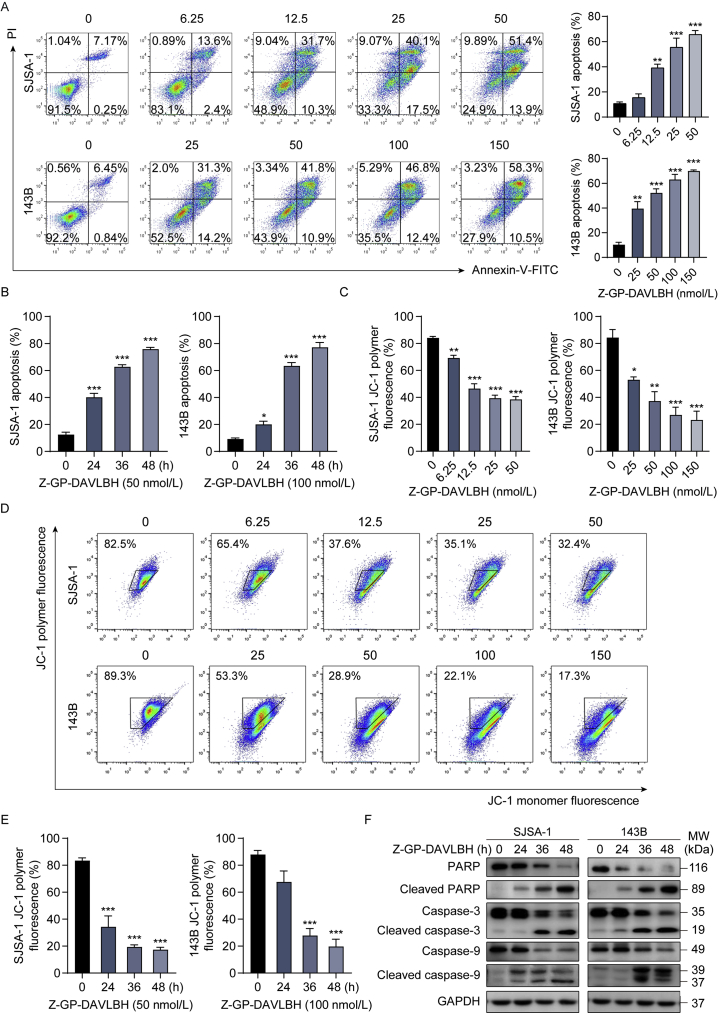
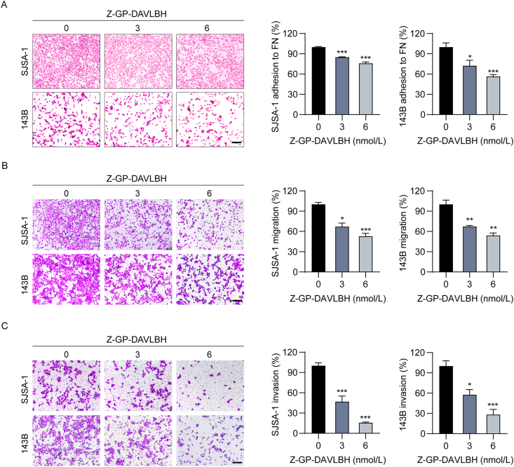
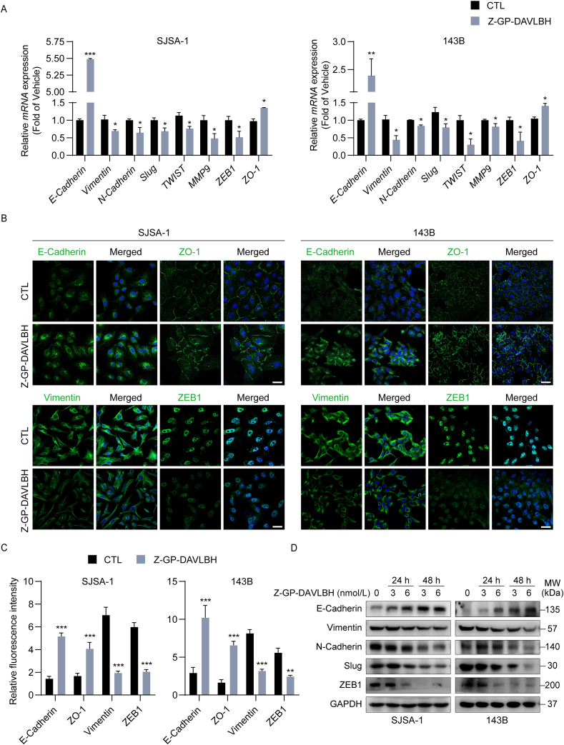
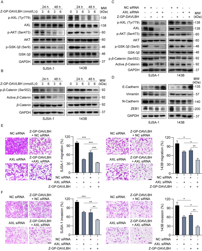
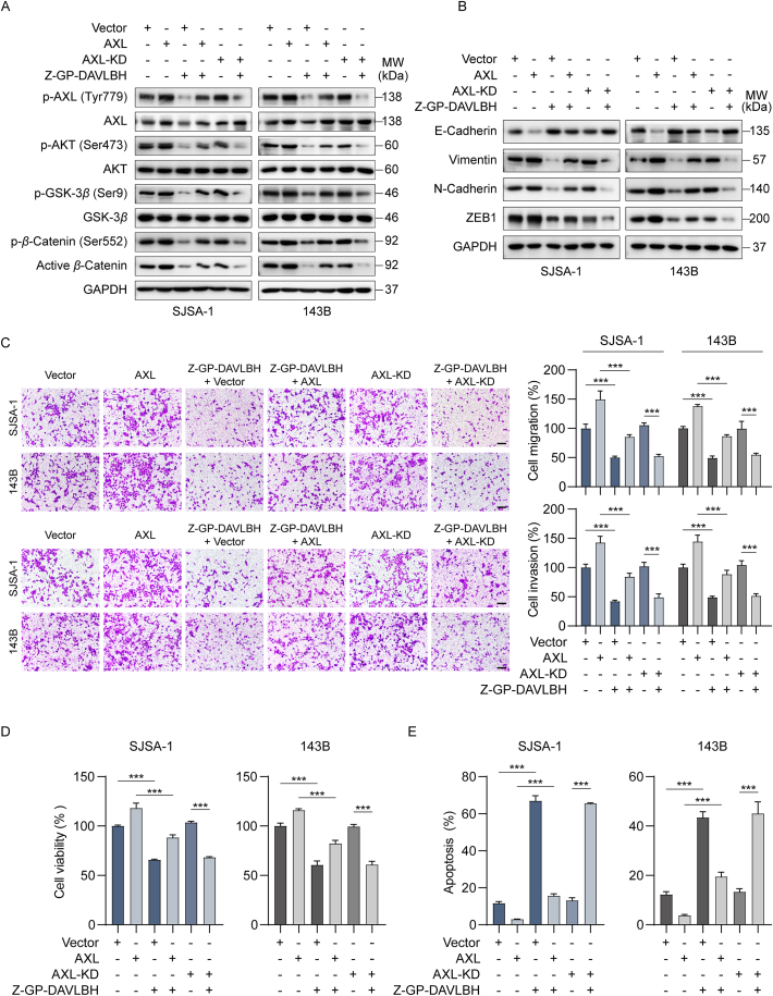
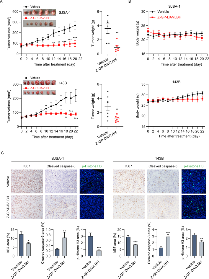
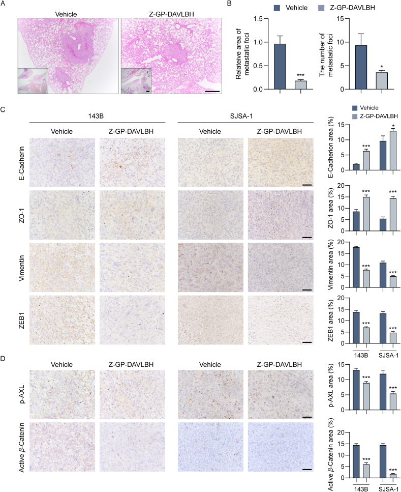
Osteosarcoma is a kind of bone tumor with highly proliferative and invasive properties, a high incidence of pulmonary metastasis and a poor prognosis. Chemotherapy is the mainstay of treatment for osteosarcoma. Currently, there are no molecular targeted drugs approved for osteosarcoma treatment, particularly effective drugs for osteosarcoma with pulmonary metastases. It has been reported that fibroblast activation protein alpha (FAPα) is upregulated in osteosarcoma and critically associated with osteosarcoma progression and metastasis, demonstrating that FAPα-targeted agents might be a promising therapeutic strategy for osteosarcoma. In the present study, we reported that the FAPα-activated vinblastine prodrug Z-GP-DAVLBH exhibited potent antitumor activities against FAPα-positive osteosarcoma cells in vitro and in vivo. Z-GP-DAVLBH inhibited the growth and induced the apoptosis of osteosarcoma cells. Importantly, it also decreased the migration and invasion capacities and reversed epithelial-mesenchymal transition (EMT) of osteosarcoma cells in vitro and suppressed pulmonary metastasis of osteosarcoma xenografts in vivo. Mechanistically, Z-GP-DAVLBH suppressed the AXL/AKT/GSK-3β/β-catenin pathway, leading to inhibition of the growth and metastatic spread of osteosarcoma cells. These findings demonstrate that Z-GP-DAVLBH is a promising agent for the treatment of FAPα-positive osteosarcoma, particularly osteosarcoma with pulmonary metastases.

Keywords: AXL; DAVLBH, desacetylvinblastine monohydrazide; EMT, epithelial–mesenchymal transition; FAPα, fibroblast activation protein alpha; Fibroblast activation protein alpha; Growth; Osteosarcoma; Pulmonary metastasis; TA-MSCs, tumor-associated mesenchymal stem cells; Vinblastine prodrug; Z-GP, N-terminal benzyloxy carbonyl-blocked (Z-blocked) GlyPro peptide; Z-GP-DAVLBH, desacetylvinblastine monohydrazide coupled to an N-terminal benzyloxy carbonyl-blocked (Z-blocked) GlyPro peptide; siRNA, small interfering RNA; β-Catenin.


Figures

Image 1
Graphical abstract
Figure 1
Figure 1
Z-GP-DAVLBH inhibits the proliferation of osteosarcoma cells in vitro. (A) The protein levels of FAPα in SJSA-1, 143B, hFOB 1.19 cells, and HBVPs were determined by Western blotting analysis. (B) SJSA-1, 143B, hFOB 1.19 cells, and HBVPs were treated with Z-GP-DAVLBH (10 μmol/L) in the presence or absence of TAL for 2 h. The hydrolysis efficiency of Z-GP-DAVLBH was analyzed by LC–MS. HBVPs serve as an FAPα-negative control cells. ND, no detection. (C) Osteosarcoma cells (SJSA-1 and 143B) were treated with various concentrations of Z-GP-DAVLBH for 24, 48, and 72 h. Cell viability was detected by an MTT assay. (D) MTT assay was conducted to determine the effect of Z-GP-DAVLBH on the viability of hFOB 1.19 cells. (E) Osteosarcoma cells (SJSA-1 and 143B) were treated with Z-GP-DAVLBH (50 nmol/L for SJSA-1 cells and 100 nmol/L for 143B cells) for 48 h in the presence or absence of TAL. Cell viability was detected by MTT assay. (F) Cell colony formation assay of SJSA-1 and 143B cells treated with the indicated concentrations of Z-GP-DAVLBH. Representative images of cell colonies are shown and clonogenicity was quantitated by normalization to the untreated group. Magnification: 100×. (G) The cell cycle distribution was detected by flow cytometry analysis. (H) Cell cycle-associated proteins were analyzed by Western blotting analysis. Data are presented as mean ± SEM, n = 3; ∗∗∗P<0.001 vs. the untreated (0 nmol/L, 0.1% DMSO) group; ###P < 0.001 vs. the Z-GP-DAVLBH-treated group.
Figure 2
Figure 2
Z-GP-DAVLBH induces apoptosis in osteosarcoma cells. (A) and (B) Osteosarcoma cells (SJSA-1 and 143B) were (A) exposed to various concentrations of Z-GP-DAVLBH for 48 h, or (B) treated with Z-GP-DAVLBH for the indicated times. Apoptosis was examined with an Annexin V-FITC apoptosis detection kit. Representative flow cytometry plots and quantification of apoptotic cells. (C)–(E) Osteosarcoma cells (SJSA-1 and 143B) were (C) treated with Z-GP-DAVLBH for the indicated times or (D) incubated with gradient concentrations of Z-GP-DAVLBH for 48 h. Mitochondrial membrane potential (MMP) was assessed with a JC-1 kit. (E) Quantification of JC-1 polymer fluorescence of (D) is shown. (F) The expression of PARP, cleaved PARP, caspase-3, cleaved caspase-3, caspase 9, and cleaved caspase-9 in SJSA-1 and 143B cells were determined by Western blotting analysis. Representative blots are shown. Data are presented as mean±SEM, n = 3; ∗P<0.05, ∗∗P<0.01, and ∗∗∗P<0.001 vs. the untreated (0 nmol/L, 0.1% DMSO) group.
Figure 3
Figure 3
Z-GP-DAVLBH decreases the adhesion, migration, and invasion capacities of osteosarcoma cells. (A) Representative images of SJSA-1 and 143B cell adhered to fibronectin after treatment with Z-GP-DAVLBH. Quantification of the number of cells adhered to fibronectin. Scale bar, 200 μm. (B) and (C) The effect of Z-GP-DAVLBH on the migration and invasion of SJSA-1 and 143B cells was determined by Transwell (B) migration and (C) invasion assays. (B) Representative images and quantification of the number of migrated cells. Scale bar, 200 μm. (C) Representative images and quantification of the number of invaded cells. Scale bar, 200 μm. Data are presented as mean ± SEM, n = 3; ∗P<0.05, ∗∗P<0.01, and ∗∗∗P<0.001 vs. the untreated (0 nmol/L, 0.1% DMSO) group.
Figure 4
Figure 4
Z-GP-DAVLBH impairs epithelial–mesenchymal transition in osteosarcoma cells. (A) SJSA-1 and 143B cells were treated with Z-GP-DAVLBH (6 nmol/L) for 48 h, and the relative mRNA expression levels of EMT-related genes were determined by RT-PCR assay. (B) SJSA-1 and 143B cells were treated with Z-GP-DAVLBH (6 nmol/L) for 48 h, and the expression of vimentin, ZEB-1, E-cadherin, and ZO-1 was evaluated by immunofluorescence staining analysis. Scale bar: 50 μm. (C) Quantification of relative fluorescence intensity in (B) is shown. (D) Osteosarcoma cells were treated with Z-GP-DAVLBH (3 or 6 nmol/L) for 24 and 48 h and then the expression of EMT-related markers was determined by Western blotting assay. Data are presented as mean±SEM, n = 3; ∗P<0.05, ∗∗P<0.01, and ∗∗∗P<0.001 vs. the CTL (0.1% DMSO) group.
Figure 5
Figure 5
Z-GP-DAVLBH inhibits the AXL/AKT/GSK-3β/β-catenin pathway in osteosarcoma cells. (A)–(B) Osteosarcoma cells were treated with vehicle (0.1% DMSO) or Z-GP-DAVLBH (6 nmol/L) for 48 h. (A) The levels of p-AXL (Tyr779), AXL, p-AKT (Ser473), AKT, p-GSK-3β (Ser9), and GSK-3β in SJSA-1 and 143B cells treated with Z-GP-DAVLBH were determined by Western blotting analysis. (B) Representative blots of β-catenin, β-catenin (Ser552), and non-phosphorylated (active) β-catenin (Ser33/37/Thr41) in osteosarcoma cells. (C) and (D) Osteosarcoma cells after transfection with either NC siRNA or AXL siRNA were treated vehicle (0.1% DMSO) or Z-GP-DAVLBH (6 nmol/L) for 48 h. (C) Western blotting analysis was conducted to evaluate the effect of Z-GP-DAVLBH on the AXL/AKT/GSK-3β/β-catenin pathway components and (D) EMT-related markers in osteosarcoma cells. (E) and (F) Osteosarcoma cells were treated with Z-GP-DAVLBH (6 nmol/L) for 24 h. Transwell assays were conducted to evaluate the effect of Z-GP-DAVLBH on the (E) migration and (F) invasion capacities of SJSA-1 and 143B cells transfected with the indicated siRNAs. Scale bar: 200 μm. Data are presented as mean±SEM, n = 3; ∗P<0.05, ∗∗P<0.01, and ∗∗∗P<0.001 vs. the indicated groups.
Figure 6
Figure 6
Ectopic expression of AXL attenuates the effect of Z-GP-DAVLBH on the malignant behaviors of osteosarcoma cells. (A) and (B) Osteosarcoma cells were treated with vehicle (0.1% DMSO) or Z-GP-DAVLBH (6 nmol/L) for 48 h. (A) Western blotting analysis of p-AXL (Tyr779), AXL, p-AKT (Ser473), AKT, p-GSK-3β (Ser9), GSK-3β, β-catenin, p-β-catenin (Ser552), and non-phosphorylated (active) β-catenin (Ser33/37/Thr41), and (B) EMT-related markers in SJSA-1 and 143B cells. (C) Osteosarcoma cells were treated with Z-GP-DAVLBH (6 nmol/L) for 24 h. Representative image of migrated (upper) and invaded (lower) osteosarcoma cells. Scale bar: 200 μm. Quantification of the number of migrated and invaded cells is shown. (D) and (E) SJSA-1 and 143B were treated with vehicle (0.1% DMSO) or Z-GP-DAVLBH (50 nmol/L for SJSA-1 cells and 100 nmol/L for 143B cells) for 48 h. (D) An MTT assay was conducted to evaluate the effect of Z-GP-DAVLBH on osteosarcoma cell viability. (E) An Annexin V-FITC apoptosis detection kit was used to evaluate the effect of Z-GP-DAVLBH on osteosarcoma cell apoptosis. Data are presented as mean±SEM, n = 3; ∗P<0.05, ∗∗P<0.01, and ∗∗∗P<0.001 vs. the indicated group.
Figure 7
Figure 7
Z-GP-DABLBH suppresses the outgrowth of xenografted osteosarcoma cells in BALB/c nude mice. (A) and (B) BALB/c nude mice bearing SJSA-1 or 143B tumors were treated with vehicle (0.9% NaCl solution containing 1% DMSO) or Z-GP-DAVLBH (2 mg/kg, i.v.) every other day. The tumor volumes of tumor-bearing mice were measured every other day. (A, right) The weights of SJSA-1 and 143B tumors is shown. Representative images of tumors are shown. Scale bar: 1 cm. (B) The body weights of tumor bearing mice were measured every other day. (C) IHC staining of Ki67 and cleaved caspase-3 and immunofluorescence staining of p-histone H3 (Ser 10) in SJSA-1 and 143B xenograft tumors. Scale bar: 100 μm for IF images or 200 μm for IHC images. (D) Quantification of IHC and IF staining in tumor tissues. Data are presented as mean±SEM, n = 6 or 7; ∗P<0.05, ∗∗P<0.01, and ∗∗∗P<0.001 vs. the Vehicle group.
Figure 8
Figure 8
Z-GP-DAVLBH inhibits epithelial–mesenchymal transition and suppresses pulmonary metastasis of osteosarcoma cells in vivo. (A) BALB/c nude mice bearing 143B tumors were treated with vehicle (0.9% NaCl solution containing 1% DMSO) or Z-GP-DAVLBH (2 mg/kg, i.v.) every other day for 22 days. Lung tissues were collected and performed by hematoxylin-eosin (H&E) staining. Scale bar: 500 μm for low magnification images or 100 μm for high magnification images. (B) Quantification of the area and number of lung metastatic foci. (C) IHC staining of EMT-related markers in SJSA-1 and 143B tumor tissues. Quantification of IHC staining of EMT-related markers is shown. (D) IHC staining of p-AXL (Tyr779) and non-phosphorylated (active) β-catenin (Ser33/37/Thr41) in SJSA-1 and 143B tumor tissues. Scale bar: 50 μm. Quantification of IHC staining is shown. Data are presented as mean±SEM, n = 6; ∗P<0.05, ∗∗P<0.01, ∗∗∗P<0.001 vs. the Vehicle group.

Similar articles

Cited by

References

    1. Mirabello L., Troisi R.J., Savage S.A. International osteosarcoma incidence patterns in children and adolescents, middle ages and elderly persons. Int J Cancer. 2009;125:229–234. - PMC - PubMed
    1. Mirabello L., Troisi R.J., Savage S.A. Osteosarcoma incidence and survival rates from 1973 to 2004: data from the surveillance, epidemiology, and end results program. Cancer. 2009;115:1531–1543. - PMC - PubMed
    1. Yang Z., Li X., Yang Y., He Z., Qu X., Zhang Y. Long noncoding RNAs in the progression, metastasis, and prognosis of osteosarcoma. Cell Death Dis. 2016;7 - PMC - PubMed
    1. Meyers P.A., Schwartz C.L., Krailo M., Kleinerman E.S., Betcher D., Bernstein M.L., et al. Osteosarcoma: a randomized, prospective trial of the addition of ifosfamide and/or muramyl tripeptide to cisplatin, doxorubicin, and high-dose methotrexate. J Clin Oncol. 2005;23:2004–2011. - PubMed
    1. Whelan J.S., Bielack S.S., Marina N., Smeland S., Jovic G., Hook J.M., et al. EURAMOS-1, an international randomised study for osteosarcoma: results from pre-randomisation treatment. Ann Oncol. 2015;26:407–414. - PMC - PubMed