Review
. 2022 Mar;12(3):1148-1162.
doi: 10.1016/j.apsb.2021.09.024. Epub 2021 Sep 30.

Engineering a folic acid-decorated ultrasmall gemcitabine nanocarrier for breast cancer therapy: Dual targeting of tumor cells and tumor-associated macrophages

Affiliations
Review

Engineering a folic acid-decorated ultrasmall gemcitabine nanocarrier for breast cancer therapy: Dual targeting of tumor cells and tumor-associated macrophages

Pearl Moharil et al. Acta Pharm Sin B. 2022 Mar.

Abstract

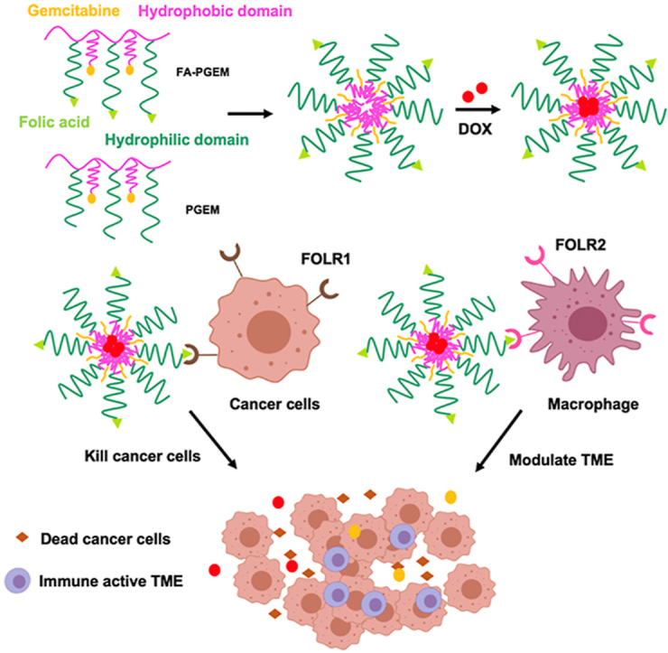
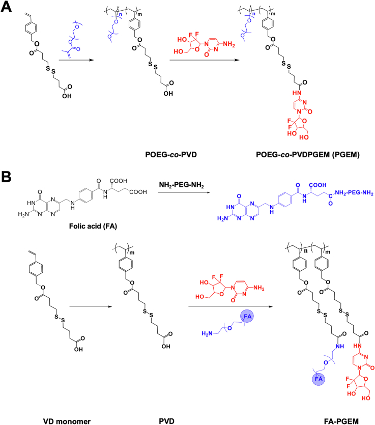
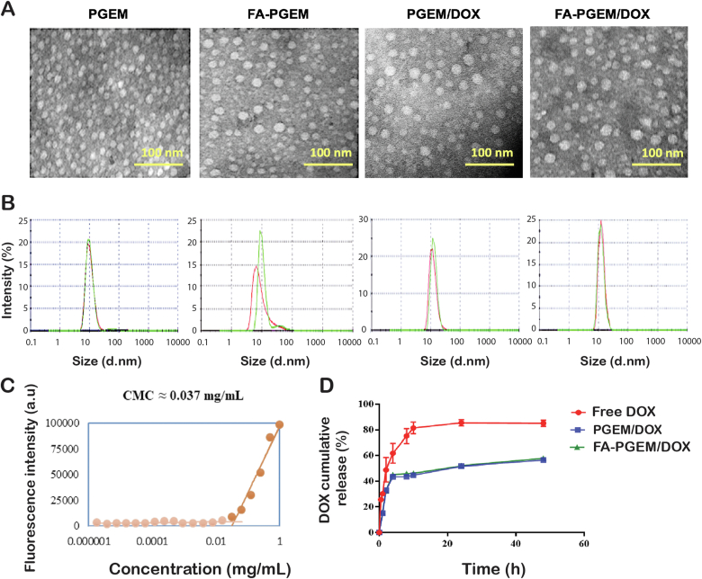
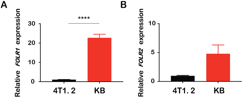
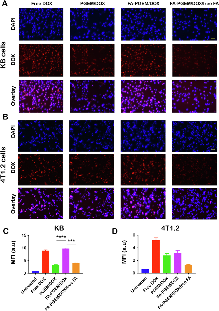
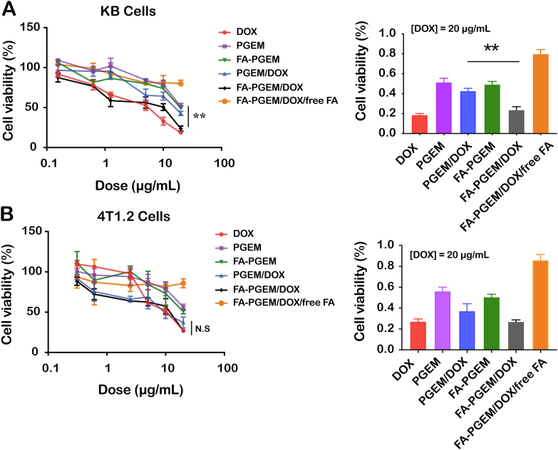
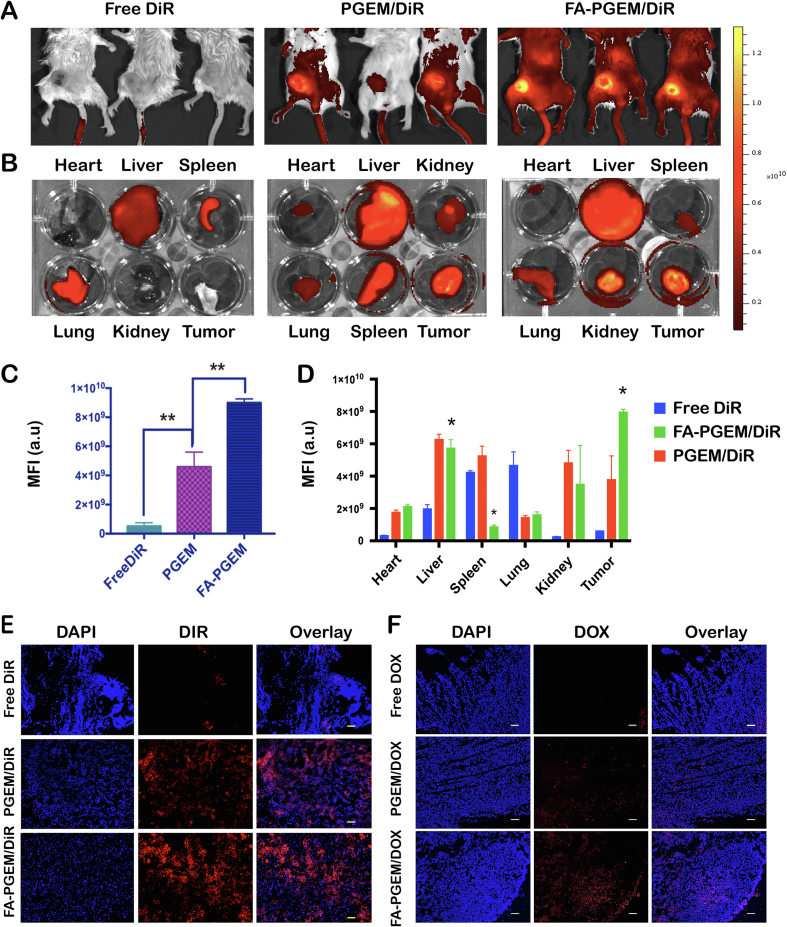
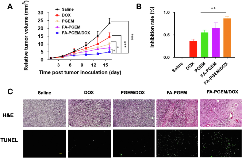
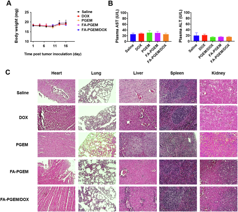
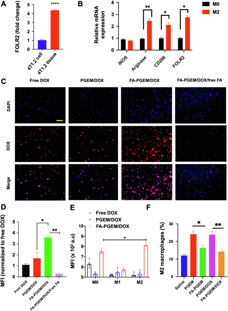
Combination of passive targeting with active targeting is a promising approach to improve the therapeutic efficacy of nanotherapy. However, most reported polymeric systems have sizes above 100 nm, which limits effective extravasation into tumors that are poorly vascularized and have dense stroma. This will, in turn, limit the overall effectiveness of the subsequent uptake by tumor cells via active targeting. In this study, we combined the passive targeting via ultra-small-sized gemcitabine (GEM)-based nanoparticles (NPs) with the active targeting provided by folic acid (FA) conjugation for enhanced dual targeted delivery to tumor cells and tumor-associated macrophages (TAMs). We developed an FA-modified prodrug carrier based on GEM (PGEM) to load doxorubicin (DOX), for co-delivery of GEM and DOX to tumors. The co-delivery system showed small particle size of ∼10 nm in diameter. The ligand-free and FA-targeted micelles showed comparable drug loading efficiency and a sustained DOX release profile. The FA-conjugated micelles effectively increased DOX uptake in cultured KB cancer cells that express a high level of folate receptor (FR), but no obvious increase was observed in 4T1.2 breast cancer cells that have a low-level expression of FR. Interestingly, in vivo, systemic delivery of FA-PGEM/DOX led to enhanced accumulation of the NPs in tumor and drastic reduction of tumor growth in a murine 4T1.2 breast cancer model. Mechanistic study showed that 4T1.2 tumor grown in mice expressed a significantly higher level of FOLR2, which was selectively expressed on TAMs. Thus, targeting of TAM may also contribute to the improved in vivo targeted delivery and therapeutic efficacy.

Keywords: Breast cancer; Doxorubicin; Dual targeting; Folic acid; Gemcitabine; Polymeric micelles; Tumor associated macrophages; Ultrasmall nanocarrier.


Figures

Image 1
Graphical abstract
Scheme 1
Scheme 1
Synthetic schemes for PGEM and FA-GEM polymers. (A) The polymer backbone POEG-co-PVD was synthesized via RAFT polymerization, followed by post conjugation with gemcitabine via EDC/HOBT coupling reaction. (B) The polymer backbone PVD was synthesized via RAFT polymerization, followed by post conjugation with gemcitabine and folic acid-modified PEG.
Scheme 2
Scheme 2
(A) Self-assembly of DOX-loaded FA-PGEM micelles. (B) FA-PGEM micelles for dual targeting of both tumor cells and M2-like TAMs. Folate receptors FOLR1 and FOLR2 are highly expressed on tumor cells and macrophages, respectively. FA-PGEM micellar carrier could enhance DOX delivery to both tumor cells and M2-like TAMs through folate-mediated active targeting effect.
Figure 1
Figure 1
In vitro biophysical characterizations of PGEM and FA-PGEM micelles. (A) TEM images of blank and DOX-loaded PGEM and FA-PGEM micelles (scale bar, 100 nm). (B) DLS analysis of micelles (carrier:drug = 10:1, w/w). (C) CMC measurement of FA-PGEM micelles by using nile red as a fluorescent probe. (D) DOX release profiles of DOX-loaded PGEM and FA-PGEM micelles with free DOX as the control. PBS was used as the release medium. Data are presented as means ± SEM (n = 3).
Figure 2
Figure 2
FOLR1 (A) and FOLR2 (B) mRNA expression levels in 4T1.2 and KB cells analyzed by quantitative real-time PCR. Relative mRNA levels were determined by the ΔΔCt method using GAPDH for internal cross-normalization. Data are presented as means ± SEM (n = 3). ∗∗∗∗P < 0.0001.
Figure 3
Figure 3
Cellular uptake of various DOX formulations. Fluorescence microscopic images of KB (A) and 4T1.2 (B) cells treated with various formulations, in the presence or absence of free folate at 37 °C for 30 min. DOX concentration was kept at 6 μL/mL and nuclei were stained with DAPI (scale bar = 50 μm); DOX uptake in KB (C) and 4T1.2 (D) cells was quantified by flow cytometry (MFI = mean fluorescence intensity, n = 3). ∗∗∗P < 0.001 ∗∗∗∗P < 0.0001.
Figure 4
Figure 4
In vitro cytotoxicity of different formulations against (A) KB and (B) 4T1.2 cells. Cells were treated with DOX, PGEM, PGEM/DOX, FA-PGEM, FA-PGEM/DOX, and FA-PGEM/DOX in the presence of free folate for 30 min, followed by incubation in drug-free medium for another 24 h. Cytotoxicity was analyzed by MTT assay. Shown in right panels are the cell viabilities of KB and 4T1.2 cells after various treatments at an equivalent DOX dose of 20 μg/mL. Data are presented as means ± SEM (n = 3) ∗∗P < 0.01.
Figure 5
Figure 5
Tissue biodistribution of various formulations in 4T1.2 tumor-bearing mice. Whole body near-infrared images (A) and ex vivo fluorescence imaging (B) of tumors and other major organs 24 h following treatment with free DiR, PGEM/DiR and FA-PGEM/DiR, respectively. (C and D) Quantitative analysis (fluorescence intensity) of the data shown in panels A and B, respectively. (E) DiR accumulation in 4T1.2 tumor sections at 24 h after injection of free DiR, PGEM/DiR, and FA-PGEM/DiR, respectively. (F) DOX accumulation in 4T1.2 tumor sections at 24 h following i.v. administration of free DOX, PGEM/DOX and FA-PGEM/DOX, respectively (magnification, 40×; Scale bar = 50 μm; DOX dose, 5 mg/kg).
Figure 6
Figure 6
In vivo evaluation of anti-tumor efficacy of various formulations. (A) Relative tumor growth curves of various formulations in 4T1.2 tumor model followed every three days for 16 days. DOX dose, 5 mg/kg. Polymer to drug ratio, 20:1 (mg/mg); (B) Inhibition rates of various treatments at the completion of study (Day 16); (C) Representative photomicrographs of H&E staining (upper) and TUNEL fluorescence staining (bottom) of tumor sections after various treatments (scale bar = 50 μm). The results are presented as means ± SEM (n = 5). ∗P < 0.05, ∗∗P < 0.01, ∗∗∗P < 0.001.
Figure 7
Figure 7
Safety/toxicity evaluations of various treatments. (A) Body weights of mice monitored every three days after various treatments; (B) Liver function assays after various treatments. Results were expressed as the mean ± SEM (n = 5); (C) Representative photomicrographs of H&E staining of heart, lung, liver, spleen and kidney (Magnification 40×, scale bar = 50 μm).
Figure 8
Figure 8
FR-mediated targeting of FA-PGEM NPs to M2 cells in vitro and in vivo. (A) FOLR2 mRNA expression levels in cultured 4T1.2 cells and 4T1.2 tumor tissues; (B) mRNA expression levels of iNOS, Arginase, CD206, and FOLR2 in M2 polarized cells as compared to M0 cells; (C) Fluorescence microscopic images of M2 cells 30 min following treatment with DOX and DOX-loaded PGEM and FA-GEM micelle, in the presence or absence of free folate at 37 °C. DOX concentration was maintained at 6 μL/mL and nuclei were stained with DAPI (Magnification 40×, scale bar = 50 μm). (D) Quantitative analysis (mean fluorescence intensity) of the data in panel C. (E) Flow cytometric analysis of DOX uptake by M0, M1 or M2 cells 30 min following treatment with various DOX formulations. (F) Flow cytometric analysis of the percentage of M2 macrophages in the tumor tissues treated with various formulations (3 dosages). Results are expressed as the mean ± SEM (n = 3); ∗P < 0.05 ∗∗P < 0.01.

Similar articles

Cited by

References

    1. Chauhan V.P., Jain R.K. Strategies for advancing cancer nanomedicine. Nat Mater. 2013;12:958–962. - PMC - PubMed
    1. Cabral H., Matsumoto Y., Mizuno K., Chen Q., Murakami M., Kimura M., et al. Accumulation of sub-100 nm polymeric micelles in poorly permeable tumours depends on size. Nat Nanotechnol. 2011;6:815. - PubMed
    1. Pluen A., Boucher Y., Ramanujan S., McKee T.D., Gohongi T., di Tomaso E., et al. Role of tumor–host interactions in interstitial diffusion of macromolecules: cranial vs. subcutaneous tumors. Proc Natl Acad Sci U S A. 2001;98:4628–4633. - PMC - PubMed
    1. Park J., Choi Y., Chang H., Um W., Ryu J.H., Kwon I.C. Alliance with EPR effect: combined strategies to improve the EPR effect in the tumor microenvironment. Theranostics. 2019;9:8073. - PMC - PubMed
    1. Hrkach J., Von Hoff D., Ali M.M., Andrianova E., Auer J., Campbell T., et al. Preclinical development and clinical translation of a PSMA-targeted docetaxel nanoparticle with a differentiated pharmacological profile. Sci Transl Med. 2012;4 128ra39. - PubMed