. 2022 Mar;12(3):1390-1405.
doi: 10.1016/j.apsb.2021.10.024. Epub 2021 Oct 30.

Inhibition of the CDK9-cyclin T1 protein-protein interaction as a new approach against triple-negative breast cancer

Affiliations

Inhibition of the CDK9-cyclin T1 protein-protein interaction as a new approach against triple-negative breast cancer

Sha-Sha Cheng et al. Acta Pharm Sin B. 2022 Mar.

Abstract

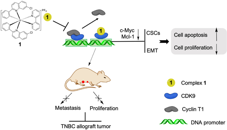
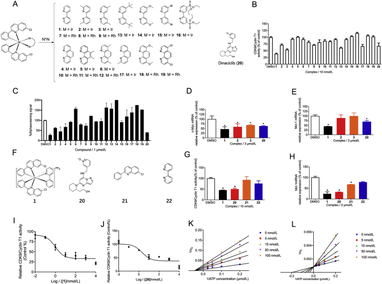
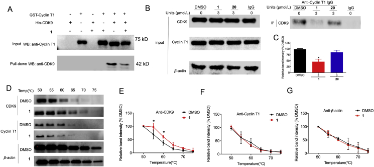
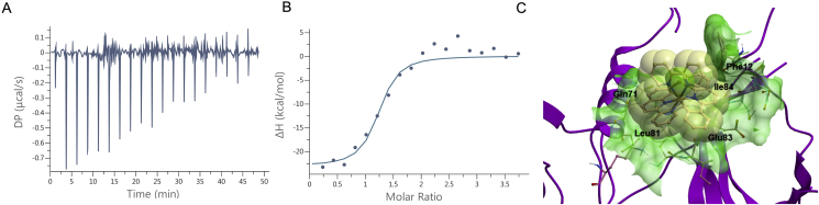
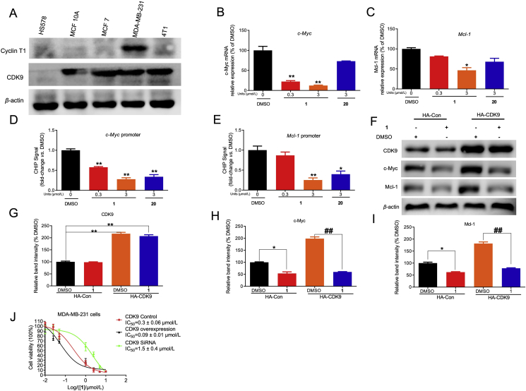
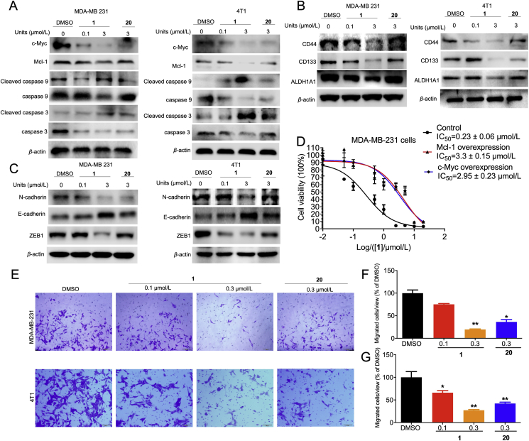
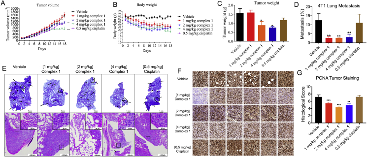
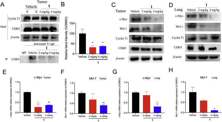
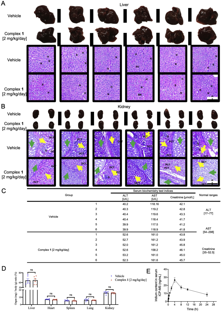
Cyclin-dependent kinase 9 (CDK9) activity is correlated with worse outcomes of triple-negative breast cancer (TNBC) patients. The heterodimer between CDK9 with cyclin T1 is essential for maintaining the active state of the kinase and targeting this protein-protein interaction (PPI) may offer promising avenues for selective CDK9 inhibition. Herein, we designed and generated a library of metal complexes bearing the 7-chloro-2-phenylquinoline CˆN ligand and tested their activity against the CDK9-cyclin T1 PPI. Complex 1 bound to CDK9 via an enthalpically-driven binding mode, leading to disruption of the CDK9-cyclin T1 interaction in vitro and in cellulo. Importantly, complex 1 showed promising anti-metastatic activity against TNBC allografts in mice and was comparably active compared to cisplatin. To our knowledge, 1 is the first CDK9-cyclin T1 PPI inhibitor with anti-metastatic activity against TNBC. Complex 1 could serve as a new platform for the future design of more efficacious kinase inhibitors against cancer, including TNBC.

Keywords: Epigenetics; Kinase inhibitor; Metal complex; Metastasis; Protein–protein interaction; Triple-negative breast cancer.


Figures

Image 1
Graphical abstract
Figure 1
Figure 1
The in cellulo and in vitro activity of complex 1 and selected complexes and ligands. (A) The chemical structures of the Ir(III) and Rh(III) complexes 119 and positive control 20 (dinaciclib) evaluated in this study. (B) Effects of 120 on CDK9/cyclin T1 activity were determined by a chemiluminescence assay. (C) Inhibition of the CDK9–cyclin T1 PPI by 120 as measured using the AlphaScreen assay (C). (D) c-Myc mRNA and (E) Mcl-1 mRNA levels by compounds (3 μmol/L) in MDA-MB-231 cells as measured by qPCR assay. (F) Structure of 1, 20, and ligands 21, 22. (G) Effect of compounds and ligands (10 nmol/L) on CDK9–cyclin T1 activity. (H) Effect of compounds and ligands (3 μmol/L) on Mcl-1 mRNA levels. (I) 1 inhibits CDK9–cyclin T1 activity in a dose-dependent manner. (J) 20 inhibits CDK9–cyclin T1 activity in a dose-dependent manner. (K) Double-reciprocal plot of initial rate against ATP concentration in the presence of complex 1 at 0, 5, 15, 50, 100 nmol/L. (L) Double-reciprocal plot of initial rate against ATP concentration in the presence of compound 20 at 0, 5, 15, 50, 100 nmol/L. ∗P < 0.05 compared to DMSO group by the t-test.
Figure 2
Figure 2
Complex 1 disrupts the interaction between CDK9 and cyclin T1 by targeting CDK9. (A) GST pull-down assay of cyclin T1 and CDK9. GST-tag cyclin T1 protein was immobilized in the gravity flow column and then incubated with 300 μL CDK9 and/or complex 1. Eluted samples were analyzed by Western blotting. (B) The CDK9–cyclin T1 PPI was examined by co-IP in MDA-MB-231 cells. MDA-MB-231 cells were treated with DMSO and 3 μmol/L 1 or 20 for 4 h, and cell lysates were subject to immunoprecipitation with the indicated antibody. (C) Relative densitometry analysis of CDK9. (D) MDA-MB-231 lysates were treated with 3 μmol/L compound 1 or DMSO for 30 min, followed by CETSA analysis to evaluate CDK9, cyclin T1, and β-actin stabilization. (E–G) Densitometry analysis of CDK9, cyclin T1, and β-actin content in the soluble fraction. ∗P < 0.05 compared to DMSO group by the t-test.
Figure 3
Figure 3
The binding model of the complex 1 against the CDK9–cyclin T1 PPI. (A–B) ITC analysis of the 1–CDK9 interaction. (A) Raw data for 19 sequential injections (2 μL per injection) of a solution of 1 (100 μmol/L) into a solution of CDK9 (5 μmol/L). (B) “Net” heat effects obtained by subtracting the dilution heat from the reaction heat, which was fitted by using the “one set of sites” binding model. N (sites) = 1.17 ± 0.05; Kd = 134 ± 68.2 nmol/L; ΔG = −9.38 kcal/mol; ΔH = −23.2 ± 1.4 kcal/mol; −TΔS = 13.4 kcal/mol. (C) Binding model of complex 1 with CDK9 (PDB code: 3BLH) which is depicted as a space-filling representation showing carbon (yellow), nitrogen (blue), chlorine (green) atoms. The binding pocket of the CDK9 is represented as a translucent green surface.
Figure 4
Figure 4
The cytotoxicity of complex 1 in CDK9 overexpressed breast cancer cells. (A) The CDK9 protein level was evaluated in different breast cancer cell lines (MCF7, 4T1, MDA-MB-231) and human normal breast cell lines (MCF 10A, HS578). CDK9, cyclin T1, and β-actin were blotted to control for total protein levels. (B–C) Inhibition effects of 1 or 20 on the c-Myc and Mcl-1 mRNA level in MDA-MB-231 cells were determined by qPCR. (D–E) Inhibition effect of 1 or 20 on the binding ability of CDK9 to c-Myc and Mcl-1 promoter in MDA-MB-231 cells by ChIP-qPCR. Indicated concentrations of 1 or 20 treated with cells for 4 h. Then cells were collected to perform ChIP-qPCR. (F) CDK9 plasmid treatment led to efficient overprotein level in MDA-MB-231 cells. CDK9, c-Myc, Mcl-1, and β-actin were blotted to control for total protein levels. (G–I) Relative densitometry analysis of CDK9 (G), c-Myc (H), and Mcl-1 (I). (J) The cytotoxicity effect of 1 on control, CDK9 knockdown, and CDK9 overexpressing MDA-MB-231 cells. 1 inhibited the growth of on control, CDK9 knockdown, overexpression-treatment MDA-MB-231 cells with an IC50 value of 0.3, 1.5 and 0.09 μmol/L, respectively. Data are presented as the mean ± SD. ∗P < 0.05, ∗∗P < 0.01 vs DMSO group, ##P < 0.01 compared to DMSO group by the t-test.
Figure 5
Figure 5
Complex 1 induced cell apoptosis and inhibited cell migration through inhibition of c-Myc and Mcl-1 protein level. (A) MDA-MB-231 and 4T1 cells were treated with the indicated concentrations of 1 or 20 treated for 4 h. Cell lysates were collected and immunoblotted with the anti-apoptosis corresponding antibodies. (B–C) The effect of 1 on CSC (B) and EMT (C) biomarkers levels in MDA-MB-231 and 4T1 cells. DMSO or indicated concentrations of 1 or 20 were treated with MDA-MB-231 and 4T1 cells for 4 h. Then cells were collected and incubated with appropriate antibodies. (D) The anti-proliferative effect of 1 in MDA-MB-231 cells after transfection with c-Myc and Mcl-1. Cells were transfected with c-Myc, Mcl-1, or control plasmid for 48 h, then treated with DMSO or 1 for 48 h. Cell viability was detected by the MTT assay. (E) 1 inhibited the migration of TNBC cell lines. Indicated concentrations of 1 or 20 treated with MDA-MB-231 and 4T1 cells for 24 h and evaluated by transwell. (F–G) Quantitative analysis of migration cells. Data are presented as the mean ± SD. ∗P < 0.05, ∗∗P < 0.01 compared to DMSO group by the t-test.
Figure 6
Figure 6
Complex 1 suppresses tumor growth, lung metastasis, and proliferation in a TNBC mouse model. Female BALB/c mice were subcutaneously inoculated with 1.0 × 106 4T1 cells, and after the tumor volume reached 60 mm3, injected intraperitoneally daily with 1 or 2 or 4 mg/kg of complex 1 or 0.5 mg/kg of cisplatin for 18 days. (A) Tumor volume of 4T1 tumor-bearing mice after control (vehicle), 1 (1, 2 or 4 mg/kg) and cisplatin (0.5 mg/kg) treatment (n = 6–8). (B) Body weight of tumor-bearing mice over the course of treatment. (C) Comparison of tumor weight after sacrifice. (D) The percentage of metastatic burden area. (E) Representative photograph images of lung metastatic tissues and H&E stained lung sections in vehicle control, complex 1, and cisplatin groups after 18 days. Scale bar = 200 μm (×40). (F) Six random PCNA-stained areas of a single tumor from each group are shown at ×40 magnification. (G) PCNA histological score is displayed. Six random areas of each tumor section are enrolled, and each dot represents the score of one area. ∗P < 0.05, ∗∗P < 0.01, ∗∗∗P < 0.001 compared to vehicle by the t-test.
Figure 7
Figure 7
1 suppressed the CDK9–cyclin T1 PPI and decreased c-Myc and Mcl-1 levels in vivo. (A–B) The CDK9–cyclin T1 PPI was examined by co-IP in 2 mg/kg and 4 mg/kg of complex 1-treated mice. (C–D) Protein levels of c-Myc, Mcl-1, cyclin T1, CDK9, and β-actin in tumor and lung tissues treated with vehicle or 2 and 4 mg/kg of 1 were measured by Western blotting. (E–H) 1 impaired c-Myc and Mcl-1 transcription in tumor and lung tissues. Data are represented as mean ± SD. ∗P < 0.05, ∗∗P < 0.01 vs Vehicle group, using Student's t-test.
Figure 8
Figure 8
The toxicity and pharmacokinetic profiles of complex 1. Twelve female mice were randomly divided into two groups and were treated with 1 (2 mg/kg/day) for 18 days. Histological examination of liver and kidney injury. (A) Liver specimens and corresponding H&E staining images indicated the hepatic architecture with nucleus (N), hepatocytes (H), and sinusoids (S) in both vehicle and 1 (2 mg/kg/day) treated group. None of significant hepatic centrilobular mononuclear cell infiltration, fatty changes, nor hepatic cell necrosis were observed, except for sporadic binucleated hepatocytes (BN) in both groups. Scale bar = 80 μm (×10). (B) Kidney specimens and corresponding H&E staining images showed the histological structure with glomerulus (G), proximal convoluted tubules (PCT), and distal convoluted tubules (DCT) in both vehicle and 1 (2 mg/kg/day) treated group. Yellow arrows indicate the renal medulla; green arrows indicate renal pelvis. None pyknotic cells, distal tubular necrosis, sloughing of tubular epithelial cells, nor proximal hemorrhage in interstitial tissue were identified. Scale bar = 40 μm (×20). (C) Serum alanine transaminase (ALT), aspartate transaminase (AST), creatinine levels, and corresponding normal ranges of vehicle and 1 (2 mg/kg/day) treated mice. (D) Normalized organ weights as percentages of body weight. Mice given vehicle and 1 (2 mg/kg/day) were sacrificed at Day 18, the normalized ratio were calculated: organs weight divided by body weight as mg/g, not significant (ns). (E) Pharmacokinetic profile of 1 at different time points. BALB/c mice were intraperitoneally injected with 1 (2 mg/kg/day). The concentrations of 1 in the serum were measured at 0, 0.08, 0.17, 0.25, 0.5, 1, 2, 4, 8, 12, and 24 h. All data were mean ± SEM from 6 mice per treatment group. Significance was determined using one-way ANOVA analysis.

Similar articles

Cited by

References

    1. Lee K.L., Kuo Y.C., Ho Y.S., Huang Y.H. Triple-negative breast cancer: current understanding and future therapeutic breakthrough targeting cancer stemness. Cancers. 2019;11:1344–1380. - PMC - PubMed
    1. Cheng S.S., Yang G.J., Wang W.H., Ma D.L., Leung C.H. Discovery of a tetrahydroisoquinoline-based CDK9‒cyclin T1 protein–protein interaction inhibitor as an anti-proliferative and anti-migration agent against triple-negative breast cancer cells. Genes Dis. 2021 https://www.sciencedirect.com/science/article/pii/S2352304221000866 Available from: - PMC - PubMed
    1. Yang G.J., Wang W., Mok S.W.F., Wu C., Law B.Y.K., Miao X.M., et al. Selective inhibition of lysine-specific demethylase 5A (KDM5A) using a rhodium (III) complex for triple-negative breast cancer therapy. Angew Chem Int Ed. 2018;57:13091–13095. - PubMed
    1. Al-Mahmood S., Sapiezynski J., Garbuzenko O.B., Minko T. Metastatic and triple-negative breast cancer: challenges and treatment options. Drug Deliv Transl Res. 2018;8:1483–1507. - PMC - PubMed
    1. McLaughlin R.P., He J., van der Noord V.E., Redel J., Foekens J.A., Martens J.W.M., et al. A kinase inhibitor screen identifies a dual cdc7/CDK9 inhibitor to sensitise triple-negative breast cancer to EGFR-targeted therapy. Breast Cancer Res. 2019;21:77–92. - PMC - PubMed

LinkOut - more resources