. 2022 Mar;12(3):1271-1287.
doi: 10.1016/j.apsb.2021.09.007. Epub 2021 Sep 11.

Targeting PFKL with penfluridol inhibits glycolysis and suppresses esophageal cancer tumorigenesis in an AMPK/FOXO3a/BIM-dependent manner

Affiliations

Targeting PFKL with penfluridol inhibits glycolysis and suppresses esophageal cancer tumorigenesis in an AMPK/FOXO3a/BIM-dependent manner

Cancan Zheng et al. Acta Pharm Sin B. 2022 Mar.

Abstract

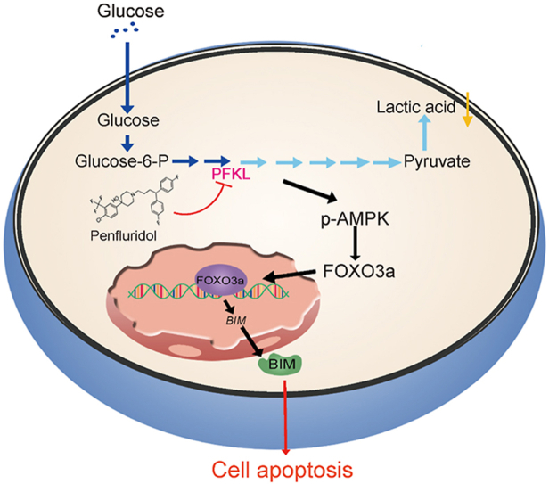
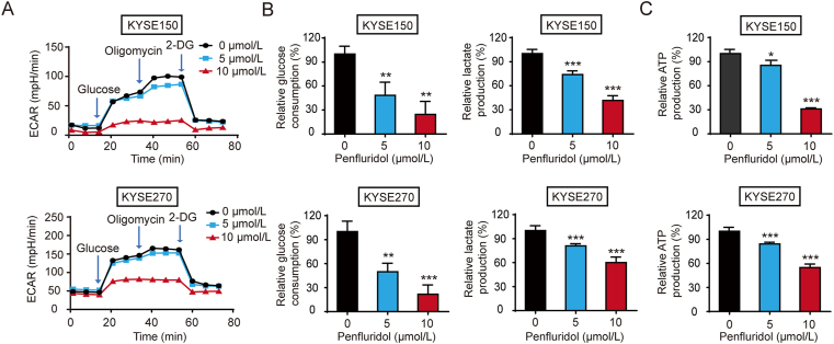
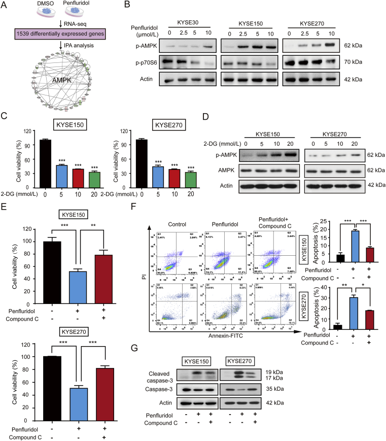
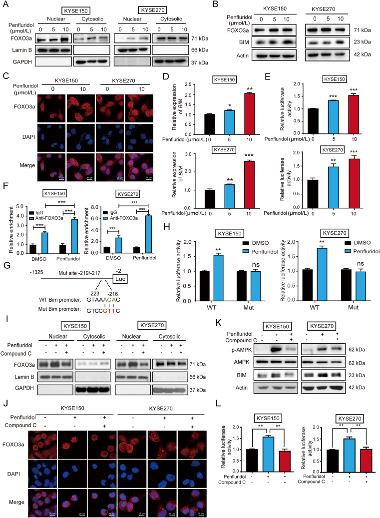
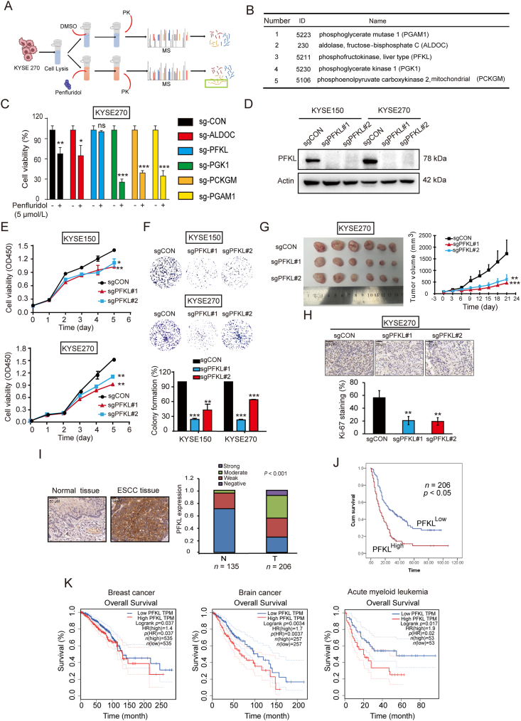
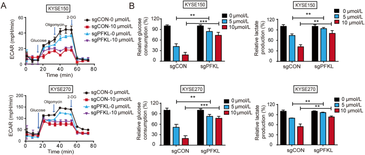
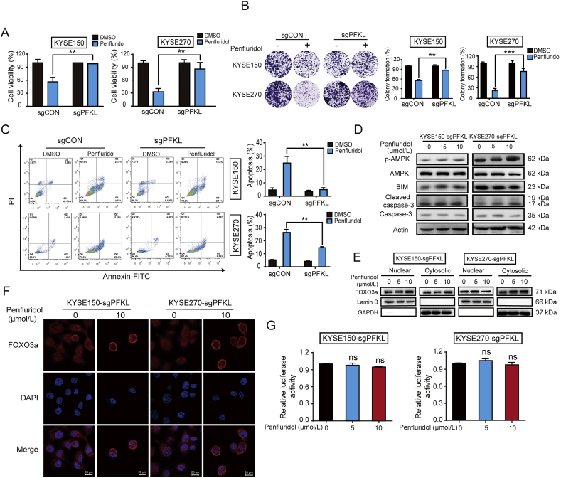
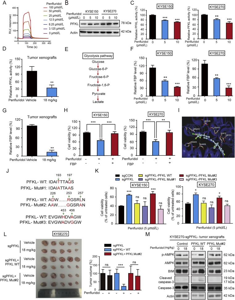
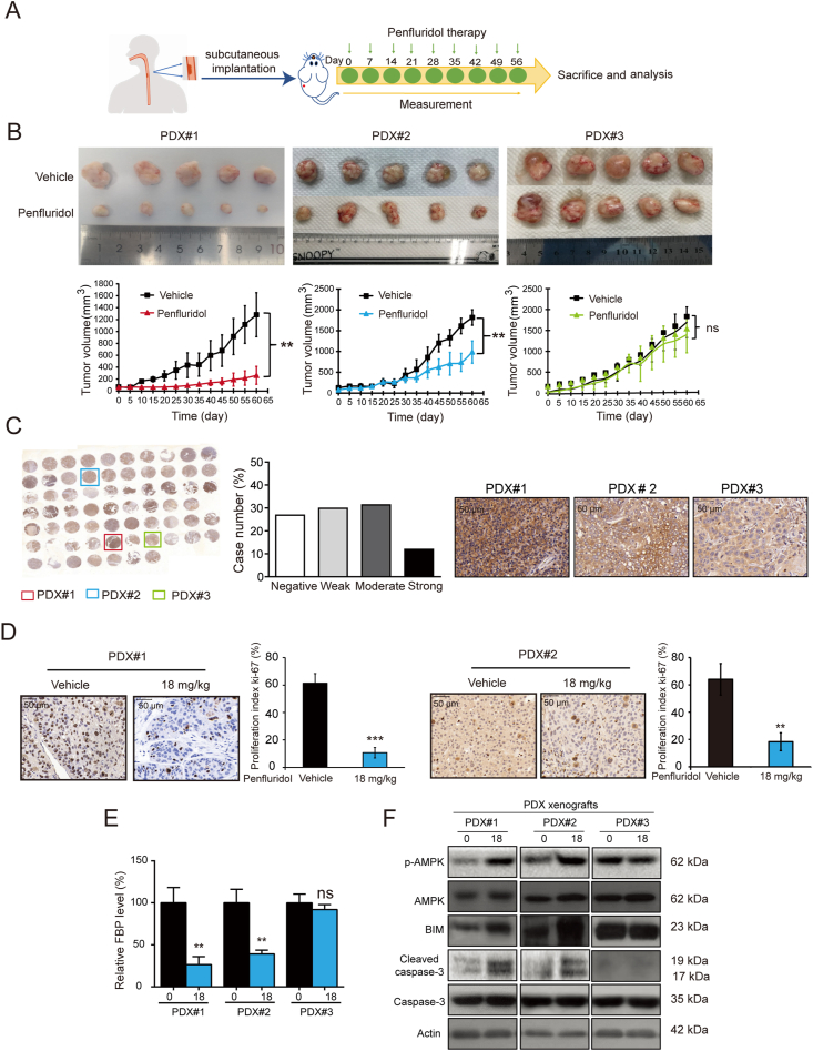
As one of the hallmarks of cancer, metabolic reprogramming leads to cancer progression, and targeting glycolytic enzymes could be useful strategies for cancer therapy. By screening a small molecule library consisting of 1320 FDA-approved drugs, we found that penfluridol, an antipsychotic drug used to treat schizophrenia, could inhibit glycolysis and induce apoptosis in esophageal squamous cell carcinoma (ESCC). Gene profiling and Ingenuity Pathway Analysis suggested the important role of AMPK in action mechanism of penfluridol. By using drug affinity responsive target stability (DARTS) technology and proteomics, we identified phosphofructokinase, liver type (PFKL), a key enzyme in glycolysis, as a direct target of penfluridol. Penfluridol could not exhibit its anticancer property in PFKL-deficient cancer cells, illustrating that PFKL is essential for the bioactivity of penfluridol. High PFKL expression is correlated with advanced stages and poor survival of ESCC patients, and silencing of PFKL significantly suppressed tumor growth. Mechanistically, direct binding of penfluridol and PFKL inhibits glucose consumption, lactate and ATP production, leads to nuclear translocation of FOXO3a and subsequent transcriptional activation of BIM in an AMPK-dependent manner. Taken together, PFKL is a potential prognostic biomarker and therapeutic target in ESCC, and penfluridol may be a new therapeutic option for management of this lethal disease.

Keywords: DARTS technology; Drug repurposing; Esophageal cancer; Glycolysis; Metabolic reprogramming; PFKL; Penfluridol.


Figures

Image 1
Graphical abstract
Figure 1
Figure 1
Penfluridol inhibits esophageal squamous cell carcinoma (ESCC) cell proliferation and tumor growth in vitro and in vivo. (A) Schematic diagram showing the approach to screen the anti-tumor drugs from the US Food and Drug Administration (FDA)-approved small molecule library. (B) KYSE270 cells were treated with 1320 drugs (10 μmol/L) individually for 48 h and subjected to WST-1 assay. (C) Cell viability of KYSE30, KYSE150, KYSE270 cells treated with indicated concentrations of penfluridol (up to 10 μmol/L) was determined. (D) Flow cytometry analysis showing the apoptosis of KYSE30, KYSE150 and KYSE270 cells in the presence of penfluridol. (E) The expression levels of caspase-3 and cleaved caspase-3 were detected in ESCC cells treated with penfluridol. (F) Mice bearing KYSE30-and KYSE270-derived tumor xenografts were given penfluridol (18 mg/kg) once a week (n = 6). (G) Penfluridol treatment significantly inhibited Ki-67 proliferation index in KYSE30 and KYSE270 tumor xenografts (n = 3). (H) Comparison of cleaved caspase-3 and caspase-3 expression in the KYSE30 and KYSE270 tumor xenografts treated with penfluridol or vehicle by Western blotting. Data are mean ± SD; ∗P < 0.05; ∗∗P < 0.01; ∗∗∗P < 0.001 compared to control group.
Figure 2
Figure 2
Penfluridol inhibits glycolysis in ESCC cells. (A) Extracellular acidification rate (ECAR) was measured by Seahorse XFe96 analyzer in KYSE150 and KYSE270 cells. (B) and (C) The glucose consumption, and production of lactate and ATP were measured in the presence or absence of penfluridol. Data are mean ± SD, n = 3; ∗P < 0.05; ∗∗P < 0.01; ∗∗∗P < 0.001 compared to control group.
Figure 3
Figure 3
Penfluridol activates AMP-activated protein kinase (AMPK) signaling in ESCC cells. (A) Ingenuity pathway analysis (IPA) suggesting AMPK pathway may be involved in the action mechanism of penfluridol in cancer cells. (B) Western blotting analysis of p-AMPK, and p-p70S6 expression in the KYSE30, KYSE150 and KYSE270 cells treated with different concentrations of penfluridol. (C) and (D) KYSE150 and KYSE270 cells were treated with 2-deoxy-D-glucose (2-DG) (0, 5, 10, and 20 mmol/L) for 24 h, WST-1 assay was taken to measure cell viability (C), and Western blotting was performed to detect expression levels of p-AMPK and AMPK (D). (E)–G) KYSE150 and KTSE270 cells were treated with penfluridol (5 μmol/L) for 48 h in the presence or absence of compound C (0.5 μmol/L), WST-1 assay was taken to measure cell viability (E), flow cytometry analysis was used to determine cell apoptosis (F), and Western blotting was performed to detect expression levels of caspase-3 and cleaved caspase-3 (G). Data are mean ± SD, n = 3; ∗P < 0.05; ∗∗P < 0.01; ∗∗∗P < 0.001 compared to control group.
Figure 4
Figure 4
The AMPK/FOXO3a/BIM regulatory axis mediates the anticancer effect of penfluridol. (A) Western blotting analysis of FOXO3a expression in the nuclear and cytoplasmic extracts of ESCC cells, with lamin B and GAPDH as internal controls. (B) KYSE150 and KYSE270 cells were exposed to penfluridol for 24 h, and Western blotting was performed to detect FOXO3a and BIM expression. (C) Immunofluorescent staining showing the subcellular localization of FOXO3a in the presence or absence of penfluridol. (D) The mRNA expression of BIM was detected by quantitative real-time polymerase chain reaction (qRT-PCR). (E) The dual luciferase reporter assay showing the effect of penfluridol on luciferase activity of BIM promoter in KYSE150 and KYSE270 cells. (F) Chromatin immunoprecipitation (ChIP) assay was used to verify the binding of FOXO3a to BIM promoter. (G) The diagram illustrating the mutation design of BIM promoter construct. (H) Dual luciferase reporter assay was used to detect the effect of penfluridol on activity of BIM promoter in ESCC cells transfected with plasmid expressing wild-type or mutated BIM promoter. (I)–(L) KYSE150 and KYSE270 cells were treated with penfluridol in the presence or absence of compound C, subcellular distribution of FOXO3a (I), as well as expression levels of p-AMPK, AMPK and BIM, were determined by Western blotting (K), the subcellular localization of FOXO3a was detected by immunofluorescence (J), and the luciferase activity of BIM promoter was measured by dual luciferase reporter (L). Data are mean ± SD, n = 3; ns, no significance; ∗P < 0.05; ∗∗P < 0.01; ∗∗∗P < 0.001 compared to control group.
Figure 5
Figure 5
High phosphofructokinase, liver type (PFKL) expression contributes to tumorigenesis and correlates with poor prognosis in ESCC. (A) Schematic diagram showing the drug affinity response target stability (DARTS) technology for identification of the target proteins of penfluridol. (B) Potential target proteins of PFKL identified by DARTS and mass spectrometry. (C) The cell viabilites of penfluridol-treated ESCC cells were determined when the potential target proteins were silenced, including fructose-bisphosphate C (ALDOC), PFKL, phosphoglycerate (PGK1), PCK2 and phosphoglycerate mutase 1 (PGAM1). (D) Successful knockout of PFKL in KYSE150 and KYSE270 cells. (E) and (F) WST-1 and colony formation assays showing the effect of PFKL silencing on ESCC cell proliferation. (G) Tumor growth curves showing the effect of PFKL knockout on growth of ESCC tumor xenografts (n = 6). (H) Immunohistochemistry was performed to compare the Ki-67 proliferation index in the tumors among groups (n = 3). (I) Representative images of PFKL staining in ESCC tumor and nontumor tissues. (J) Kaplan–Meier analysis showing the suivival of 206 ESCC patients based on tumor PFKL expression. (K) Tumor PFKL expression is correlated with the survival of patients with breast cancer, brain cancer and acute myeloid leukemia. Data are mean ± SD; ns, no significance; ∗P < 0.05; ∗∗P < 0.01; ∗∗∗P < 0.001 compared to control group.
Figure 6
Figure 6
PFKL is important for the function of penfluridol in inhibiting glycolysis. (A) ECAR was measured in the PFKL-deficient KYSE150 and KYSE270 cells treated with penfluridol. (B) Comparison of glucose consumption and lactate production in PFKL-knockout cells exposed to penfluridol. Data are mean ± SD, n = 3; ∗∗P < 0.01; ∗∗∗P < 0.001 compared to control group.
Figure 7
Figure 7
PFKL is required for the bioactivity of penfluridol in regulating AMPK/FOXO3a/BIM signaling and tumorigenesis. (A)–(C) Cell viability assay (A), colony formation assay (B), and flow cytometry analysis (C) were performed to determine the effect of penfluridol on proliferation and apoptosis in PFKL-deficent KYSE150 and KYSE270 cells. (D)–(G) PFKL-deficient KYSE30 and KYSE270 cells were treated with penfluridol, Western blotting (D and E), immunofluorescence (F), and dual luciferase reporter assays (G) were used to detect the subcellular distribution of FOXO3a, expression levels of p-AMPK, AMPK, BIM, cleaved caspase-3, caspase-3, and luciferase activity of BIM promoter. Data are mean ± SD, n = 3; ns, no significance; ∗∗P < 0.01; ∗∗∗P < 0.001 compared to control group.
Figure 8
Figure 8
Asp-226, Arg-253, and Leu-257 sites in PFKL are the binding sites required for anticancer bioactivity of penfluridol. (A) Surface plasmon resonance (SPR) result showing that binding of PFKL protein to penfluridol. (B) Western blotting analysis of PFKL expression in KYSE150 and KYSE270 cells treated with penfluridol. (C) Penfluridol inhibited the activity of PFKL in KYSE150 and KYSE270 cells in a dose-dependent manner. (D) Penfluridol inhibited the activity of PFKL in ESCC tumor xenografts (n = 3). (E) The diagram showing the process of glycolysis. (F) and (G) Penfluridol reduced fructose 1,6-bisphosphate (FBP) level in ESCC cells (F) and tumor xenografts (n = 3) (G). (H) WST-1 assay was used to determine the cell viability in KYSE150 and KYSE270 cells exposed to penfluridiol (5 μmol/L) in the presence or absence of FBP (0.5 μmol/L) for 24 h. (I) The homology modeling structure predicting the binding sites of penfluridol on PFKL protein. (J) Schematic diagram of PFKL mutation. (K) The PFKL-deficient KYSE150 and KYSE270 cells re-overexpressed with the plasmid expressing PFKL-WT, PFKL-Mut#1, PFKL-Mut#2 or PFKL-Mut#3, respectively, were treated with penfluridol for 24 h and cell activity was detected by WST-1 assasy. (L) The pictures and tumor volume showing that the antitumor effect of penfluridol was recovered when the PFKL-deficient ESCC cells were re-overexpressed with wild-type PFKL, but not the mutant PFKL (n = 5). (M) Western blotting analysis of p-AMPK, AMPK, BIM, cleaved caspase-3, caspase-3 expression in the indicated tumor xenografts treated with penfluridol or vehicle. Data are mean ± SD; ns, no significance; ∗P < 0.05; ∗∗P < 0.01; ∗∗∗P < 0.001 compared to control group.
Figure 9
Figure 9
Penfluridol exerts antitumor activity in the Patient-derived xenograft (PDX) models with high PFKL expression. (A) and (B) Three patient-derived xenografts were established in immunodeficient mice, and the mice were given penfluridol (18 mg/kg) or vehicle by oral administration weekly. Penfluridol significantly inhibited the growth of PDX#1 and PDX#2 tumors, but has no effect on PDX#3 (n = 5). (C) The expression of PFKL was examined by immunohistochemistry in a tissue microarray consisting of 67 cases ESCC tissues. (D) Quantification of Ki-67 proliferaton index in PDX#1 and PDX#2 tumors (n = 3). (E) FBP level in PDX tumors among groups (n = 3). (F) Western blot showing the expression of p-AMPK, AMPK, BIM, cleaved caspase-3 and caspase-3 in PDX tumors. Data are mean ± SD; ns, no significance; ∗∗P < 0.01; ∗∗∗P < 0.001 compared to control group.
Figure 10
Figure 10
Schematic diagram summarizing how penfluridol suppresses glycolysis and tumorigenesis.

Similar articles

Cited by

References

    1. Morandi A., Indraccolo S. Linking metabolic reprogramming to therapy resistance in cancer. Biochim Biophys Acta Rev Cancer. 2017;1868:1–6. - PubMed
    1. Koppenol W.H., Bounds P.L., Dang C.V. Otto Warburg's contributions to current concepts of cancer metabolism. Nat Rev Cancer. 2011;11:325–337. - PubMed
    1. Feng Y., Zhang Y., Cai Y., Liu R., Lu M., Li T., et al. A20 targets PFKL and glycolysis to inhibit the progression of hepatocellular carcinoma. Cell Death Dis. 2020;11:89. - PMC - PubMed
    1. Zhao Y., Hu X., Liu Y., Dong S., Wen Z., He W., et al. ROS signaling under metabolic stress: cross-talk between AMPK and AKT pathway. Mol Cancer. 2017;16:79. - PMC - PubMed
    1. Gwinn D.M., Shackelford D.B., Egan D.F., Mihaylova M.M., Mery A., Vasquez D.S., et al. AMPK phosphorylation of raptor mediates a metabolic checkpoint. Mol Cell. 2008;30:214–226. - PMC - PubMed