Review
. 2022 Apr 15;12(4):1436-1455.
eCollection 2022.

Targeting 2-oxoglutarate dehydrogenase for cancer treatment

Affiliations
Review

Targeting 2-oxoglutarate dehydrogenase for cancer treatment

Ling-Chu Chang et al. Am J Cancer Res. .

Abstract

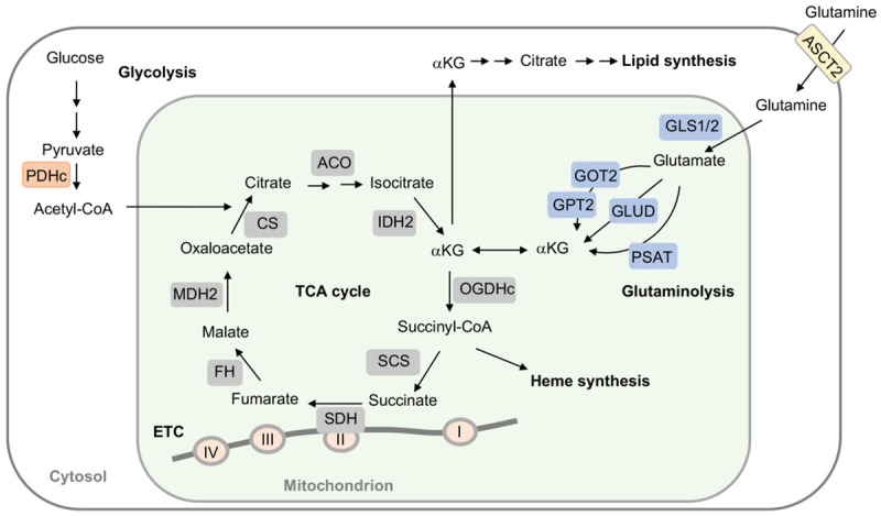
Tricarboxylic acid (TCA) cycle, also called Krebs cycle or citric acid cycle, is an amphoteric pathway, contributing to catabolic degradation and anaplerotic reactions to supply precursors for macromolecule biosynthesis. Oxoglutarate dehydrogenase complex (OGDHc, also called α-ketoglutarate dehydrogenase) a highly regulated enzyme in TCA cycle, converts α-ketoglutarate (αKG) to succinyl-Coenzyme A in accompany with NADH generation for ATP generation through oxidative phosphorylation. The step collaborates with glutaminolysis at an intersectional point to govern αKG levels for energy production, nucleotide and amino acid syntheses, and the resources for macromolecule synthesis in cancer cells with rapid proliferation. Despite being a flavoenzyme susceptible to electron leakage contributing to mitochondrial reactive oxygen species (ROS) production, OGDHc is highly sensitive to peroxides such as HNE (4-hydroxy-2-nonenal) and moreover, its activity mediates the activation of several antioxidant pathways. The characteristics endow OGDHc as a critical redox sensor in mitochondria. Accumulating evidences suggest that dysregulation of OGDHc impairs cellular redox homeostasis and disturbs substrate fluxes, leading to a buildup of oncometabolites along the pathogenesis and development of cancers. In this review, we describe molecular interactions, regulation of OGDHc expression and activity and its relationships with diseases, specifically focusing on cancers. In the end, we discuss the potential of OGDHs as a therapeutic target for cancer treatment.

Keywords: 2-oxoglutarate dehydrogenase; cancer metabolism; reactive oxygen species; tricarboxylic acid cycle; α-ketoglutarate dehydrogenase complex.


Conflict of interest statement

None.

Figures

Figure 1
Figure 1
The role of OGDH and αKG in bioenergy synthesis. αKG is generated from glucose-derived citrate, namely glycolysis and glutaminolysis. Glutaminolysis initiates from deamination of glutamine via glutaminases (GLS1/GLS2) to produce glutamate and ammonia. Glutamate is then converted to αKG by glutamate dehydrogenase (GLUD) or through transamination by glutamate-oxaloacetate transaminase (GOT) and glutamate-pyruvate transaminase 2 (GPT2), as well as, phosphoserine transaminase (PSAT). αKG is oxidatively decarboxylated by 2-oxoglutarate dehydrogenase complex (OGDHc) to succinyl-CoA and CO2, followed by succinyl-CoA synthetase (SCS) converting to succinate. Succinyl-CoA is also forwarded to heme synthesis. Succinate is then oxidized to fumarate by succinate dehydrogenase (SDH). SDH is a part of both tricarboxylic acid (TCA) cycle and electron transport chain (ETC), where SDH is a component of Complex II. However, when pyruvate dehydrogenase complex (PDHc) activity is impeded, αKG is converted to citrate for lipid generation. ACO, aconitase; CS, citrate synthase; FH, fumarate hydratase; IDH2, isocitrate dehydrogenase 2; MDH2, malate dehydrogenase 2.
Figure 2
Figure 2
Regulation of OGDH complex. A. OGDH complex comprises three components, 2-oxoglutarate dehydrogenase (E1 OGDH), dihydrolipoamide succinyltransferase (E2 DLST), and dihydrolipoamide dehydrogenase (E3 DLDH), carrying out the coupled reaction to decarboxylate α-ketoglutarate to succinyl-CoA. E1 OGDH subunit is α-ketoglutarate decarboxylase that uses thiamine pyrophosphate (TPP) as a cofactor to decarboxylate α-ketoglutarate (αKG) for CO2 production and succinylation of TPP. The succinyl-moiety is then transferred to lipoamide in the E2 DLST subunit where yields succinyl-CoA and dihydrolipoamide. The dihydrolipoamide is re-oxidized by E3 DLDH subunit. Here, free electrons are transferred from FAD to reduce NAD+ to generate NADH. OGDH complex contains two susceptible sites for ROS generation (marked by red spots). All of the 3 subunits can be S-glutathionylation. Activity of OGDH complex is regulated by TPP, lipoic acid, CoA, FAD, NAD+, succinyl-CoA, and ROS. The susceptible target by ROS is the prosthetic lipoic acid of the E2 DLST subunit. B. The isoforms of OGDH, OGDH-like (OGDHL), and circ-OGDH. There are three major variants of OGDH in human, OGDH1, OGDH2 and OGDH3. All three OGDH isoforms contain TPP binding domain. OGDH2 lacks of transketolase (TK) domain. Circ-OGDH is the product of reverse splice of OGDH gene, 295 bp.
Figure 3
Figure 3
The role of OGDH complex in redox homeostasis. A. OGDH complex (OGDHc) is a potential ROS generator. E1 OGDH and E3 DLDH are two generation sites for ROS production (marked by red spots) within the OGDHc. E3 DLDH-derived NADH promotes ROS generation by Complex I located in electron transport chain (ETC). The glutathionylation (-S-SG) of OGDHc and Complex I enhances ROS generation, which can be suppressed by glutathione reductase 2 (Grx2) and thioredoxin 2 (Trx). OGDHc-generated ROS is also increased by α-ketoglutarate (αKG). B. OGDHc contributes to antioxidant defense. ROS generated from E3 DLDH can inhibit E1 OGDH and itself activities. ROS-enhanced glutathionylation of E2 DLSH protects peroxidative effects by ROS. The capability of ROS generation of E3 DLDH are regulated in response to cellular oxidative status, and thus, E3 DLDH-generated ROS is inhibited when a higher level of Complex III-derived ROS occurs.
Figure 4
Figure 4
OGDH regulates malate-asparate shuttle activity. In OGDH-dependent or mutated catalytic subunit of phosphatidylinositol 3-kinase cancer cells, a lower OGDH activity leads to α-ketoglutarate (αKG) accumulation, which in turns to ameliorate the asparate and malate-asparate shuttle activity due to the depletion of asparate levels and disruption of NAD+/NADH homeostasis. AGC, asparate/glutamate carrier; GAPDH, glyceraldehyde-3-P-dehydrogenase; Glu, glutamate; GOT1/2, glutamate oxaloacetate transaminase 1/2 (asparate transaminase); MDH1/2, malate dehydrogenase 1/2; OGC, oxoglutarate/malate carrier.
Figure 5
Figure 5
The role of αKG and oncometabolites in epigenetic regulation. In cancer cells, αKG can be converted to L-2-hydroxyglutarate (L-2-HG) through lactate dehydrogenase A (LDHA) and malate dehydrogenase 2 (MDH2) under hypoxia. Mutated IDH1/2 (isocitrate dehydrogenase1 and 2) can constitutively convert αKG to D-2-hydroxyglutarate (D-2-HG). Both L-2-HG and D-2-HG inhibit αKG-dependent dioxygenase activities, including histone lysine demethylases (KDMs), ten-eleven translocation proteins (TETs), and AlkB homologues (ALKBHs), which are involved in the transcriptional suppression of pro-oncogenes by maintaining the hypermethylation status of DNA and histones through epigenetic regulation.
Figure 6
Figure 6
OGDHs serves as a target for cancer treatment. Engineered DLDH or ferroptosis inducers such as sorafenib acting on xCT can activate OGDH to promote ROS production and cancer cell death. Derivatives of succinate and lipoate induce cancer cell death via OGDH inhibition-mediated ROS provocation. Supplementation with exogenous αKG or αKG derivatives attenuates cancer cell growth by elevating αKG/2-hydroxyglutarate (2-HG) ratio followed by inhibiting DNA/histone methylation and/or HIF-1α stability.

Similar articles

Cited by

References

    1. Maas E, Bisswanger H. Localization of the alpha-oxoacid dehydrogenase multi-enzyme complexes within the mitochondrion. FEBS Lett. 1990;277:189–190. - PubMed
    1. Sumegi B, Srere PA. Complex I binds several mitochondrial NAD-coupled dehydrogenases. J Biol Chem. 1984;259:15040–15045. - PubMed
    1. Burch JS, Marcero JR, Maschek JA, Cox JE, Jackson LK, Medlock AE, Phillips JD, Dailey HA Jr. Glutamine via α-ketoglutarate dehydrogenase provides succinyl-CoA for heme synthesis during erythropoiesis. Blood. 2018;132:987–998. - PMC - PubMed
    1. Mailloux RJ, Craig Ayre D, Christian SL. Induction of mitochondrial reactive oxygen species production by GSH mediated S-glutathionylation of 2-oxoglutarate dehydrogenase. Redox Biology. 2016;8:285–297. - PMC - PubMed
    1. Yang L, Venneti S, Nagrath D. Glutaminolysis: a hallmark of cancer metabolism. Annu Rev Biomed Eng. 2017;19:163–194. - PubMed

LinkOut - more resources