

Published Erratum
doi: 10.3389/fonc.2022.911817.
eCollection 2022.
Corrigendum: MELK Inhibition Effectively Suppresses Growth of Glioblastoma and Cancer Stem-Like Cells by Blocking AKT and FOXM1 Pathways
Affiliations
- PMID: 35530328
- PMCID: PMC9074389
- DOI: 10.3389/fonc.2022.911817
Item in Clipboard
Published Erratum
Corrigendum: MELK Inhibition Effectively Suppresses Growth of Glioblastoma and Cancer Stem-Like Cells by Blocking AKT and FOXM1 Pathways
Front Oncol.
.
Abstract
[This corrects the article DOI: 10.3389/fonc.2020.608082.].
Keywords: OTSSP167; glioblastoma multiforme; glioblastoma stem-like cells; maternal embryonic leucine-zipper kinase; targeted therapy.
Copyright © 2022 Zhang, Wang, Wang, Liu, Li, Yu, Wu, Liang, Yu and Liu.
Figures
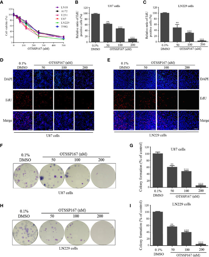
OTSSP167 inhibits GBM cell proliferation and colony formation. (A) CCK-8 viability analysis of cells treated with six OTSSP167 concentrations, including 0 nM, 31.25 nM, 62.5 nM, 125 nM, 250 nM, and 500 nM for 72 h. (B–E) The U87 and LN229 cells were treated with indicated concentrations of OTSSP167 for 24 h, and the EdU assay was performed to assess cell proliferation. Panels (B, C) show the results of the quantitative analysis of the EdU test; panels (D, E) show the representative images of EdU analysis after OTSSP167 treatment of the U87 and LN229 cells. (F–I) OTSSP167 inhibits colony formation in U87 and LN229 cells in a dose-dependent manner. Quantitative analysis of the results of the colony formation experiment was performed. All the Data are presented as means ± SEM. **P < 0.01, ***P < 0.001 compared with the 0.1% DMSO treated group.
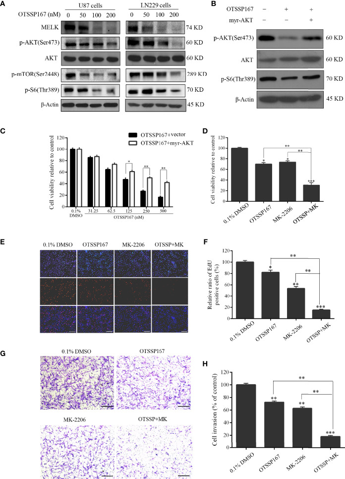
OTSSP167 reduces MELK protein expression and blocks AKT pathway activation, thereby inhibiting the proliferation and invasion of GBM cells. (A) U87 and LN229 cells were treated with OTSSP167 for 24 h. Western blotting showing the expression levels of MELK, p-AKT(Ser473), AKT, p-mTOR(Ser2448), and p-S6(Thr389) proteins. (B) U87 cells transfected with myr-AKT plasmid were treated with OTSSP167, followed by western blotting to assess changes in p-AKT(Ser473), AKT, and p-S6(Thr389) expression. (C) CCK-8 assay shows the effects of OTSSP167 treatment on U87 cells transfected with myr-AKT plasmid compared to the control group. (D) CCK-8 assay showing the viability of U87 cells treated with 50 nM OTSSP167 and 1 μM MK-2206 (AKT inhibitor) alone or combined OTSSP167 and MK-2206 treatment for 72 h. (E) Measurement of cell proliferation after treating with 50 nM OTSSP167 and 1 μM MK-2206 alone or their combinations by EdU incorporation assay. (F, H) Quantitative analysis of proliferative and invading cell numbers. The numbers of proliferative and invading cells were normalized to that of the control group. (G) U87 cells were incubated with 50 nM OTSSP167 and 1 μM MK-2206 alone or their combinations. Cell invasive abilities were evaluated by transwell assay. Results were expressed as means ± SEM of three independent experiments. *P < 0.05, **P < 0.01 and ***P < 0.001 compared with control group.
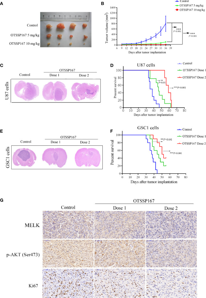
OTSSP167 suppresses tumor growth in vivo and increases the survival of animals bearing intracranial GBM. (A) Representative tumors isolated from the control and OTSSP167-treated groups of subcutaneous tumor model. (B) The mean tumor volumes were assessed at the indicated numbers of days after tumor implantation. (C) Mice bearing U87 xenograft tumor were treated with OTSSP167 (5 μL of 1 μM (dose 1) or 2 μM (dose 2) OTSSP167 in 1% DMSO in PBS per mouse) or vehicle control by intratumoral injection once a week for 4 weeks. Representative images of H&E staining of whole brain sections from control group and OTSSP167 treatment group. (D) Kaplan-Meier survival curves of mice implanted with U87 cells (n=10, **P < 0.01, ***P < 0.001). In vivo animal studies to investigate the effect of OTSSP167 administration on the growth of GSC-driven tumor. Tumor size (E) and survival time (F) were analyzed by using the above same treatment. The survival time of tumor-bearing mice was counted by the end of the 55th day after tumor implantation. (G) Representative IHC staining images of p-AKT(Ser473) and Ki67 expression in U87 xenograft tumor of control and OTSSP167 treatment groups. Sections were counterstained with hematoxylin.
Erratum for
-
MELK Inhibition Effectively Suppresses Growth of Glioblastoma and Cancer Stem-Like Cells by Blocking AKT and FOXM1 Pathways.Front Oncol. 2021 Jan 14;10:608082. doi: 10.3389/fonc.2020.608082. eCollection 2020. Front Oncol. 2021. PMID: 33520717 Free PMC article.
Similar articles
-
MELK Inhibition Effectively Suppresses Growth of Glioblastoma and Cancer Stem-Like Cells by Blocking AKT and FOXM1 Pathways.Front Oncol. 2021 Jan 14;10:608082. doi: 10.3389/fonc.2020.608082. eCollection 2020. Front Oncol. 2021. PMID: 33520717 Free PMC article.
-
Inhibition of maternal embryonic leucine zipper kinase with OTSSP167 displays potent anti-leukemic effects in chronic lymphocytic leukemia.Oncogene. 2018 Oct;37(41):5520-5533. doi: 10.1038/s41388-018-0333-x. Epub 2018 Jun 12. Oncogene. 2018. PMID: 29895969
-
Maternal embryonic leucine zipper kinase: key kinase for stem cell phenotype in glioma and other cancers.Mol Cancer Ther. 2014 Jun;13(6):1393-8. doi: 10.1158/1535-7163.MCT-13-0764. Epub 2014 May 2. Mol Cancer Ther. 2014. PMID: 24795222 Free PMC article. Review.
-
Maternal embryonic leucine zipper kinase is a key regulator of the proliferation of malignant brain tumors, including brain tumor stem cells.J Neurosci Res. 2008 Jan;86(1):48-60. doi: 10.1002/jnr.21471. J Neurosci Res. 2008. PMID: 17722061
-
Enigmatic MELK: The controversy surrounding its complex role in cancer.J Biol Chem. 2020 Jun 12;295(24):8195-8203. doi: 10.1074/jbc.REV120.013433. Epub 2020 Apr 29. J Biol Chem. 2020. PMID: 32350113 Free PMC article. Review.
Publication types
LinkOut - more resources
Full Text Sources
Miscellaneous