. 2022 May 5;12(22):13645-13652.
doi: 10.1039/d2ra00993e.

Fabrication of gelatin Bi2S3 capsules as a highly sensitive X-ray contrast agent for gastrointestinal motility assessment in vivo

Affiliations

Fabrication of gelatin Bi2S3 capsules as a highly sensitive X-ray contrast agent for gastrointestinal motility assessment in vivo

Ya Wen et al. RSC Adv. .

Abstract

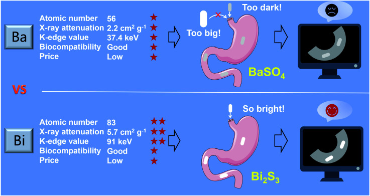
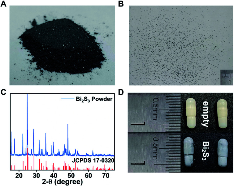
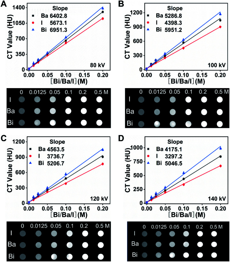
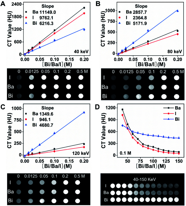
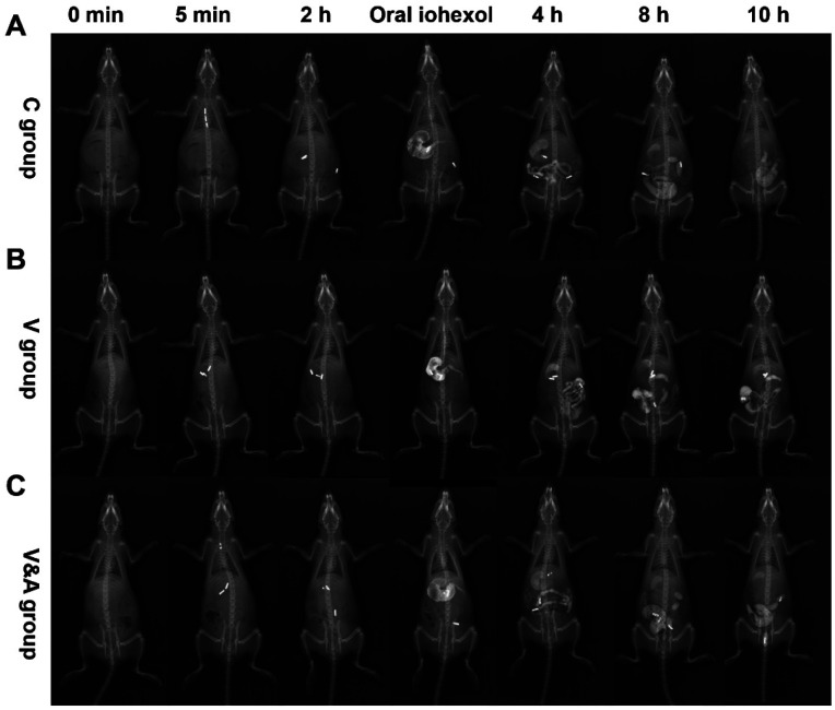
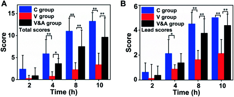
Tiny BaSO4 rod-based X-ray imaging is the most frequently-used method for clinical diagnosis of gastrointestinal motility disorders. The BaSO4 rods usually have a small size to pass through the gastrointestinal tract smoothly, but suffer from unavoidably low sensitivity. Herein, we developed Bi2S3 capsules as a high-performance X-ray contrast agent for gastrointestinal motility assessment for the first time. The Bi2S3 capsules were synthesized by the encapsulation of commercial Bi2S3 powder into commercial gelatin capsules and subsequent coating of ultraviolet-curable resin. The prepared Bi2S3 capsules showed excellent biocompatibility in vitro and in vivo and superior X-ray attenuation ability due to the large atomic number and high K-edge value of Bi. The developed Bi2S3 capsules can serve as a small but highly sensitive X-ray contrast agent to quantitatively assess gastrointestinal motility in a vincristine-induced gastrointestinal motility disorder model in vivo by X-ray, CT and spectral CT imaging successfully, solving the intrinsic drawbacks of clinically used BaSO4.


Conflict of interest statement

The authors declare no competing financial interest.

Figures

Scheme 1
Scheme 1. Schematic illustration of synthesis of Bi2S3 capsules as a X-ray contrast agent for gastrointestinal tract visualization in vivo.
Fig. 1
Fig. 1. Characterization of Bi2S3 powder and capsules. (A) The photo of commercial Bi2S3 powder. (B) The photo of dispersed Bi2S3 power in millimeter scale. (C) X-ray diffraction pattern of Bi2S3 powder. (D) The photo of empty gelatin capsule (upper panel) and Bi2S3 capsules (lower panel).
Fig. 2
Fig. 2. HU curves and CT images of Bi2S3, BaSO4 and iohexol suspensions with different concentrations of radiopaque elements (0.0125, 0.025, 0.05, 0.1, and 0.2 M Bi, Ba or I) at (A) 80 kV, (B) 100 kV, (C) 120 kV, (D) 140 kV.
Fig. 3
Fig. 3. HU curves and spectral CT images of Bi2S3, BaSO4 and iohexol suspensions at different concentrations of radiopaque elements (0.0125, 0.025, 0.05, 0.1, and 0.2 M Bi, Ba or I) at (A) 40 keV, (B) 80 keV, (C) 120 keV. (D) Spectral CT HU curves and images of Bi2S3, BaSO4 and iohexol (0.1 M Bi, Ba or I) at different monochromatic energies.
Fig. 4
Fig. 4. Spectral CT images and HU values of full of Bi2S3, BaSO4 and iohexol capsules.
Fig. 5
Fig. 5. X-ray images of rats after various treatments at different time points (0 min, 5 min, 2 h, 4 h, 8 h, 10 h) in vivo. (A) Control group (C group), (B) vincristine-treated group (V group), (C) vincristine & AM251 treated group (V&A group).
Fig. 6
Fig. 6. The total scores (A) and lead capsule scores (B) of X-ray imaging of rats after various treatments at different time points. These data were shown as means ± SD, n = 8, evaluated by one-way analysis of variance, *p < 0.05, **p < 0.01.
Fig. 7
Fig. 7. Three-dimensional reconstructed images of rats under different monochromatic energies after various treatments at different time points (0 min, 5 min, 2 h, 4 h, 8 h, 10 h) in vivo. (A) Control group (C group), (B) vincristine-treated group (V group), (C) vincristine & AM251 treated group (V&A group).

Similar articles

References

    1. McQuade R. M. Stojanovska V. Abalo R. Bornstein J. C. Nurgali K. Front. Pharmacol. 2016;7:414. - PMC - PubMed
    1. Bytzer P. Talley N. J. Leemon M. Arch. Intern. Med. 2001;161:1989–1996. - PubMed
    1. Richards E. M. Pepine C. J. Raizada M. K. Kim S. Curr. Hypertens. Rep. 2017;19:36. - PMC - PubMed
    1. Fu X. Li Z. Na Z. Yu H. Liu J. Nutr. Metab. 2014;11:3. - PMC - PubMed
    1. Yeh B. M. FitzGerald P. F. Edic P. M. Lambert J. W. Colborn R. E. Marino M. E. Evans P. M. Roberts J. C. Wang Z. J. Wong M. J. Bonitatibus Jr P. J. Adv. Drug Delivery Rev. 2017;113:201–222. - PMC - PubMed