Characterization of Immunogenicity of Malignant Cells with Stemness in Intrahepatic Cholangiocarcinoma by Single-Cell RNA Sequencing
- PMID: 35530414
- PMCID: PMC9076354
- DOI: 10.1155/2022/3558200
Characterization of Immunogenicity of Malignant Cells with Stemness in Intrahepatic Cholangiocarcinoma by Single-Cell RNA Sequencing
Abstract
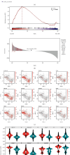
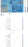
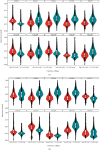
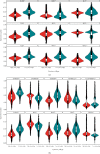
Cancer stem cells (CSCs) are responsible for long-term maintenance of tumors and thought to play a role in treatment resistance. The interaction between stemness and immunogenicity of CSCs in the intrahepatic cholangiocarcinoma (iCCA) is largely unknown. Here, we used single-cell transcriptomic data to study immunogenicity of malignant cells in human iCCA. Using an established computerized method CytoTRACE, we found significant heterogeneity in stemness/differentiation states among malignant cells. We demonstrated that the high stemness malignant cells express much lower levels of major histocompatibility complex II molecules when compared to low stemness malignant cells, suggesting a role of immune evasion in high stemness malignant cells. In addition, high stemness malignant iCCA cells exhibited significant expression of certain cytokine members, including CCL2, CCL20, CXCL1, CXCL2, CXCL6, CXCL8, TNFRSF12A, and IL6ST, indicating communication with surrounding immune cells. These results indicate that high stemness malignant cells retain their intrinsic immunological feature that facilitate the escape of immune surveillance.
Copyright © 2022 Jing Bian et al.
Conflict of interest statement
The authors declare that there is no conflict of interest regarding the publication of this paper.
Figures


Similar articles
-
Cancer Stem Cells Are Possible Key Players in Regulating Anti-Tumor Immune Responses: The Role of Immunomodulating Molecules and MicroRNAs.Cancers (Basel). 2021 Apr 2;13(7):1674. doi: 10.3390/cancers13071674. Cancers (Basel). 2021. PMID: 33918136 Free PMC article. Review.
-
Cancer Stem Cells in Intrahepatic Cholangiocarcinoma; Their Molecular Basis, and Therapeutic Implications.Front Physiol. 2022 Jan 17;12:824261. doi: 10.3389/fphys.2021.824261. eCollection 2021. Front Physiol. 2022. PMID: 35111082 Free PMC article. Review.
-
CAFs shape myeloid-derived suppressor cells to promote stemness of intrahepatic cholangiocarcinoma through 5-lipoxygenase.Hepatology. 2022 Jan;75(1):28-42. doi: 10.1002/hep.32099. Epub 2021 Dec 5. Hepatology. 2022. PMID: 34387870
-
Single-cell transcriptomic architecture and intercellular crosstalk of human intrahepatic cholangiocarcinoma.J Hepatol. 2020 Nov;73(5):1118-1130. doi: 10.1016/j.jhep.2020.05.039. Epub 2020 Jun 5. J Hepatol. 2020. PMID: 32505533
-
Whole-exome sequencing reveals the origin and evolution of hepato-cholangiocarcinoma.Nat Commun. 2018 Mar 1;9(1):894. doi: 10.1038/s41467-018-03276-y. Nat Commun. 2018. PMID: 29497050 Free PMC article.
Cited by
-
CXCL6: A potential therapeutic target for inflammation and cancer.Clin Exp Med. 2023 Dec;23(8):4413-4427. doi: 10.1007/s10238-023-01152-8. Epub 2023 Aug 23. Clin Exp Med. 2023. PMID: 37612429 Review.
-
Morphomolecular Pathology and Genomic Insights into the Cells of Origin of Cholangiocarcinoma and Combined Hepatocellular-Cholangiocarcinoma.Am J Pathol. 2025 Mar;195(3):345-361. doi: 10.1016/j.ajpath.2024.08.014. Epub 2024 Sep 26. Am J Pathol. 2025. PMID: 39341365 Review.
-
Multi-omics analysis of the oncogenic value of copper Metabolism-Related protein COMMD2 in human cancers.Cancer Med. 2023 May;12(10):11941-11959. doi: 10.1002/cam4.5320. Epub 2022 Oct 7. Cancer Med. 2023. PMID: 36205192 Free PMC article.
-
Interplay of Cardiometabolic Syndrome and Biliary Tract Cancer: A Comprehensive Analysis with Gender-Specific Insights.Cancers (Basel). 2024 Oct 9;16(19):3432. doi: 10.3390/cancers16193432. Cancers (Basel). 2024. PMID: 39410050 Free PMC article. Review.
-
Integration of single-cell RNA-seq and bulk RNA-seq to construct liver hepatocellular carcinoma stem cell signatures to explore their impact on patient prognosis and treatment.PLoS One. 2024 Apr 18;19(4):e0298004. doi: 10.1371/journal.pone.0298004. eCollection 2024. PLoS One. 2024. PMID: 38635528 Free PMC article.
References
LinkOut - more resources
Full Text Sources

