Case Reports
. 2022 Apr 1;16(4):1-10.
doi: 10.3941/jrcr.v16i4.4474. eCollection 2022 Apr.

An uncommon case of traumatic pisiform dislocation with triquetral fracture

Affiliations
Case Reports

An uncommon case of traumatic pisiform dislocation with triquetral fracture

Parveen Sulthana Mohamed Ali et al. J Radiol Case Rep. .

Abstract

The pisiform is a sesamoid bone that acts as one of the key medial stabilizers of the wrist. We present a case of a 35-year-old gentleman who presented with medial wrist pain following a fall while rollerblading. Radiographs and Magnetic resonance imaging (MRI) revealed a rare combination of an acute pisiform dislocation with associated triquetral fracture. Subsequently, he was successfully treated with excision of the pisiform. Pisiform dislocation is an uncommon injury and can easily be missed in an acute emergency presentation. Therefore, it is important to be aware of the characteristic imaging appearance to avoid a delay in diagnosis and treatment.

Keywords: Pisiform; dislocation; excision; fracture; triquetrum.


Figures

Figure 1
Figure 1
35-year-old male with pisiform dislocation in the left wrist. Findings: (a) Frontal posteroanterior view radiograph of the left wrist shows the distal and medial dislocation of the pisiform bone (asterisk). The oval white circle indicates the normally expected location of the pisiform bone, projected over the triquetrum. (b) Frontal posteroanterior view radiograph of the normal left wrist for comparison shows normal expected location of the pisiform bone (yellow asterisk) projected over the triquetrum. Technique: Frontal posteroanterior view radiograph of the left wrist with settings at a kVp of 40–45 and mAs of 5–6.
Figure 2
Figure 2
35-year-old male with pisiform dislocation in the left wrist. Findings: (a) Sagittal proton density-weighted (PD) MRI image of the left wrist shows distal dislocation of the pisiform (white asterisk) with relation to the triquetrum (T). (b) Sagittal proton density-weighted (PD) MRI image of the normal left wrist in a different patient (given for comparison) shows normal pisotriquetral articulation. The pisiform is indicated by a white asterisk and the triquetrum is labeled as the letter ‘T’ in white. Technique: Sagittal proton density-weighted (PD) MRI of left wrist in a 3T scanner-Philips Ingenia. TR=2917.66, TE=40.00, Flip Angle=90.00 and slice thickness=1.4mm.
Figure 3
Figure 3
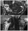
35-year-old male with pisiform dislocation in the left wrist. Findings: (a) Coronal STIR (Short Tau Inversion Recovery) MRI image of the left wrist shows a minimally displaced fracture of the triquetrum (white arrow) with bone marrow edema. Mild marrow edema is also noted in the medial aspect of the lunate due to contusion (white asterisk). The partly imaged base of the first metacarpal appears hyperintense due to incomplete fat suppression (artefact). (b) Coronal STIR (Short Tau Inversion Recovery) MRI image of the left wrist shows bone marrow edema in the pisiform bone (asterisk) and surrounding soft tissue edema. The pisohamate ligament is intact (white arrow). The partly imaged base of the first metacarpal appears hyperintense due to incomplete fat suppression.
Figure 4
Figure 4
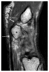
35-year-old male with pisiform dislocation in the left wrist. Findings: Sagittal proton density-weighted (PD) MRI image of the left wrist shows mild hyperintense signal at the distal insertion of intact FCU tendon (dotted white arrows) into the pisiform (black asterisk) in keeping with mild strain. The partly imaged pisometacarpal ligament is intact (white arrow). Technique: Sagittal proton density-weighted (PD) MRI image of the left wrist in a 3T scanner-Philips Ingenia. TR=2917.66, TE=40.00, Flip Angle=90.00 and slice thickness=1.4mm.
Figure 5
Figure 5
35-year-old male with pisiform dislocation in the left wrist. Findings: Radiographs of both wrists obtained in flexion show reduced mid carpal flexion on the left side. The capitolunate angle measurements in both wrists are as shown. Capitolunate angle is the angle between the long axis of the capitate and mid axis of the lunate. The capitolunate angle in the left wrist measured 12 degrees compared to 30 degrees on the right indicating reduced flexion at the left capito-lunate joint.
Figure 6
Figure 6
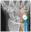
Normal pisiform attachments Findings: Graphic illustration of the pisiform attachments projected over the normal frontal posteroanterior view radiograph of the right wrist (Illustration by Dr Joe Francis). The oval white circle indicates normal expected location of pisiform bone, projected over the triquetrum. The FCU inserts onto the palmar aspect of the pisiform bone. The pisiform also receives attachments from pisohamate, pisometacarpal, and pisotriquetral ulnar ligaments. P- Pisiform, H- Hamate, MC- 5th metacarpal, FCU- Flexor carpi ulnaris (blue), PHL (pink)-Pisohamate ligament, PML (light green)- Pisometacarpal ligament, PTUL(dark green)-Pisotriquetral ulnar ligament. The red line represents the ulnar artery and the yellow line represents the ulnar nerve.
Figure 7
Figure 7
Special radiographic projections of a normal wrist Findings: (a) Normal Carpal tunnel view radiograph of the left wrist in an adult shows the pisiform distinctly from the rest of the carpal bones. Pisiform fractures can be well seen in this view. (b) Normal Semi supinated anteroposterior oblique radiograph of the left wrist in an adult shows the pisotriquetral joint in profile. Pisiform fracture, subluxation, dislocation and pisotriquetral joint disorders can be well assessed in this view. Technique: (a) The palmar surface of the patient’s hand is placed on the cassette with the wrist dorsiflexed. The x-ray tube is angled 25–30 degrees to the long axis of the hand. Settings - kVp: 40–45 and mAs: 3–5. (b) The patient’s wrist is placed in partial supination at an angle of 45 degrees. The central beam is directed at the dorsal aspect of the mid carpal region. Settings - kVp: 40–45 and mAs: 3–5.

Similar articles

Cited by

References

    1. Cohen I. Dislocation of the pisiform. Ann Surg. 1922;75:238–9. - PMC - PubMed
    1. Petrou IG, Savioz-Leissing C, Gray A. Traumatic Dislocation of the Pisiform Bone. J Hand Microsurg. 2018;10(1):37–40. - PMC - PubMed
    1. Hurni Y, Fusetti C, De Rosa V. Fracture dislocation of the pisiform bone in children: a case report and review of the literature. J Pediatr Orthop B. 2015;24(6):556–60. - PubMed
    1. Blum AG, Zabel JP, Kohlmann R, et al. Pathologic conditions of the hypothenar eminence: evaluation with multidetector CT and MR imaging. Radiographics. 2016;26(4):1021–44. - PubMed
    1. Gan LP, Satkunanantham M, Sreedharan S, Chew WY. Triquetral fracture with associated pisiform subluxation. Singapore Med J. 2015;56(3):e39–41. - PMC - PubMed

Publication types