

Synthesis of calcium carbonate microcapsules as self-healing containers
- PMID: 35530589
- PMCID: PMC9069483
- DOI: 10.1039/c9ra03804c
Synthesis of calcium carbonate microcapsules as self-healing containers
Abstract
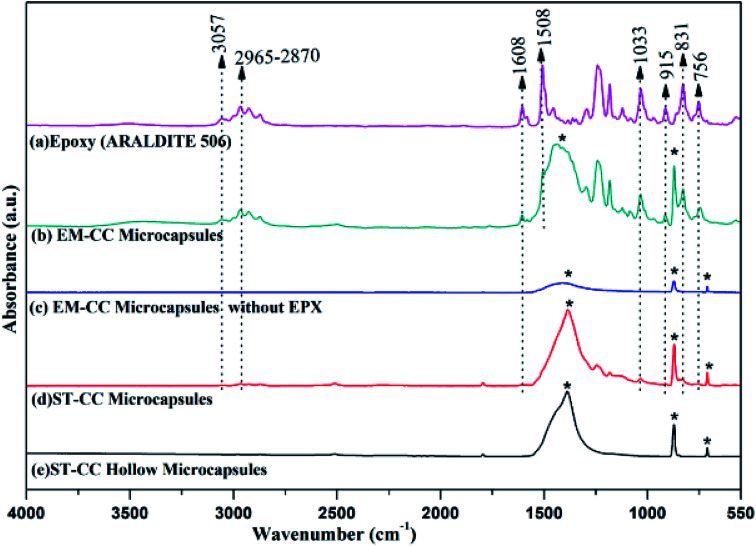
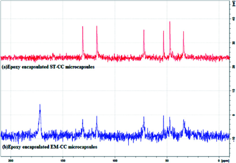
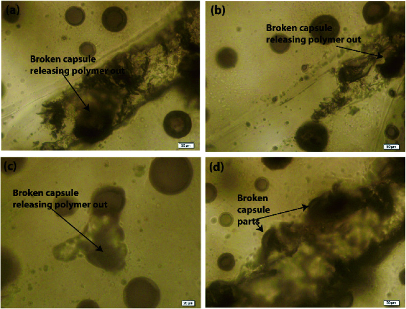
Contemporary studies of self-healing polymer composites are based on microcapsules synthesized using synthetic and toxic polymers, biopolymers, etc. via methods such as in situ polymerization, electrospraying, and air atomization. Herein, we synthesized a healing agent, epoxy (EPX) encapsulated calcium carbonate (CC) microcapsules, which was used to prepare self-healing EPX composites as a protective coating for metals. The CC microcapsules were synthesized using two facile methods, namely, the soft-template method (STM) and the in situ emulsion method (EM). Microcapsules prepared using the STM (ST-CC) were synthesized using sodium dodecyl sulphate (SDS) surfactant micelles as the soft-template, while the microcapsules prepared using the EM (EM-CC) were synthesized in an oil-in-water (O/W) in situ emulsion. These prepared CC microcapsules were characterized using light microscopy (LMC), field emission scanning electron microscopy (FE-SEM), fourier transform infrared spectroscopy (FTIR), nuclear magnetic resonance spectroscopy (NMR), and thermogravimetric analysis (TGA). The synthesized ST-CC microcapsules were spherical in shape, with an average diameter of 2.5 μm and an average shell wall thickness of 650 nm, while EM-CC microcapsules had a near-spherical shape with an average diameter of 3.4 μm and an average shell wall thickness of 880 nm. The ST-CC capsules exhibited flake-like rough surfaces while EM-CC capsules showed smooth bulgy surfaces. The loading capacity of ST-CC and EM-CC microcapsules were estimated using TGA and found to be 11% and 36%, respectively. The FTIR and NMR spectra confirmed the EPX encapsulation and the unreactive nature of the loaded EPX with the inner walls of CC microcapsules. The synthesized CC microcapsules were further incorporated into an EPX matrix to prepare composite coatings with 10 (w/w%), 20 (w/w%), and 50 (w/w%) capsule loadings. The prepared EPX composite coatings were scratched and observed using FE-SEM and LMC to evaluate the release of encapsulated EPX inside the CC capsules, which is analogous to the healing behaviour. Moreover, EPX composite coatings with 20 (w/w%) and 50 (w/w%) of ST-CC showed better healing performances. Thus, it was observed that ST-CC microcapsules outperformed EM-CC. Additionally, the EPX/CC coatings showed remarkable self-healing properties by closing the gaps of the scratch surfaces. Thus, these formaldehyde-free, biocompatible, biodegradable, and non-toxic CC based EPX composite coatings hold great potential to be used as a protective coating for metal substrates. Primary results detected significant corrosion retardancy due to the self-healing coatings under an accelerated corrosion process, which was performed with a salt spray test.
This journal is © The Royal Society of Chemistry.
Conflict of interest statement
The authors declare no conflict of interest.
Figures


Similar articles
-
In Situ Evaluation of Epoxy Self-Healing Coating by Encapsulated Linseed Oil in Poly(Urea-Formaldehyde-Melamine) Microcapsules.Materials (Basel). 2025 Apr 23;18(9):1906. doi: 10.3390/ma18091906. Materials (Basel). 2025. PMID: 40363410 Free PMC article.
-
High linoleic waste sunflower oil: A distinctive recycled source of self-healing agent for smart metal coatings.Heliyon. 2023 Apr 14;9(4):e15364. doi: 10.1016/j.heliyon.2023.e15364. eCollection 2023 Apr. Heliyon. 2023. PMID: 37151619 Free PMC article.
-
Facile preparation of Artemisia argyi oil-loaded antibacterial microcapsules by hydroxyapatite-stabilized Pickering emulsion templating.Colloids Surf B Biointerfaces. 2013 Dec 1;112:96-102. doi: 10.1016/j.colsurfb.2013.08.002. Epub 2013 Aug 13. Colloids Surf B Biointerfaces. 2013. PMID: 23973909
-
Progress in the Preparation and Applications of Microcapsules for Protective Coatings Against Corrosion.Int J Mol Sci. 2025 Feb 10;26(4):1473. doi: 10.3390/ijms26041473. Int J Mol Sci. 2025. PMID: 40003938 Free PMC article. Review.
-
Self-Repairing Composites for Corrosion Protection: A Review on Recent Strategies and Evaluation Methods.Materials (Basel). 2019 Aug 27;12(17):2754. doi: 10.3390/ma12172754. Materials (Basel). 2019. PMID: 31461982 Free PMC article. Review.
References
-
- Khoee S. Payandeh S. H. Jafarzadeh P. Asadi H. Smart Mater. Struct. 2016;25:084014. doi: 10.1088/0964-1726/25/8/084014. - DOI
-
- Wu D. Y. Meure S. Solomon D. Prog. Polym. Sci. 2008;33:479–522. doi: 10.1016/j.progpolymsci.2008.02.001. - DOI
-
- Hia I. L. Vahedi V. Pasbakhsh P. Polym. Rev. 2016;56:225–261. doi: 10.1080/15583724.2015.1106555. - DOI
LinkOut - more resources
Full Text Sources
Miscellaneous