. 2019 Jul 30;9(41):23658-23665.
doi: 10.1039/c9ra03404h. eCollection 2019 Jul 29.

A spherical poly(acrylic acid) brush-enzyme block with high catalytic capacity for signal amplification in digital biological assays

Affiliations

A spherical poly(acrylic acid) brush-enzyme block with high catalytic capacity for signal amplification in digital biological assays

Yibei Wang et al. RSC Adv. .

Abstract

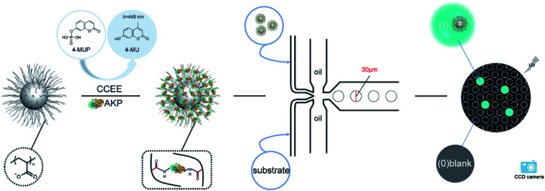
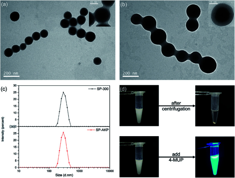
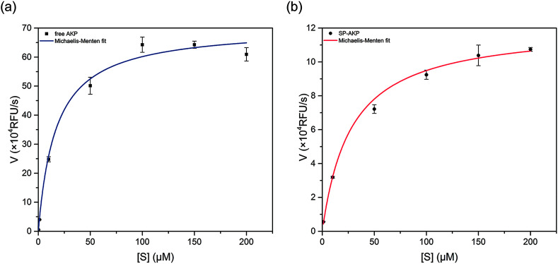
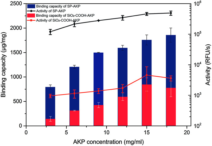
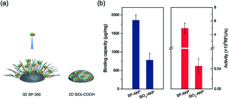
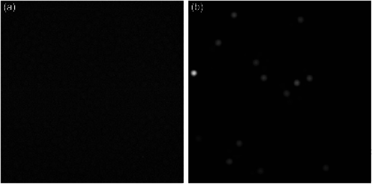
Ultrasensitive determination of some ultra-low abundance biological molecules closely related to diseases is currently a wide concern and urgent issue to be addressed. Here, a spherical poly(acrylic acid)-alkaline phosphatase (SP-AKP) signal amplification block using spherical poly(acrylic acid) brush nanoparticles (SP) as the immobilized carriers was designed and synthesized optimally first. The results show that a single SP-AKP with high enzyme binding capacity and high catalytic ability (up to about 4800 effective free AKP per SP-AKP) has much greater fluorescence signal amplification ability than a single free AKP or SiO2-COOH-AKP. Then, a droplet generation microfluidic chip was prepared successfully, and the SP-AKP was loaded and confined in a 14 pL droplet by adjusting its concentration to ensure at most one SP-AKP was encapsulated in each droplet according to Poisson's theory. Finally, the fluorescence signals produced by 4-methylumbelliferyl phosphate (4-MUP) catalyzed via SP-AKP within 6 min were sufficient to be detected by a fluorescence microscope. Thus, the digital signal distribution of "1/0" (signal/background) was obtained, making this SP-AKP signal amplification block a promising enzyme label for potential high sensitivity digital biological detection applications.


Conflict of interest statement

There are no conflicts to declare.

Figures

Scheme 1
Scheme 1. Illustration of the protein immobilization and digital detection.
Fig. 1
Fig. 1. TEM images of SP (a) before and (b) after covalent immobilization with AKP by CCEE (with a close-up of one particle at top left). (c) The corresponding DLS size distributions of particles SP-300 and SP–AKP. (d) Photos of the SP–AKP in aqueous suspension (left), after centrifugation and the fluorescent products (right).
Fig. 2
Fig. 2. Dependence of the catalytic rate of (a) free AKP and (b) SP–AKP on 4-MUP concentration. The activity was measured in the presence of 0.1 M Tris–HCl and 0.5 mM MgCl2 at pH 9.0. Data points were fitted to the Michaelis–Menten equation to give a Km of free AKP and the SP–AKP of 10.96 μM and 16.84 μM respectively.
Fig. 3
Fig. 3. Binding capacity and activity of SP–AKP and SiO2–COOH–AKP and in dependence of concentration of AKP. Histogram shows binding capacity and line chart shows activity.
Fig. 4
Fig. 4. (a) Comparison on protein immobilization mechanisms between 3D SP-300 and 2D SiO2–COOH. (b) Comparison of SP–AKP and SiO2–COOH–AKP in terms of protein binding capacity and activity.
Fig. 5
Fig. 5. The fluorescence images of (a) free AKP (λ = 0.1) and (b) the SP–AKP (λ = 0.1) in 14 pL droplets after a 6 min incubation with 0.25 mM 4-MUP. The exposure time was 100 ms. The brightness and contrast of two images were automatically adjusted by ImageJ software to clearly distinguish all droplets.

Similar articles

References

    1. Lequin R. M. Clin. Chem. 2005;51:2415–2418. doi: 10.1373/clinchem.2005.051532. - DOI - PubMed
    1. Kuo H. T. Yeh J. Z. Jiang C. M. Wu M. C. J. Immunol. Methods. 2012;381:32–40. doi: 10.1016/j.jim.2012.04.006. - DOI - PubMed
    1. Macdonald M. J. Gapinski J. P. Metab., Clin. Exp. 1989;38:450–452. doi: 10.1016/0026-0495(89)90197-2. - DOI - PubMed
    1. Ohwovoriole A. E. Wilkin T. J. J. Immunol. Methods. 1988;115:99–104. doi: 10.1016/0022-1759(88)90315-8. - DOI - PubMed
    1. Tsitsilonis O. E. Thrasyvoulides A. Balafas A. Voutsas J. F. Papamichail M. Lymberi P. J. Pharm. Biomed. Anal. 2004;34:811–822. doi: 10.1016/S0731-7085(03)00563-6. - DOI - PubMed