

3D orientation and kinematic characteristics of zygapophyseal joints while sitting
- PMID: 35530947
- PMCID: PMC9073805
- DOI: 10.21037/atm-22-969
3D orientation and kinematic characteristics of zygapophyseal joints while sitting
Abstract
Background: Orientation of the lumbar facet joints (FJs) in the transverse plane is associated with degenerative lumbar spine disease. However, there is a lack of measurements of the sagittal and coronal facet angles, and the effect of 3D facet angles on joint motion in the sitting position is unknown. The present study was to investigate the 3D orientation and in vivo motion characteristics of the FJ in the sitting position.
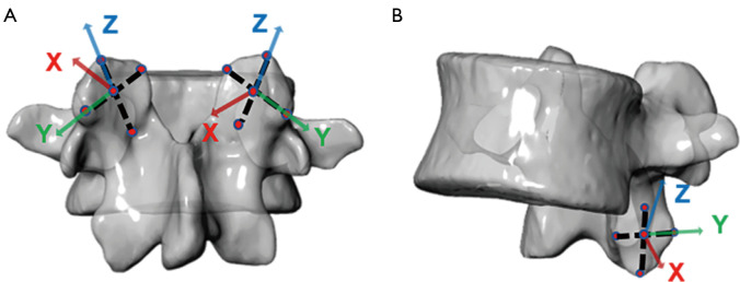
Methods: Dual fluoroscopic imaging system and computed tomography (CT) were used to determine the 3D orientation and kinematic characteristics of FJs. L3-S1 segments were studied in 10 asymptomatic participants (5 male and 5 female, age: 25-35 years, body mass index: 22.4±1.8). Angles of the facet in the sagittal, coronal, and axial planes, and the range of motion of the FJs in seated flexion and extension movements were measured.
Results: The difference in sagittal facet angles between the 2 sides of the L3-S1 facet joints was not significant. The superior coronal facet angle on the left side of L5 was significantly smaller than that on the right side by 6.4° (P=0.01). The inferior transverse facet angle on the left side of L5 was greater than that on the right side by 7.1; the results were not statistically significantly different. In the sitting position, the range of motion of the left and right sides of L5-S1 differed significantly, with the right side being 5.5° (P=0.004) and 11.7° (P=0.026) greater than the left side in the sagittal and coronal planes, respectively. There was a correlation between mobility and the 3D orientation angle of the FJs in each segment.
Conclusions: Quantification of the 3D orientation of the lumbar spine FJs provides new perspectives to study the kinematics of the lumbar spine and the etiology of lumbar degenerative diseases. In sitting flexion and extension movements, there is a significant difference in the left-right lateral mobility of the FJs of the L5-S1 segments. With the exception of the transverse facet angle of the lumbar spine FJs, the sagittal and coronal facet angles also have an effect on lumbar spine mobility.
Keywords: 3D orientation; Zygapophyseal joint; facet tropism (FT); sitting.
2022 Annals of Translational Medicine. All rights reserved.
Conflict of interest statement
Conflicts of Interest: All authors have completed the ICMJE uniform disclosure form (available at https://atm.amegroups.com/article/view/10.21037/atm-22-969/coif). The authors have no conflicts of interest to declare.
Figures


Similar articles
-
Influence of weight-bearing on the 3D movement of lumbar facet joints in the sitting position.BMC Musculoskelet Disord. 2023 Jul 10;24(1):561. doi: 10.1186/s12891-023-06698-y. BMC Musculoskelet Disord. 2023. PMID: 37430257 Free PMC article.
-
3D kinematic characteristics of lumbar facet joints in sitting position.Surg Radiol Anat. 2022 Sep;44(9):1289-1295. doi: 10.1007/s00276-022-03005-7. Epub 2022 Aug 13. Surg Radiol Anat. 2022. PMID: 35962832
-
Kinematic Characteristics and Biomechanical Changes of Lower Lumbar Facet Joints Under Different Loads.Orthop Surg. 2021 May;13(3):1047-1054. doi: 10.1111/os.12894. Epub 2021 Mar 11. Orthop Surg. 2021. PMID: 33709625 Free PMC article.
-
Facet Tropism in Lumbar Spine and Cervical Spine: A Systematic Review and Meta-Analysis.World Neurosurg. 2021 Mar;147:47-65. doi: 10.1016/j.wneu.2020.11.171. Epub 2020 Dec 9. World Neurosurg. 2021. PMID: 33309642
-
Amniotic membrane and/or umbilical cord tissue for treatment of facet joint syndrome: a narrative review.J Orthop Surg Res. 2023 Oct 2;18(1):744. doi: 10.1186/s13018-023-04241-2. J Orthop Surg Res. 2023. PMID: 37784162 Free PMC article. Review.
Cited by
-
Influence of weight-bearing on the 3D movement of lumbar facet joints in the sitting position.BMC Musculoskelet Disord. 2023 Jul 10;24(1):561. doi: 10.1186/s12891-023-06698-y. BMC Musculoskelet Disord. 2023. PMID: 37430257 Free PMC article.
-
Effect of different loads on facet joint motion during lumbar lateral bending in sitting position.J Orthop Surg Res. 2024 Jan 13;19(1):61. doi: 10.1186/s13018-024-04533-1. J Orthop Surg Res. 2024. PMID: 38218824 Free PMC article.
-
The Helical Compliance Vector: Utility for Quantifying Spinal Mechanics.JOR Spine. 2025 Jun 19;8(2):e70088. doi: 10.1002/jsp2.70088. eCollection 2025 Jun. JOR Spine. 2025. PMID: 40538464 Free PMC article.
References
LinkOut - more resources
Full Text Sources