. 2022 Apr;10(7):400.
doi: 10.21037/atm-21-7015.

Regulating mitochondrial homeostasis and inhibiting inflammatory responses through Celastrol

Affiliations

Regulating mitochondrial homeostasis and inhibiting inflammatory responses through Celastrol

Zhenyu Tao et al. Ann Transl Med. 2022 Apr.

Abstract

Background: The high morbidity and mortality rate of coronary heart disease poses a serious threat to human health. Atherosclerosis, a chronic inflammation of the blood vessel wall, is a significant pathological process leading to coronary heart disease. Macrophage inflammation plays a crucial role in the occurrence and development of atherosclerosis.

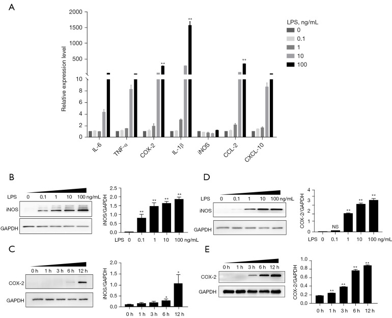
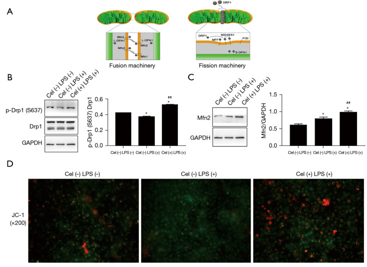
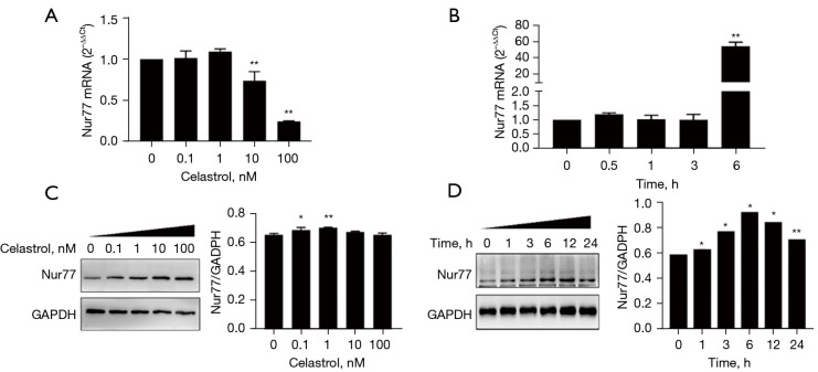
Methods: Macrophage inflammation model was constructed by lipopolysaccharide (LPS), and macrophages were treated with Celastrol at different concentrations (0, 0.1, 1, 10, 100 ng/mL) and different time points (0, 1, 3, 6, 12 h). Real-time quantitative PCR (qPCR) and Western Blot were used to detect the expression of Nur77 mRNA and protein. Macrophages were then pretreated with 100 nmol/L tripterine for 40min and co-cultured with 100 ng/mL LPS. The expression levels of inflammatory factors and chemokines, phosphorylation of phospho-dynamin-related protein 1 (p-Drp1) at Ser637 and expression of mitochondrial fusion protein mitochondrial fusion protein mitofusin-2 (Mfn2) were detected by qPCR, Western blot and ELISA, respectively. The changes of mitochondrial membrane potential were detected by JC-1 probe.

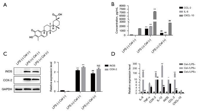
Results: 100 nmol/L Celastrol can significantly inhibit LPS-induced inflammatory responses and down-regulate the expression levels of cytokines such as inducible nitric oxide synthase (iNOS), cyclooxygenase-2 (COX2), tumor necrosis factor-α (TNF-α), chemokines (CCL-2, and CXCL-10), as well as chemokines. And Celastrol could regulate mitochondrial fission and fusion by promoting the phosphorylation of the Drp1 at the Ser637 site, thereby inhibiting mitochondrial fission. At the same time, by up-regulating the level of the Mfn2, Celastrol also promoted mitochondrial fusion. In addition, we found that the nuclear factor-k-gene binding (NF-κB), extracellular signal-regulated kinase 1/2 (ERK1/2), and p38 signaling pathways aided the drug's anti-inflammatory effects. We also explored the relationship between Celastrol and the nuclear receptor Nur77 and found that it could up-regulate the expression of Nur77.

Conclusions: Our study found that Celastrol could reduce inflammation by regulating Drp1 dependent mitochondrial fission and fusion, as well as the ERK1/2, p38, NF-κB signaling pathways. This finding provides a strong direction for the development of new anti-inflammatory drugs for atherosclerosis.

Keywords: Celastrol; Nur77; dynamin-related protein 1 (Drp1); inflammation; mitochondrial fission and fusion.


Conflict of interest statement

Conflicts of Interest: All authors have completed the ICMJE uniform disclosure form (available at https://atm.amegroups.com/article/view/10.21037/atm-21-7015/coif). The authors have no conflicts of interest to declare.

Figures

Figure 1
Figure 1
Expressions of IL-6, TNF-α, COX-2, IL-1β, iNOS, CCL-2, and CXCL-10. (A) The level of IL-6, TNF-α, COX-2, IL-1β, iNOS, CCL-2, and CXCL-10 were detected by qPCR; (B) the expressions of iNOS were detected by 0–100 ng/mL LPS induced; (C) the expressions of COX 2 were detected by 0–100 ng/mL LPS induced; (D) the expressions of iNOS were detected by 0, 1, 3, 6, 12 h LPS induced; (E) the expressions of COX2 were detected by 0, 1, 3, 6, 12 h LPS induced. *, P<0.05 compared with 0 h; **, P<0.01 compared with 0 h; NS represents P>0.05. IL, interleukin; TNF-α, tumor necrosis factor-α; COX-2, cyclooxygenase-2; IL-1β, interleukin-1β; iNOS, inducible nitric oxide synthase; CCL-2, chemokine (C-C motif) ligand 2; CXCL-10, chemokine (C-X-C motif) ligand 10; LPS, lipopolysaccharide; qPCR, real-time quantitative PCR.
Figure 2
Figure 2
Celastrol inhibits LPS-induced inflammatory response. (A) The structure of Celastrol; (B) expressions of IL-6, CCL-2, and CXCL-10; (C) expressions of iNOS and COX-2; (D) expressions of IL-6, TNF-α, COX-2, IL-1β, iNOS, CCL-2, and CXCL-10. ##, P<0.01 compared with LPS without Cel; **, P<0.01 compared without LPS and Cel; NS represents P>0.05 compared with LPS without Cel. IL, interleukin; TNF-α, tumor necrosis factor-α; COX-2, cyclooxygenase-2; IL-1β, interleukin-1β; iNOS, inducible nitric oxide synthase; CCL-2, chemokine (C-C motif) ligand 2; CXCL-10, chemokine (C-X-C motif) ligand 10; LPS, lipopolysaccharide.
Figure 3
Figure 3
Study on the mechanism of Celastrol in regulating mitochondrial morphology and function. (A) Study on the mechanism of Celastrol in regulating mitochondrial morphology and function; (B) expressions of p-Drp1 and Drp1; (C) the expression of Mfn2; (D) immunofluorescence expression of JC-1 (×100). ##, P<0.01 compared with LPS without Cel; *, P<0.05 compared without LPS and Cel. p-Drp1, phospho-dynamin-related protein 1; Mfn2, mitochondrial fusion protein mitofusin-2; LPS, lipopolysaccharide; GAPDH, glyceraldehyde-3-phosphate dehydrogenase.
Figure 4
Figure 4
Research on the mechanism of Celastrol regulating related inflammatory signaling pathways. (A) Expressions of p-ERK/ERK, p-JNK/JNK, p-P38/P38; (B) expressions of p-P65/P65; (C) expression of p65. ##, P<0.01 compared with LPS without Cel; *, P<0.05 compared without LPS and Cel; **, P<0.01 compared without LPS and Cel. ERK, extracellular signal-regulated kinase; LPS, lipopolysaccharide; GAPDH, glyceraldehyde-3-phosphate dehydrogenase.
Figure 5
Figure 5
The effect of Celastrol on the expression of the nuclear receptor Nur77. (A) The expression of Nur77 after treating with different concentrations of Celastrol by qPCR; (B) the expression of Nur77 after treating with Celastrol in different time by qPCR; (C) the expression of Nur77 after treating with different concentrations of Celastrol by Western blot; (D) the expression of Nur77 after treating with Celastrol in different time by Western blot. *, P<0.05 compared with 0 h; **, P<0.01 compared with 0 h. qPCR, real-time quantitative PCR; Nur77, nerve growth factor-induced gene B NGFI-B; GAPDH, glyceraldehyde-3-phosphate dehydrogenase.

Similar articles

Cited by

References

    1. Heller DJ, Coxson PG, Penko J, et al. Evaluating the Impact and Cost-Effectiveness of Statin Use Guidelines for Primary Prevention of Coronary Heart Disease and Stroke. Circulation 2017;136:1087-98. 10.1161/CIRCULATIONAHA.117.027067 - DOI - PMC - PubMed
    1. Hansson GK. Inflammation, atherosclerosis, and coronary artery disease. N Engl J Med 2005;352:1685-95. 10.1056/NEJMra043430 - DOI - PubMed
    1. Patel MJ, Blazing MA. Inflammation and atherosclerosis: disease modulating therapies. Curr Treat Options Cardiovasc Med 2013;15:681-95. 10.1007/s11936-013-0268-z - DOI - PubMed
    1. Shao Q, Han F, Peng S, et al. Nur77 inhibits oxLDL induced apoptosis of macrophages via the p38 MAPK signaling pathway. Biochem Biophys Res Commun 2016;471:633-8. 10.1016/j.bbrc.2016.01.004 - DOI - PubMed
    1. Kavurma MM, Rayner KJ, Karunakaran D. The walking dead: macrophage inflammation and death in atherosclerosis. Curr Opin Lipidol 2017;28:91-8. 10.1097/MOL.0000000000000394 - DOI - PMC - PubMed