Review
. 2022 May 2:17:1927-1950.
doi: 10.2147/IJN.S357598. eCollection 2022.

Nanomedicine: An Emerging Novel Therapeutic Strategy for Hemorrhagic Stroke

Affiliations
Review

Nanomedicine: An Emerging Novel Therapeutic Strategy for Hemorrhagic Stroke

Yating Xu et al. Int J Nanomedicine. .

Abstract

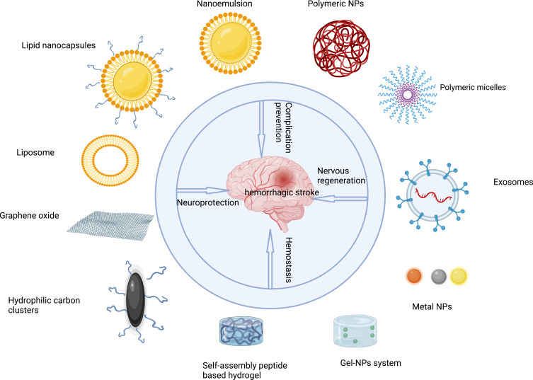
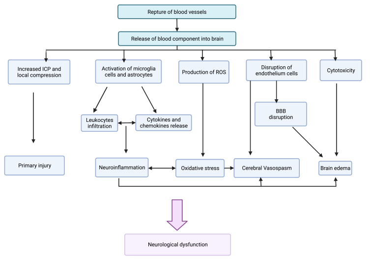
Hemorrhagic stroke is one of the most devastating diseases worldwide due to a high rate of disability and mortality with few effective treatments. Recent advances in nanomedicines to promote hemostasis, drug delivery, neuroprotection, and nerve regeneration may provide insight into hemorrhagic stroke treatment. In this review, we first view the pathophysiology and conventional therapeutics of hemorrhagic stroke. Second, we comprehensively summarize the current nanomedicines applied in hemorrhagic stroke, including inorganic nanomaterials, polymer-based nanomaterials, lipid-based nanomaterials, self-assembling peptide-based hydrogel, exosomes, and gel systems. Finally, the challenges, opportunities, and future perspectives of nanomedicines for hemorrhagic stroke are discussed. Thus, this review promotes greater exploration of effective therapies for hemorrhagic stroke with nanomedicines.

Keywords: hemorrhagic stroke; intracerebral hemorrhage; nanomedicine; subarachnoid hemorrhage; therapy.


Conflict of interest statement

The authors report no conflicts of interest in this work.

Figures

Figure 1
Figure 1
The nanomaterials used for therapy of hemorrhagic stroke. Created with Biorender.com.
Figure 2
Figure 2
The pathophysiology of hemorrhagic stroke. Created with Biorender.com.
Figure 3
Figure 3
ICH treatment with DEF-HCC-PEG. (A) The mechanism of DEF-PEG-HCC in combating ICH. Hemin can lead to senescence of neurons by inducing nuclear and mitochondrial DNA damage. Although PEG-HCC therapy can reduce senescence and DNA damage after exposure to hemin, it simultaneously increases iron-mediated ferroptosis. In contrast, DEF-HCC-PEG can prevent ferroptosis, DNA damage, and senescence with simultaneous iron chelation and ROS scavenging ability. (B) PEG-HCC-DEF restores the viability of hemin-treated neurons beyond that of PEG-HCC or DEF alone. The cultured neurons were treated with 5 μM hemin and received corresponding drugs. Total cell death was measured by MTT assay. (C) DEF-PEG-HCC exhibited higher efficiency in preventing hemin-induced DNA damage. The cultured neurons were treated with 5 μM of hemin and received corresponding drugs. IB was used to measure the level of γH2AX and p-53BP1 as biomarkers of genome damage. The histogram shows the quantitation results of IB. (D and E) The IB quantitation result of (D) GPX4 or (E) MDA levels in cultured neurons treated with 100 μM FeSO4 and in the presence or absence of hemin, PEG-HCC, DEF, or DEF-HCC-PEG. The reduction of GPX4 or increase of MDA levels can be markers of ferroptosis, respectively. Results are represented as mean ± SEM from three independent experiments. Significant differences: *p < 0.01; **p < 0.05. Reprinted with permission from American Chemical Society: ACS Nano, Pervasive Genomic Damage in Experimental Intracerebral Hemorrhage: Therapeutic Potential of a Mechanistic-Based Carbon Nanoparticle, Dharmalingam P, Talakatta G, Mitra J et al. Copyright 2020 American Chemical Society.
Figure 4
Figure 4
The treatment of ICH animals with micelles attenuated the neurological deficits, BBB damage, apoptosis, ferroptosis, and neuro-inflammation after ICH. (A) The DRC release profile of DPM in the presence or absence of 5 mM Fe2+. The results of (B) modified neurological severity score assessment, (C) brain water content measurement, (D) Evans blue extravasation assay, (E) Western blot of GPX-4 and Bcl-2, (F) Western blot of Bax and caspase-3, and (G) immunostaining of (a) Iba-1, (b) GFAP, as well as (c) MPO in each group. Craniotomy was used to set up the sham group. The ICH groups were set up by autologous whole blood double infusion in mice treated with vehicle (0.9% saline), free DRC, blank micelles (PM), or DPM. The mice were assessed 24 h after drug administration. Higher brain water content and Evans blue leakage are used as markers of BBB disruption. The depletion of GPX-4 is a marker of ferroptosis. The lower level of Bcl-2/Bax ratio and a higher level of caspase-3 indicate apoptosis. Iba-1, GFAP, and MPO are specifically expressed in microglia, astrocytes, and neutrophils, respectively. Values are presented as mean ± SD. *p < 0.05 was considered as statistically significant. Reprinted with permission from Springer Nature: Journal of Nanobiotechnology, Metal ion-responsive nanocarrier derived from phosphorated calix[4]arenes for delivering dauricine specifically to sites of brain injury in a mouse model of intracerebral hemorrhage, Li M, Liu G, Wang K et al. Copyright © 2020, The Author(s)..
Figure 5
Figure 5
Dual-function nanoscavenger targeting iron chelation and ROS scavenging for hemorrhagic stroke therapy. (A) Dual-functional nanoscavenger consists of DEF units and Cat moieties. The results of (B) iron staining, (C) total iron content measurement, (D) ROS staining, (E) ROS level measured by flow cytometry, (F) SOD measurement, (G) MDA measurement, (H) total GSH content measurement, and (I) brain cell viability measurement in each group. ICH mouse model was set up by collagenase injection. Each group received saline, free DEF (50 mg/kg), or P3 nanoscavenger (50 mg/kg equivalent DEF) twice a day. The normal group received no intervention. The mice were sacrificed on the 4th day for evaluation. P3 refers to poly(DEF-PEG0.42)8. Values are presented as mean ± SD. Significant differences: *p < 0.05, **p < 0.01, and ***p < 0.001. Adapted with permission from American Chemical Society: ACS Appl Mater Interfaces, Efficient Iron and ROS Nanoscavengers for Brain Protection after Intracerebral Hemorrhage, Fang Zhu, Liu Zi, Peng Yang et al. Copyright 2021 American Chemical Society.

Similar articles

Cited by

References

    1. Smith SD, Eskey CJ. Hemorrhagic stroke. Radiol Clin North Am. 2011;49(1):27–45. doi:10.1016/j.rcl.2010.07.011 - DOI - PubMed
    1. Yang Q, Tong X, Schieb L, et al. Vital signs: recent trends in stroke death rates—United States, 2000–2015. Morb Mortal Wkly Rep. 2017;66(35):933. doi:10.15585/mmwr.mm6635e1 - DOI - PMC - PubMed
    1. Chen S, Zeng L, Hu Z. Progressing haemorrhagic stroke: categories, causes, mechanisms and managements. J Neurol. 2014;261(11):2061–2078. doi:10.1007/s00415-014-7291-1 - DOI - PMC - PubMed
    1. Caceres JA, Goldstein JN. Intracranial hemorrhage. Emerg Med Clin North Am. 2012;30(3):771–794. doi:10.1016/j.emc.2012.06.003 - DOI - PMC - PubMed
    1. Mendelow AD, Gregson BA, Fernandes HM, et al. Early surgery versus initial conservative treatment in patients with spontaneous supratentorial intracerebral haematomas in the International Surgical Trial in Intracerebral Haemorrhage (STICH): a randomised trial. Lancet. 2005;365(9457):387–397. doi:10.1016/S0140-6736(05)70233-6 - DOI - PubMed