Review
. 2022 May 2:17:1951-1970.
doi: 10.2147/IJN.S357980. eCollection 2022.

Biomedical Applications of Quantum Dots: Overview, Challenges, and Clinical Potential

Affiliations
Review

Biomedical Applications of Quantum Dots: Overview, Challenges, and Clinical Potential

Ahmed A H Abdellatif et al. Int J Nanomedicine. .

Abstract

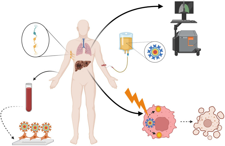
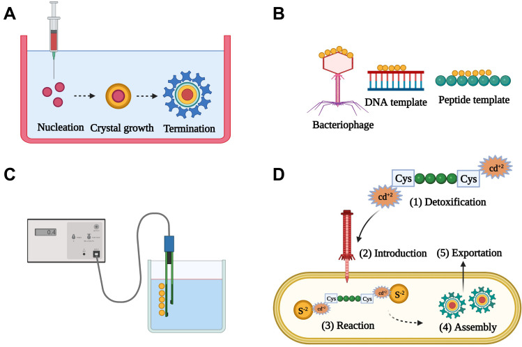
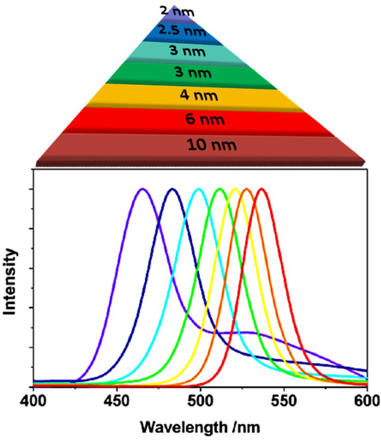
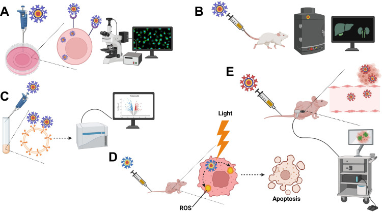
Despite the massive advancements in the nanomedicines and their associated research, their translation into clinically-applicable products is still below promises. The latter fact necessitates an in-depth evaluation of the current nanomedicines from a clinical perspective to cope with the challenges hampering their clinical potential. Quantum dots (QDs) are semiconductors-based nanomaterials with numerous biomedical applications such as drug delivery, live imaging, and medical diagnosis, in addition to other applications beyond medicine such as in solar cells. Nevertheless, the power of QDs is still underestimated in clinics. In the current article, we review the status of QDs in literature, their preparation, characterization, and biomedical applications. In addition, the market status and the ongoing clinical trials recruiting QDs are highlighted, with a special focus on the challenges limiting the clinical translation of QDs. Moreover, QDs are technically compared to other commercially-available substitutes. Eventually, we inspire the technical aspects that should be considered to improve the clinical fate of QDs.

Keywords: biosensors; clinical translation; clinical trials; in vivo imaging; photodynamic therapy; quantum dots.


Conflict of interest statement

The authors report no conflict of interests associated with this work.

Figures

None
Graphical abstract
Figure 1
Figure 1
A scheme of some commonly-used methods for the preparation of QDs. (A) Colloidal synthesis. Precursors are injected into organic or aqueous system under high temperature to facilitate their conversion into the molecular state, with a subsequent assembly into QDs. The major reaction steps include nucleation, crystal growth, and termination, which can be manipulated to control the physico-chemical properties of the produced QDs. (B) Biotemplate-based synthesis. Biological entities such as bacteriophages, genetically-engineered viruses, DNA, or peptides are used as templates to assemble the precursors into QDs. (C) Electrochemical assembly. Electrochemical driving force is used to assemble the precursor ions into QDs at the electrolyte-metal interface. (D) Biogenic synthesis. Heavy metal ions are detoxified via binding to cysteine-terminated peptides, followed by their introduction into microorganisms such as E. coli, where they react with the endogenous co-precursors (eg, sulfide ions) and assemble into QDs, which are subsequently exported out of the microorganism. Created by BioRender.com.
Figure 2
Figure 2
Impact of the particle size of CdSe QDs on their emission spectra upon irradiation with UV light. The emission wavelength is directly proportional to the particle size. Adapted from Bera D, Qian L, Tseng T-K, Holloway PH. Quantum Dots and Their Multimodal Applications: A Review. 2010;3(4):2260–2345.Copyright © 2010 by the authors; licensee Molecular Diversity Preservation International, Basel, Switzerland. This article is an open-access article distributed under the terms and conditions of the Creative Commons Attribution license (http://creativecommons.org/licenses/by/3.0/).
Figure 3
Figure 3
Some biomedical applications of QDs. (A) Intracellular imaging. QDs decorated with specific targeting moieties are used as fluorescent labels for intracellular visualization by fluorescence microscopy or confocal laser scanning microscopy (CLSM). (B) In vivo imaging. Following administration, QDs modified with tissue-specific targeting moieties can be used for visualization of certain organs in question using in vivo imaging systems (IVIS). (C) Fluorescence-activated cell sorting (FACS). QDs decorated with cell-specific ligands can be used as fluorescent probes for cell sorting during flow cytometry. Thanks to numerous advantages, QDs have a better capacity in polychromatic cell sorting compared to conventional organic dyes. (D) Photodynamic therapy (PDT). Following irradiation, QDs can act as photosensitizers or energy donors to other photosensitizers to generate reactive oxygen species (ROS) in situ leading to apoptotic cell death during cancer treatment. (E) Traceable drug delivery vehicles. QDs can be used as drug carriers to various tissues with high extravasation and tissue penetration capabilities thanks to their ultra-fine particle sizes. Owing to their quantum properties, QDs accumulation in the target tissues can be easily tracked. Created by BioRender.com.
Figure 4
Figure 4
Some common routes by which functionalized QDs internalize into the target cells. (A) Clathrin-mediated endocytosis. Following concentration of QDs at the receptor region, QDs are engulfed into clathrin-coated cell membrane invaginations, forming clathrin-coated vesicles. Dynamin mediates the dissociation of the aforementioned vesicles from the cell membrane into the cytosol leading to the formation of endosome. Subsequently, QDs should escape from the endosome and be released into the cytosol to avoid lysosomal degradation. (B) Caveolae-mediated endocytosis. The bindings of functionalized QDs to certain receptors mediates their internalization into the target cells via cholesterol-rich flask-shaped membrane invaginations, called caveolae, which subsequently dissociate from the cell membrane, forming caveosomes. Caveosomes are thought to be less destructive than endosomes. (C) Macropinocytosis. The binding of functionalized QDs to certain receptors activates the formation of cell membrane ruffles that engulf QDs into the cytosol forming macropinosomes, which subsequently leak their cargo into the cytosol. Created by BioRender.com.
Figure 5
Figure 5
Common challenges hampering the clinical translation of QDs. Stability issues include the high liability for degradation and aggregation during storage. Industrial issues arise from the complex scale-up procedures and the environmental hazards rendered by the heavy metal components. In vivo issues include the substantial loss of the injected dose via renal clearance owing to the ultra-fine particle size, poor selectivity due to non-specific interactions with tissues and cellular membranes, and intracellular toxicity caused by the generation of ROS or DNA damage. Created by BioRender.com.
Figure 6
Figure 6
Some potential applications of QDs in clinics. (A) Diagnostic imaging. QDs modified with specific targeting ligands can be injected intravenously to accumulate into the target organ, allowing its visualization. (B) Sentinel lymph node (SLN) detection. QDs can substitute the currently-used blue dye or radionuclide methods for the intraoperative detection of SLNs. Following peritumoral injection, QDs diffuse to the affected SLN(s) allowing accurate and safe detection of SLNs metastases. (C) Detection of micrometastases. Blood samples from cancer patients are fractioned by gradient centrifugation to collect PBMC and CTCs, followed by their mixing with magnetic nanoparticles that are labeled with anti-pan-ck antibody to enrich lung cancer epithelial cells. The micrometastatic cells conjugated to the magnetic nanoparticles are separated using a magnetic field, incubated with double antibody-labelled QDs, and detected under a fluorescence microscope. The figure (6C) is reproduced from Wang Y, Zhang Y, Du Z, Wu M, Zhang G. Detection of micrometastases in lung cancer with magnetic nanoparticles and quantum dots. Int J Nanomedicine. 2012;7:2315–2324. With a permission from Dove Medical Press, copyright 2012. (D) Cancer photodynamic therapy. Following administration of QDs, they tend to accumulate into the tumor site via passive or active targeting. Local irradiation of the tumor site induces the generation of ROS that kill the tumor cells. Created by BioRender.com.

Similar articles

Cited by

References

    1. Younis MA, Tawfeek HM, Abdellatif AAH, Abdel-Aleem JA, Harashima H. Clinical translation of nanomedicines: challenges, opportunities, and keys. Adv Drug Deliv Rev. 2022;181:114083. doi:10.1016/j.addr.2021.114083 - DOI - PubMed
    1. Yan ZP, Yang M, Lai CL. COVID-19 vaccines: a review of the safety and efficacy of current clinical trials. Pharmaceuticals. 2021;14:5. - PMC - PubMed
    1. Khalil IA, Younis MA, Kimura S, Harashima H. Lipid nanoparticles for cell-specific in vivo targeted delivery of nucleic acids. Biol Pharm Bull. 2020;43(4):584–595. doi:10.1248/bpb.b19-00743 - DOI - PubMed
    1. Younis MA, Khalil IA, Elewa YHA, Kon Y, Harashima H. Ultra-small lipid nanoparticles encapsulating sorafenib and midkine-siRNA selectively-eradicate sorafenib-resistant hepatocellular carcinoma in vivo. J Control Release. 2021;331:335–349. doi:10.1016/j.jconrel.2021.01.021 - DOI - PubMed
    1. Younis MA, Khalil IA, Abd Elwakil MM, Harashima H. A multifunctional lipid-based nanodevice for the highly specific codelivery of sorafenib and midkine siRNA to hepatic cancer cells. Mol Pharm. 2019;16(9):4031–4044. doi:10.1021/acs.molpharmaceut.9b00738 - DOI - PubMed