. 2022 Apr 21:10:832460.
doi: 10.3389/fcell.2022.832460. eCollection 2022.

Identification and Characterization of a Novel Long Noncoding RNA that Regulates Osteogenesis in Diet-Induced Obesity Mice

Affiliations

Identification and Characterization of a Novel Long Noncoding RNA that Regulates Osteogenesis in Diet-Induced Obesity Mice

Zhekai Hu et al. Front Cell Dev Biol. .

Abstract

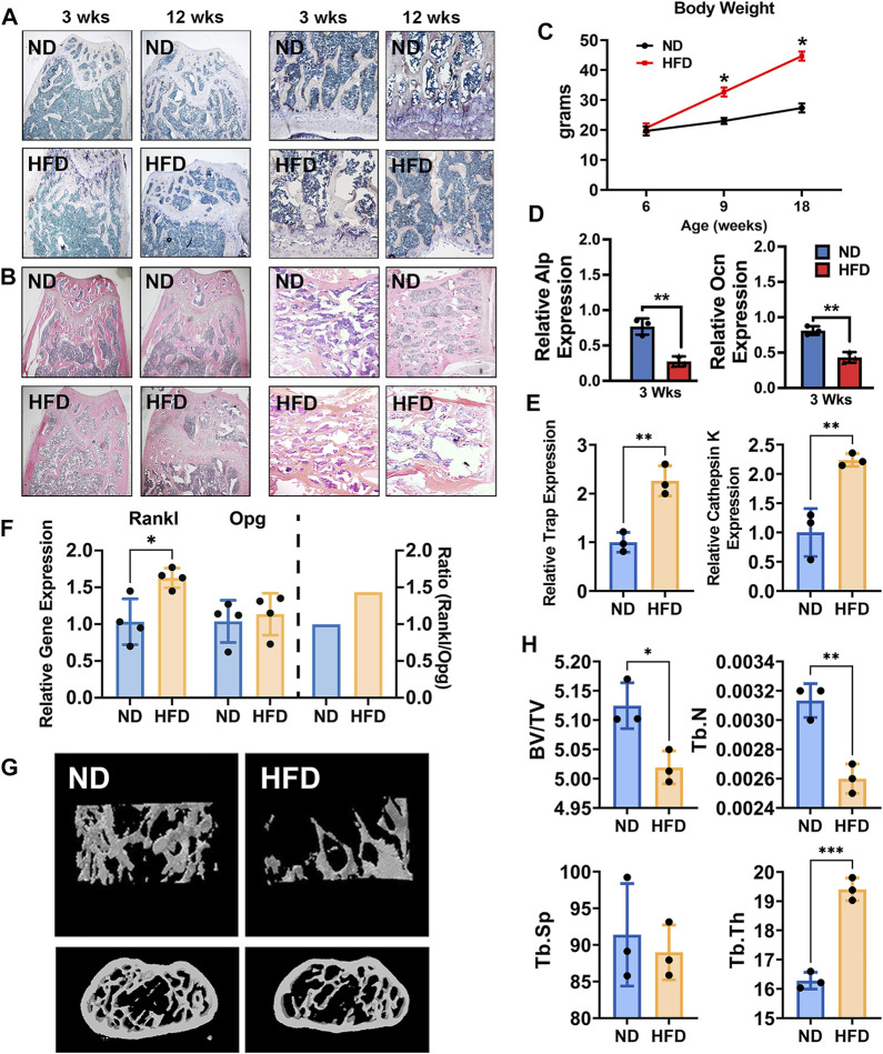
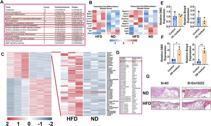
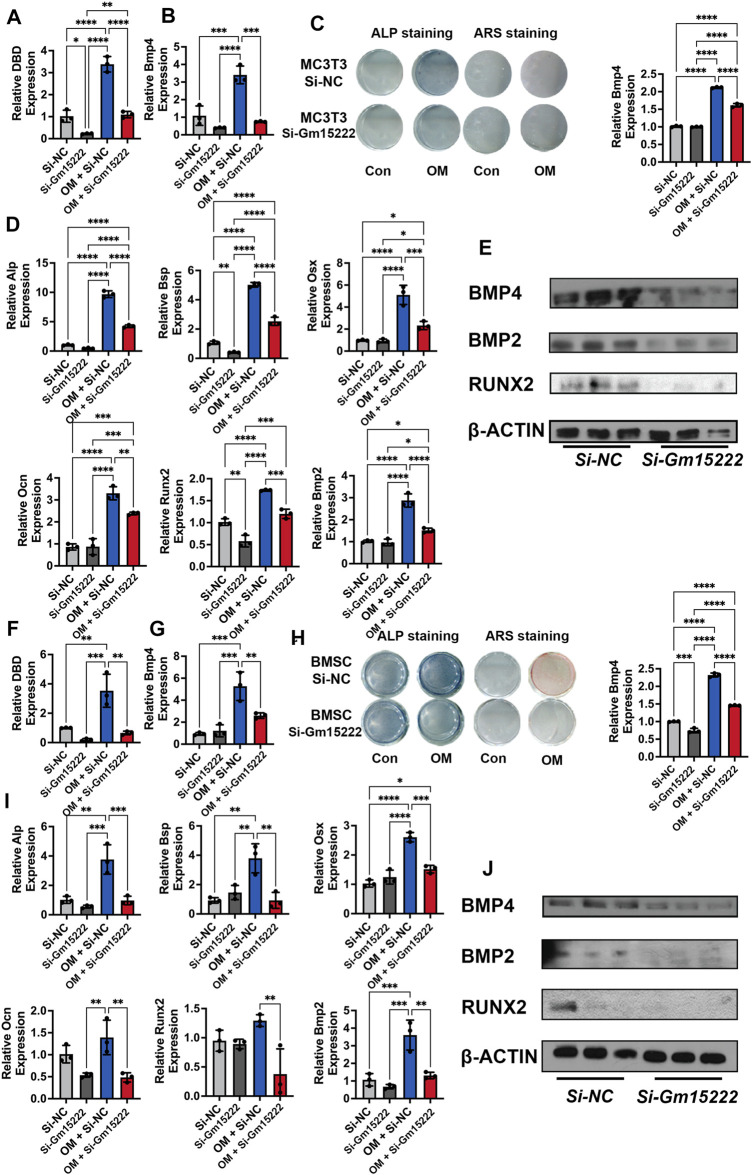
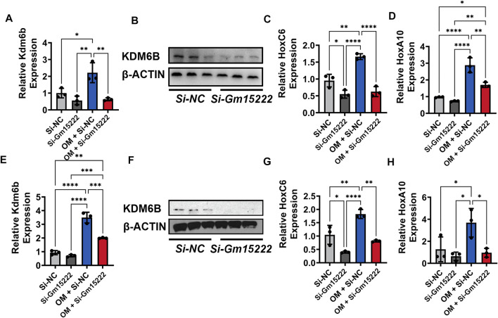
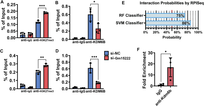
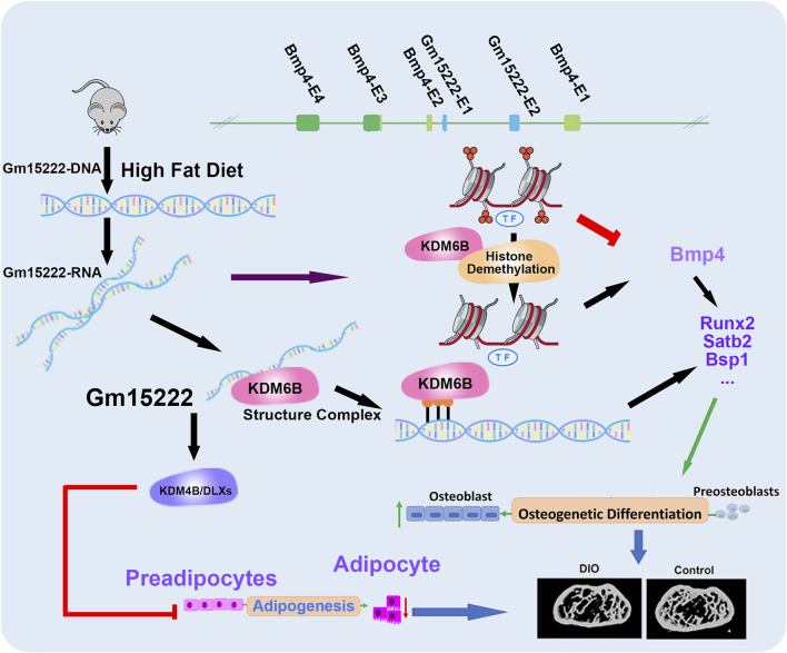
As a precursor to type 2 diabetes mellitus (T2D), obesity adversely alters bone cell functions, causing decreased bone quality. Currently, the mechanisms leading to alterations in bone quality in obesity and subsequently T2D are largely unclear. Emerging evidence suggests that long noncoding RNAs (lncRNAs) participate in a vast repertoire of biological processes and play essential roles in gene expression and posttranscriptional processes. Mechanistically, the expression of lncRNAs is implicated in pathogenesis surrounding the aggregation or alleviation of human diseases. To investigate the functional link between specific lncRNA and obesity-associated poor bone quality and elucidate the molecular mechanisms underlying the interaction between the two, we first assessed the structure of the bones in a diet-induced obese (DIO) mouse model. We found that bone microarchitecture markedly deteriorated in the DIO mice, mainly because of aberrant remodeling in the bone structure. The results of in vitro mechanistic experiments supported these observations. We then screened mRNAs and lncRNAs from DIO bones and functionally identified a specific lncRNA, Gm15222. Further analyses demonstrated that Gm15222 promotes osteogenesis and inhibits the expression of adipogenesis-related genes in DIO via recruitment of lysine demethylases KDM6B and KDM4B, respectively. Through this epigenetic pathway, Gm15222 modulates histone methylation of osteogenic genes. In addition, Gm15222 showed a positive correlation with the expression of a neighboring gene, BMP4. Together, the results of this study identified and provided initial characterization of Gm15222 as a critical epigenetic modifier that regulates osteogenesis and has potential roles in targeting the pathophysiology of bone disease in obesity and potential T2D.

Keywords: BMP4; LncR-DBD; diabetic bone diseases; diet-induced obese; epigenetics; long noncoding RNAs.


Conflict of interest statement

The authors declare that the research was conducted in the absence of any commercial or financial relationships that could be construed as a potential conflict of interest.

Figures

FIGURE 1
FIGURE 1
High-fat diet (HFD) leads to bone loss and alters bone metabolism in adult mice. (A) TRAP staining of femurs (left two panels) and vertebrae (right panels) of normal diet (ND) mice (18-week-old with ND) and HFD mice (18-week-old with 12 weeks of HFD). (B) H&E staining of femurs (left two panels) and vertebrae (right panels) of ND mice (18-week-old with ND) and HFD mice (18-week-old with 12 weeks of HFD) at 18 weeks of age. (C) The body weight curve. Male C57BL/6J DIO mice were fed HFD (60 kcal% fat) and control mice were fed normal diet (ND, 10 kcal% fat) between the ages of 6 and 18 weeks n = 4. Data were shown as mean ± S.D. (D) The mRNA expression of ALP (upper) and Ocn (lower), as detected by qRT-PCR in femurs of HFD and ND mice (3 weeks of HFD, n = 3). (E) The mRNA expression of TRAP and cathepsin K in femurs of HFD and ND mice (12 weeks of HFD, n = 3). (F) The mRNA expression of Opg and Rankl, as detected by qRT-PCR in femurs of HFD and ND mice (12 weeks of HFD, n = 4). (G) Representative images as detected and reconstructed by micro-CT and Ctan software in the femur of HFD and ND mice (12 weeks of HFD). (H) Histomorphometric analysis of micro-CT reconstruction of distal femurs, as measured by Ctan software, n = 3. Data were shown as mean ± S.D., two-tail t-test was used to test for those data between two groups. *p < 0.05; **p < 0.01; ***p < 0.001; ****p < 0.0001.
FIGURE 2
FIGURE 2
Identification of Gm15222 which has a distinct function in bone regeneration in the HFD mouse bones. (A) Upregulated genes annotated to a gene ontology (GO) term and example genes by gene ontology enrichment analysis of each component for ND mice and HFD mice. (B) Relative gene expression of osteogenesis (left) and osteoclastogenesis (right) markers ND and HFD mice. (C) The cluster heat map shows differentially expressed genes with expression change of more than two-fold from microarray data (three biological replicates per group, p < 0.05). Selected lncRNA has been highlighted in the red box. (D) lncRNAs classification and subgroup analysis showed the significantly expressed lncRNAs and their significantly expressed nearby genes. (E) BMMs were treated with or without osteoclasts differentiation medium [M-CSF (10 ng/ml) and RANKL (50 ng/ml)] for 5 days. The relative expression of Gm15222 and its related gene, Bmp4, were examined, n = 3. (F) BMSCs were treated with or without a mineralization medium [OM, ascorbic acid (50 μg/ml), 5 mM β-glycerophosphate] for 5 days, the relative expression of Gm15222 and its related gene Bmp4 were examined, n = 3. (G) Gm15222 deficiency inhibits bone regeneration in cranial bone of wild-type (WT) mice and high-fat-diet (HFD) mice. New bone was shown and highlighted with blue dotted lines. Data were shown as mean ± S.D., two-tail t-test was used to test for those data between two groups. *p < 0.05; **p < 0.01; ***p < 0.001; ****p < 0.0001.
FIGURE 3
FIGURE 3
lncRNA Gm15222 upregulates the expression of Bmp4 and promotes osteoblast differentiation. (A) The interference efficacy of si-Gm15222, as detected by qRT-PCR in MC3T3-E1 cell line treated with or without mineralization medium, n = 3 [OM, ascorbic acid (50 μg/ml), 5 mM β-glycerophosphate]. Si-NC, siRNA negative control; Si-Gm15222, Gm15222 siRNA; OM, osteogenic medium. (B) The silence of Gm15222 inhibited its neighboring gene Bmp4 expression, as detected by qRT-PCR in MC3T3-E1 cell line treated with or without mineralization medium and si-Gm15222, n = 3. (C) The silence of Gm15222 impaired its neighboring gene Bmp4 expression as detected by ALP staining and Alizarin red R staining in MC3T3-E1 cell line treated with or without mineralization medium: the left two columns of plates were detected by alkaline phosphatase (ALP) staining; and the right two columns of plates were detected by Alizarin Red S (ARS) staining; the right column plot was the quantification of ARS staining, n = 3. (D) The silence of Gm15222 impaired osteogenic genes Alp, Bsp, Osx, Ocn, Runx2, and Bmp2 mRNA expression as detected by qRT-PCR in MC3T3-E1 cell line treated with or without mineralization medium and Si-Gm15222, n = 3. (E) The silence of Gm15222 impaired osteogenic genes Bmp4, Runx2, and Bmp2 protein expression as detected by Western blot in MC3T3-E1 cell line treated with or without Si-Gm15222, n = 3. (F) The interference efficacy of si-Gm15222 as detected by qRT-PCR in BMSCs cell line treated with or without mineralization medium and Si-Gm15222, n = 3. (G) The silence of Gm15222 impaired gene Bmp4 expression as detected by ALP staining and Alizarin red R staining in BMSCs cell line treated with or without mineralization medium and Si-Gm15222, n = 3. (H) The silence of Gm15222 inhibited the osteogenesis of BMSCs treated with or without mineralization medium and Si-GM15222: the left two columns of plates were detected by ALP staining, and the right two were detected by ARS staining; the right column plot was the quantification of ARS staining, n = 3. (I) The silence of Gm15222 impaired osteogenic genes Alp, Bsp, Osx, Ocn, Runx2, and Bmp2 expression, as detected by qRT-PCR in BMSCs cell line treated with or without mineralization medium, n = 3 [OM, ascorbic acid (50 μg/ml), 5 mM β-glycerophosphate] and Si-Gm15222. (J) The silence of Gm15222 impaired osteogenic Bmp4, Runx2, and Bmp2 protein expression, as detected by Western blot in BMSCs cell line treated with or without Si-Gm15222, n = 3. Data were shown as mean ± S.D., one-way ANOVA with Tukey’s post hoc test was used to test those data. *p < 0.05; **p < 0.01; ***p < 0.001; ****p < 0.0001.
FIGURE 4
FIGURE 4
Gm15222 regulates the expression of Kdm6b and its downstream Hox gene family. (A) The silence of Gm15222 inhibited KDM6B expression, as detected by qRT-PCR in MC3T3-E1 cell line treated with or without mineralization medium and Si-Gm15222, n = 3. (B) The silence of Gm15222 impaired KDM6B protein expression in the process of mineralization, as detected by Western blot in MC3T3-E1 cell line treated with or without mineralization medium and Si-GM15222. (C) The silence of Gm15222 impaired HOXC6 expression, as detected by qRT-PCR in MC3T3-E1 cell line treated with or without mineralization medium and Si-Gm15222. (D) The silence of Gm15222 impaired HOXA10 gene expression, as detected by qRT-PCR in MC3T3-E1 cell line treated with or without mineralization medium and Si-Gm15222, n = 3. (E) The silence of Gm15222 inhibited KDM6B expression, as detected by qRT-PCR in BMSCs treated with or without mineralization medium and Si-GM15222, n = 3. (F) The silence of Gm15222 impaired KDM6B expression in the process of mineralization, as detected by Western blot in BMSCs treated with or without Si-Gm15222, n = 3. (G) The silence of Gm15222 impaired HOXC6 gene expression, as detected by qRT-PCR in BMSCs treated with or without mineralization medium and Si-Gm15222. (H) The silence of Gm15222 impaired HOXA10 expression, as detected by qRT-PCR in BMSCs treated with or without mineralization medium and Si-Gm15222, n = 3. Data were shown as mean ± S.D., one-way ANOVA with Tukey’s post hoc test was used to test those data. *p < 0.05; **p < 0.01; ***p < 0.001; ****p < 0.0001.
FIGURE 5
FIGURE 5
Gm15222 recruits KDM6B and alters the histone methylation in the promoters of BMP4 and HOXC6. (A) Quantification of BMP4 from retrieved Chromatin of BMSCs in the process of mineralization treated with or without si-Gm15222 in ChIP assay using H3K27me3 antibody, n = 3. SiNC, siRNA negative control. (B) Quantification of Bmp4 from retrieved Chromatin of BMSCs in the process of mineralization treated with or without si-Gm15222 in ChIP assay using Kdm6B antibody. (C) Quantification of HoxC6 from retrieved Chromatin of BMSCs in the process of mineralization treated with or without si-Gm15222 in ChIP assay using H3K27me3 antibody, n = 3. (D) Quantification of HoxC6 from retrieved DNAs of BMSCs in the process of mineralization treated with or without si-Gm15222 in ChIP assay using Kdm6B antibody, n = 3. (E) The interaction possibilities of Gm15222 and Kdm6b from RPISeq. (F) Quantification of Gm15222 from retrieved RNAs of BMSCs in RIP assay using Kdm6b antibody, n = 4. Data were shown as mean ± S.D., a two-tail t-test was used to test for those data between two groups, and a one-way ANOVA with Tukey’s post hoc test was used to test those data between more than two groups. *p < 0.05; **p < 0.01; ***p < 0.001; ****p < 0.0001.
FIGURE 6
FIGURE 6
A schematic diagram showing our newly identified Gm15222 and the epigenetic mechanisms and potential of how Gm15222 targets the pathophysiology of diabetic bone disease (DBD). In detail, Bmp4 contains four exons (E1-E4), and Gm15222 contains two exons (E1-E2). lnR-Gm15222 can recruit KDM6B and reducing the modification of H3K27me3, thereby leading to the expression of osteogenic genes; meanwhile, to overcome the adipogenesis, Gm15222 might participate the KDM4B associated adipogenic inhibition.

Similar articles

Cited by

References

    1. Agger K., Christensen J., Cloos P. A., Helin K. (2008). The Emerging Functions of Histone Demethylases. Curr. Opin. Genet. Dev. 18, 159–168. 10.1016/j.gde.2007.12.003 - DOI - PubMed
    1. Andersen R. E., Hong S. J., Lim J. J., Cui M., Harpur B. A., Hwang E., et al. (2019). The Long Noncoding RNA Pnky Is a Trans-acting Regulator of Cortical Development In Vivo . Develop. Cel 49, 632–642. 10.1016/j.devcel.2019.04.032 - DOI - PMC - PubMed
    1. Bouillon R. (1991). Diabetic Bone Disease. Calcif Tissue Int. 49, 155–160. 10.1007/bf02556109 - DOI - PubMed
    1. Bouxsein M. L., Boyd S. K., Christiansen B. A., Guldberg R. E., Jepsen K. J., Müller R. (2010). Guidelines for Assessment of Bone Microstructure in Rodents Using Micro-computed Tomography. J. Bone Miner Res. 25, 1468–1486. 10.1002/jbmr.141 - DOI - PubMed
    1. Brownlee M. (2001). Biochemistry and Molecular Cell Biology of Diabetic Complications. Nature 414, 813–820. 10.1038/414813a - DOI - PubMed