. 2022 Apr 21:10:857230.
doi: 10.3389/fcell.2022.857230. eCollection 2022.

Retinoic Acid is Required for Normal Morphogenetic Movements During Gastrulation

Affiliations

Retinoic Acid is Required for Normal Morphogenetic Movements During Gastrulation

Michal Gur et al. Front Cell Dev Biol. .

Abstract

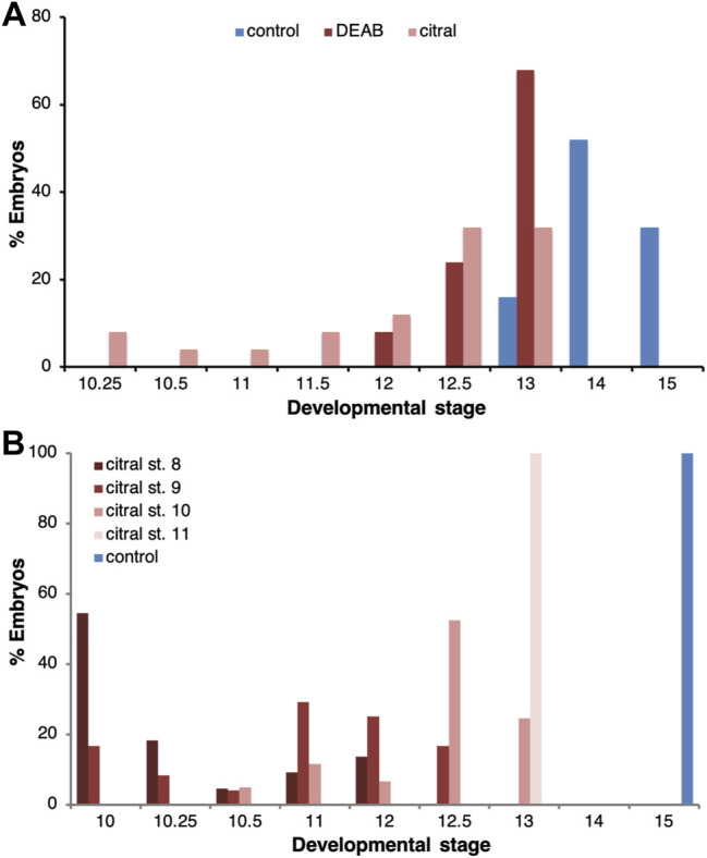
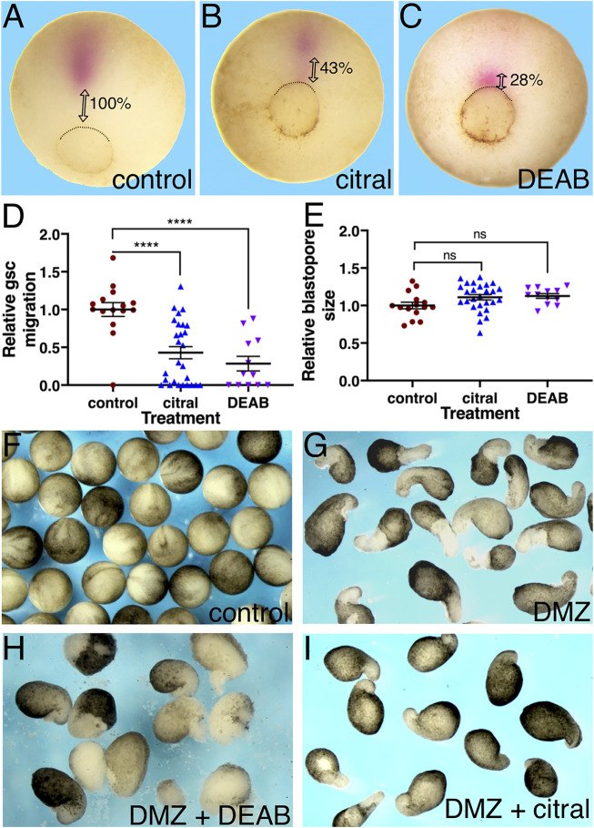
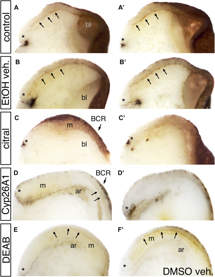
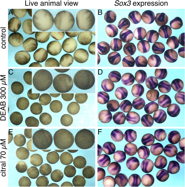
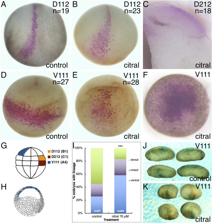
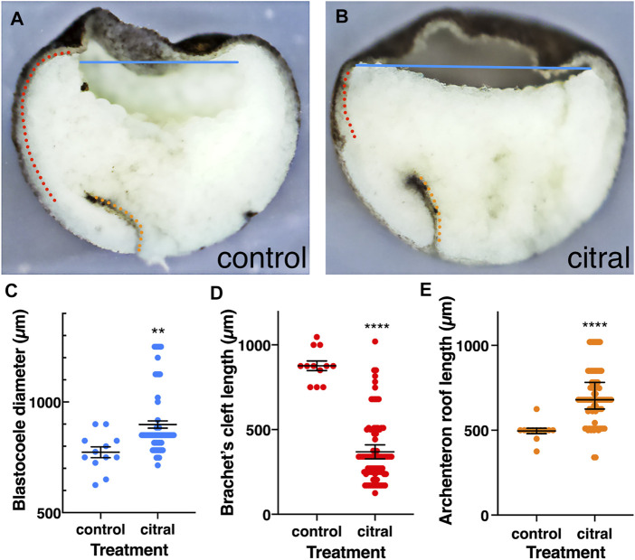
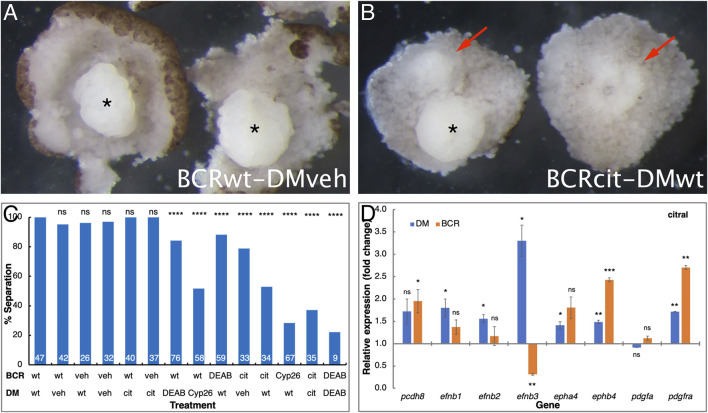
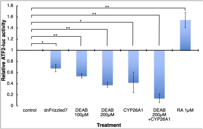
Retinoic acid (RA) is a central regulatory signal that controls numerous developmental processes in vertebrate embryos. Although activation of Hox expression is considered one of the earliest functions of RA signaling in the embryo, there is evidence that embryos are poised to initiate RA signaling just before gastrulation begins, and manipulations of the RA pathway have been reported to show gastrulation defects. However, which aspects of gastrulation are affected have not been explored in detail. We previously showed that partial inhibition of RA biosynthesis causes a delay in the rostral migration of some of the earliest involuting cells, the leading edge mesendoderm (LEM) and the prechordal mesoderm (PCM). Here we identify several detrimental gastrulation defects resulting from inhibiting RA biosynthesis by three different treatments. RA reduction causes a delay in the progression through gastrulation as well as the rostral migration of the goosecoid-positive PCM cells. RA inhibition also hampered the elongation of explanted dorsal marginal zones, the compaction of the blastocoel, and the length of Brachet's cleft, all of which indicate an effect on LEM/PCM migration. The cellular mechanisms underlying this deficit were shown to include a reduced deposition of fibronectin along Brachet's cleft, the substrate for their migration, as well as impaired separation of the blastocoel roof and involuting mesoderm, which is important for the formation of Brachet's cleft and successful LEM/PCM migration. We further show reduced non-canonical Wnt signaling activity and altered expression of genes in the Ephrin and PDGF signaling pathways, both of which are required for the rostral migration of the LEM/PCM, following RA reduction. Together, these experiments demonstrate that RA signaling performs a very early function critical for the progression of gastrulation morphogenetic movements.

Keywords: Brachet’s cleft; Xenopus embryo; embryo development; gastrulation delay; morphogenetic movements; retinoic acid signaling; tissue separation.


Conflict of interest statement

The authors declare that the research was conducted in the absence of any commercial or financial relationships that could be construed as a potential conflict of interest.

Figures

FIGURE 1
FIGURE 1
Delayed progression through gastrulation as a result of RA biosynthesis inhibition. (A) The progression of embryos through gastrulation treated with DEAB (300 μM; n = 38) or citral (70 μM; n = 39) to inhibit RA biosynthesis. When the majority of control embryos reached early neurula (st. 14/15; n = 38), the stage distribution of the treated samples was determined. The percentage of embryos at each stage in each treatment is shown. (B) The temporal sensitivity window for the delay in gastrulation induced by inhibition of RA biosynthesis was studied by initiating the citral treatment at different developmental stages (8, n = 22; 9, n = 24; 10, n = 61; 11, n = 24). When control embryos reached early neurula (st. 15), the stage distribution of the treated samples was determined. The percentage of embryos at each stage in each treatment is shown.
FIGURE 2
FIGURE 2
RA inhibition delays the rostral migration of the PCM. (A–E) Embryos were treated with citral or DEAB to inhibit the biosynthesis of RA. The extent of migration of the PCM during involution was determined by measuring their distance from the dorsal lip of the blastopore. The PCM cells were identified by gsc in situ hybridization. (A) Control embryo (n = 15). (B) Citral (70 µM) treated embryo (n = 27). (C) DEAB (300 µM) treated embryo (n = 12). (D) Relative PCM migration distribution. The migration distance was normalized to the embryo diameter. (E) Relative blastopore size distribution indicates embryos were at the same developmental stage. For each embryo, we measured its diameter and the diameter of the blastopore and calculated the ratio between them. (F–H) The inhibitory effect of reduced RA signaling was studied in explanted DMZs. RA biosynthesis was inhibited in explanted DMZs by incubation in citral or DEAB until control embryos reached st. 18 (F). (G) Control DMZ explants all had elongated columns of unpigmented mesoderm (n = 34) whereas in DEAB-treated DMZ explants (n = 16) (H) and citral-treated DMZ explants (n = 27) (I) these were missing or stunted. ****, p < 0.0001; ns, not significant.
FIGURE 3
FIGURE 3
The effect of reduced RA signaling on fibronectin (FN) deposition. Embryos were treated with DEAB or citral or injected with RNA encoding CYP26A1 to reduce RA levels. (A,A′) Two examples of untreated sibling control embryos. Arrows point to fibronectin deposited along Brachet’s cleft. * indicates the dorsal lip of the blastopore; bl, blastocoel. (B,B′) Two examples of embryos treated with EtOH vehicle only. FN deposition was equivalent to untreated controls. (C,C′) Two examples of embryos treated with citral. FN is detected in the ectoderm of the blastocoel roof (BCR) and mesoderm (m), but is not deposited along Brachet’s cleft. (D,D′) Two examples of embryos injected with cyp26a1 mRNA. FN is not detected in the BCR, the mesoderm (m), or Brachet’s cleft (arrows). ar, archenteron. (E) Embryo treated with DEAB showed reduced FN staining along Brachet’s cleft compared to a sibling embryo treated only with DMSO vehicle (F).
FIGURE 4
FIGURE 4
Reduced RA signaling affects the apparent location of the neural plate. Embryos were treated with DEAB or citral to inhibit the biosynthesis of RA. At st. 15, embryos were analyzed for the formation of the neural plate while alive by visual examination (A,C,E), or after in situ hybridization with the neural plate marker, sox3 (B,D,F). (A,B) Control embryos (n = 25). (C,D) Embryos treated with DEAB (300 μM; n = 23). (E,F) Treatment of embryos with citral (70 μM; n = 26).
FIGURE 5
FIGURE 5
Reduced RA signaling does not alter the dorsal-ventral axis. Single 32-cell blastomeres were injected with lineage tracer mRNA and labeled clones mapped at different developmental stages. (A,B) At stage 15, the D112/B1 clone (pink cells) occupies the medial and anterior neural plate in both untreated control (A) and citral treated (B) embryos. Dorso-anterior views. (C) The D212/C1 blastomere that normally contributes to the involuting dorsal mesoderm at gastrulation (st 10.5; Bauer et al., 1984) also does so in citral-treated embryos. Mid-sagittal section with anterior to the left and dorsal to the top. (D) At stage 17, the V111/A4 clone in untreated control embryos forms a coherent ventrolateral clone. Ventral view with anterior to the top. (E) In citral-treated sibling embryos, the stage 17 V111/A4 clone remains medial, i.e., has not expanded laterally, and the cells are more dispersed. Ventral view with anterior to the top. (F) At gastrula stages (st 10.5), the V111/A4 clone in citral-treated embryos occupies the blastocoel roof, similar to control embryos (cf. H). (G) Location of the 32-cell blastomeres that were lineage traced. Dorsal to the right, animal to the top. (H) Location of the V111/A4 clone (blue cells) at early gastrulation, modified from Bauer et al. (1984). Animal to the top and dorsal to the right. (I) Percentage of embryos in which the V111/A4 clone was located predominantly in the dorsal, ventral, or both (mixed) epidermis at tailbud stages. (J) Examples of control embryos showing V111/A4 descendants (blue cells) mostly in the dorsolateral epidermis. Dorsal to the top, anterior to the left. (K) Examples of citral treated embryos showing V111/A4 descendants (blue cells) mostly in the ventral and ventrolateral epidermis. Dorsal to the top, anterior to the left.
FIGURE 6
FIGURE 6
Internal reorganization of the embryo as a result of reduced RA signaling. Control (n = 12) and citral-treated (n = 84) embryos were incubated to mid-gastrula (st. 11) and bisected sagittally. (A,B) Control and citral-treated bisected embryos. Blastocoele diameter, blue line; Brachet’s cleft; dotted red line; Archenteron roof, dotted orange line. (C) Distribution of the blastocoel diameter in control and citral-treated embryos. (D) Measurement of Brachet’s cleft length in citral-treated and control embryos. (E) Length of the archenteron roof in citral-treated and control embryos. Citral-treated embryos were compared to the control values. **, p < 0.01; ****, p < 0.0001.
FIGURE 7
FIGURE 7
RA regulates the tissue separation behavior across Brachet’s cleft. Embryos were manipulated to reduce RA signaling levels by treatment with DEAB, citral, or injected with mRNA encoding CYP26A1. For controls, embryos were treated with the diluent alone (veh) for the DEAB or citral dilutions or left untreated (wt). The explanted BCR or DM regions were analyzed separately or conjugated for the separation behavior assay. (A) Control examples of separation behavior assay conjugates between control (wt) BCR and vehicle (veh) treated DM (asterisk). (B) Loss of separation behavior (arrows) when control DM was placed onto BCR treated with citral. * indicates a DM that did remain separated. (C) The extent of separation behavior (%) in all the assays performed in all combinations. # separation/# total: 47/47 DMwt/BCRwt; 40/42 DMveh/BCRwt; 25/26 DMwt/BCRveh; 31/32 DMveh/BCRveh; 40/40 DMcit/BCRwt; 37/37 DMcit/BCRveh; 64/76 DMDEAB/BCRwt; 30/58 DMCyp26/BCRwt; 52/59 DMwt/BCRDEAB; 26/33 DMveh/BCRcit; 18/34 Dmwt/BCRcit; 19/67 DMwt/BCRCyp26; 13/35 DMcit/BCRcit; 2/9 DMDEAB/BCRDEAB. (D) qPCR analysis of pcdh8, efnb1, efnb2, efnb3, epha4, ephb4, pdgfa, and pdgfra gene expression changes in the DM and BCR following citral treatment. **, p < 0.01; ***, p < 0.001; ****, p < 0.0001; ns, not significant.
FIGURE 8
FIGURE 8
RA signaling is necessary for normal Wnt/PCP activity. Embryos were injected with the ATF2-based non-canonical Wnt signaling reporter plasmid and subjected to DEAB or RA treatments, or co-injected with mRNA encoding dominant-negative Frizzled 7 or CYP26A1. During late gastrula (st. 12) embryos were collected and analyzed and the effect on the reporter activity (luciferase) was determined. *, p < 0.05; **, p < 0.01.

Similar articles

Cited by

References

    1. Ang H. L., Duester G. (1999). Stimulation of Premature Retinoic Acid Synthesis in Xenopus Embryos Following Premature Expression of Aldehyde Dehydrogenase ALDH1. Eur. J. Biochem. 260, 227–234. 10.1046/j.1432-1327.1999.00139.x - DOI - PubMed
    1. Barua D., Nagel M., Winklbauer R. (2021). Cell-cell Contact Landscapes in Xenopus Gastrula Tissues. Proc. Natl. Acad. Sci. U.S.A. 118. 10.1073/pnas.2107953118 - DOI - PMC - PubMed
    1. Bauer D. V., Huang S., Moody S. A. (1994). The Cleavage Stage Origin of Spemann's Organizer: Analysis of the Movements of Blastomere Clones before and during Gastrulation in Xenopus. Development 120, 1179–1189. 10.1242/dev.120.5.1179 - DOI - PubMed
    1. Begemann G., Schilling T. F., Rauch G.-J., Geisler R., Ingham P. W. (2001). The Zebrafishnecklessmutation Reveals a Requirement Forraldh2in Mesodermal Signals that Pattern the Hindbrain. Development 128, 3081–3094. 10.1242/dev.128.16.3081 - DOI - PubMed
    1. Blaner W. S. (2019). Vitamin A Signaling and Homeostasis in Obesity, Diabetes, and Metabolic Disorders. Pharmacol. Ther. 197, 153–178. 10.1016/j.pharmthera.2019.01.006 - DOI - PMC - PubMed

LinkOut - more resources