. 2022 May;10(9):e15301.
doi: 10.14814/phy2.15301.

Liver vitronectin release into the bloodstream increases due to reduced vagal muscarinic signaling after cerebral stroke in female mice

Affiliations

Liver vitronectin release into the bloodstream increases due to reduced vagal muscarinic signaling after cerebral stroke in female mice

Matthew P Keasey et al. Physiol Rep. 2022 May.

Abstract

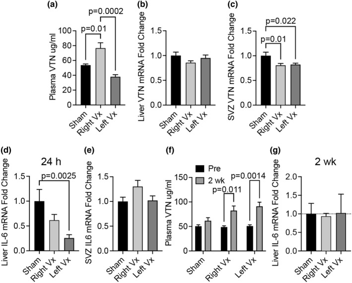
Vitronectin (VTN) is a glycoprotein enriched in the blood and activates integrin receptors. VTN blood levels increase only in female mice 24 h after an ischemic stroke and exacerbate brain injury through IL-6-driven inflammation, but the VTN induction mechanism is unknown. Here, a 30 min middle cerebral artery occlusion (MCAO) in female mice induced VTN protein in the liver (normally the main source) in concert with plasma VTN. Male mice were excluded as VTN is not induced after stroke. MCAO also increased plasma VTN levels after de novo expression of VTN in the liver of VTN-/- female mice, using a hepatocyte-specific (SERPINA1) promoter. MCAO did not affect SERPINA1 or VTN mRNA in the liver, brain, or several peripheral organs, or platelet VTN, compared to sham mice. Thus, hepatocytes are the source of stroke-induced increases in plasma VTN, which is independent of transcription. The cholinergic innervation by the parasympathetic vagus nerve is a potential source of brain-liver signaling after stroke. Right-sided vagotomy at the cervical level led to increased plasma VTN levels, suggesting that VTN release is inhibited by vagal tone. Co-culture of hepatocytes with cholinergic neurons or treatment with acetylcholine, but not noradrenaline (sympathetic transmitter), suppressed VTN expression. Hepatocytes have muscarinic receptors and the M1/M3 agonist bethanechol decreased VTN mRNA and protein release in vitro via M1 receptors. Finally, systemic bethanechol treatment blocked stroke-induced plasma VTN. Thus, VTN translation and release are inhibited by muscarinic signaling from the vagus nerve and presents a novel target for lessening detrimental VTN expression.

Keywords: blood protein; cholinergic; ischemic stroke; mice; vagus nerve; vitronectin.


Conflict of interest statement

None.

Figures

FIGURE 1
FIGURE 1
Plasma and liver VTN protein expression increases after stroke. (a) A 30 min ischemic stroke by MCAO in female C57BL/6 mice caused an increase in plasma VTN by 24 h, but not 3 h, after cessation of the stroke relative to naïve uninjured mice as measured by ELISA. Data = mean + SEM, n = 3,3,5. *p < 0.05, **p < 0.01, ****p < 0.0001 by ANOVA. (b) VTN mRNA levels in the liver were induced at 24 h, but not at 3 h, after MCAO relative to VTN levels in the livers of uninjured mice as measured by qPCR. Data are expressed as fold of naïve. (c) VTN protein expression in the liver was also increased at 24 h as shown by western blots of individual mice and quantified by densitometry of the VTN band as a ratio of the GAPDH loading control (d)
FIGURE 2
FIGURE 2
Plasma and liver VTN expression increase after stroke in VTN−/− female mice with restored liver VTN. (a) Mouse VTN or GFP (control) were cloned downstream of a hepatocyte specific alpha antitrypsin promoter (hAAT, Serpina A1) within an AAV backbone. (b) AAV‐GFP vectors were injected into the peritoneal cavity of VTN−/− female mice. After 6 weeks, GFP expression showed strong fluorescence in liver, but not brain sections. (c) VTN−/− female mice injected with AAV‐VTN under a hepatocyte‐specific Serpina 1A (Alpha antitrypsin; hAAT) promoter had VTN plasma levels at increasing concentrations over a 6‐week period to within physiological range, as determined by ELISA. An AAV‐GFP construct in VTN−/− females was used as a control. (d) AAV‐VTN females had a substantial increase in plasma VTN levels at 24 h after MCAO compared to their baseline levels before the stroke, as shown by ELISA. (e) The newly expressed VTN activated FAK in the liver as shown by pFAK in female mice at 24 h after an MCAO, as compared to AAV‐GFP controls as shown by Western blotting followed by densitometry (f)
FIGURE 3
FIGURE 3
MCAO does not induce VTN mRNA relative to sham surgery. (a) At 24 h after MCAO, mRNA expression of the hepatocyte‐specific gene, Serpina 1A, in the liver was unchanged in the AAV‐VTN infected VTN−/− mice. (b) In C57BL/6 mice, VTN mRNA expression has not induced the liver (Liv) or in other organs including forebrain (FoB), ovaries (Ova), uterus (Ute), stomach (Sto), jejunum (Jej), colon (Col), kidney (Kid), lung (Lun) and heart (Hea) at 24 h after MCAO relative to sham operated mice. (c) VTN protein content of platelets were not affected at 24 h post MCAO as measured by ELISA
FIGURE 4
FIGURE 4
Vagotomy increases VTN release into the bloodstream. (a) Naïve wildtype C57BL/6 female mice with a unilateral right, but not left, vagotomy in the neck area have increased plasma VTN levels 24 h later, compared to sham operated females, as shown by ELISA. N = 5,5,5. *p < 0.05, **p < 0.01, ***p < 0.001 by ANOVA. (b) In these same mice, expression of liver VTN mRNA was not affected but was slightly reduced in the subventricular zone (SVZ) region of the brain in both left and right vagotomized mice relative to sham operated mice at 24 h (c). (d) IL‐6 expression was decreased by the left‐sided vagotomy. (e) No changes were observed in the medial striatum containing the SVZ area of the brain. (f) Two weeks after right or left vagotomy, plasma levels of VTN protein were increased compared to pre‐vagotomy levels as shown by ELISA. N = 5, 4, 5. (g) Liver IL‐6 mRNA expression was not affected
FIGURE 5
FIGURE 5
VTN expression is regulated by acetylcholine through muscarinic receptors. (a) Co‐cultured differentiated cholinergic SH‐SY neuronal cells had a large inhibitory effect on VTN mRNA expression by AML hepatocytes, as shown by specific qPCR for mouse VTN in AML and not the human SH‐SY cells (3 independent experiments each). (b) Acetylcholine (ACh), but not noradrenaline (NA), reduced VTN within 4 h in AML cells (n = 4 each concentration). (c) The M1/3 muscarinic agonist bethanechol (BTH) suppressed VTN expression with increased efficacy at higher concentrations (n = 3 per concentration). (d) SiRNA mediated knockdown of the M1 but not M3 muscarinic receptors in AML cells showed increased VTN mRNA under BTH conditions which suppress VTN expression (n = 4 each). (e) M1 siRNA increased the amount of VTN in the medium of AML cells in the presence of BTH (n = 4 each). (f) siRNA against M1 or M3 receptors had no effect on fibronectin (FN) mRNA expression in these BTH‐treated AML cells. (g) Acetylcholine levels in the plasma were not affected at 24 h after an MCAO compared to naïve or sham operated female mice. (n = 4, 4, 4)
FIGURE 6
FIGURE 6
Bethanechol suppresses stroke induced plasma VTN but does not improve outcome. (a) Bethanechol (BTH, 20 mg/kg/day) was infused by Alzet osmotic pump placed subcutaneously for 24 h after an ischemic stroke by a 30 min MCAO in female C57BL/6 mice reduced VTN plasma levels at 24 h as shown by ELISA (n = 5,4). *p < 0.05, **p < 0.01, t‐test. (b) Liver VTN mRNA was not altered. (c) Similarly, striatal VTN mRNA was not significantly different after following BTH treatment relative to vehicle in MCAO mice. (d) Mice were infused with BTH (20 mg/kg/day) via osmotic pumps placed subcutaneously for 3 days post MCAO. Motor performance after stroke was not affected by BTH treatment, as measured by a grid‐walking test (n = 4,7). (e) Body weight was also not affected by BTH. (f) Histological assessment at 28 days after MCAO of tissue loss after stroke showed no effect of the earlier BTH treatment as measured in GFAP‐stained sections. (g, h) Inflammation was also not significantly affected by the earlier BTH treatment when measuring CD68 positive staining in injured brain tissue at 28 days. (9) At 24 h, liver IL‐6 mRNA was reduced in the liver or BTH treated MCAO mice in (A) relative to vehicle treated counterparts. (10) In contrast, striatal forebrain IL‐6, but not (11) TNF or (12) IL‐10, was robustly increased at 24 h after BTH treatment, as was (13) serum IL‐6 (n = 5,4)

Similar articles

Cited by

References

    1. Abhishekh, H. A. , Nisarga, P. , Kisan, R. , Meghana, A. , Chandran, S. , Trichur, R. , & Sathyaprabha, T. N. (2013). Influence of age and gender on autonomic regulation of heart. Journal of Clinical Monitoring and Computing, 27, 259–264. 10.1007/s10877-012-9424-3 - DOI - PubMed
    1. Ahnstedt, H. , McCullough, L. D. , & Cipolla, M. J. (2016). The importance of considering sex differences in translational stroke research. Translational Stroke Research, 7, 261–273. 10.1007/s12975-016-0450-1 - DOI - PMC - PubMed
    1. Akmayev, I. G. , Fidelina, O. V. , & Vikhreva, O. V. (1994). The rat dorsal vagal nucleus: Features of cellular and synaptic structure. Acta Biologica Hungarica, 45, 143–153. - PubMed
    1. Alessi, M.‐C. , Nicaud, V. , Scroyen, I. , Lange, C. , Saut, N. , Fumeron, F. , Marre, M. , Lantieri, O. , Fontaine‐Bisson, B. , Juhan‐Vague, I. , Balkau, B. , Tregouet, D.‐A. , & Morange, P.‐E. (2011). Association of vitronectin and plasminogen activator inhibitor‐1 levels with the risk of metabolic syndrome and type 2 diabetes mellitus. Results from the D.E.S.I.R. prospective cohort. Thrombosis and Haemostasis, 106, 416–422. 10.1160/TH11-03-0179 - DOI - PubMed
    1. Al‐Qudah, Z. A. , Yacoub, H. A. , & Souayah, N. (2015). Disorders of the autonomic nervous system after hemispheric cerebrovascular disorders: An update. Journal of Vascular and Interventional Neurology, 8, 43–52. - PMC - PubMed