. 2022 Oct 28;31(21):3683-3693.
doi: 10.1093/hmg/ddac103.

Functional analysis of rare genetic variants in complement factor I in advanced age-related macular degeneration

Affiliations

Functional analysis of rare genetic variants in complement factor I in advanced age-related macular degeneration

Anuja Java et al. Hum Mol Genet. .

Abstract

Factor I (FI) is a serine protease inhibitor of the complement system. Heterozygous rare genetic variants in complement factor I (CFI) are associated with advanced age-related macular degeneration (AMD). The clinical impact of these variants is unknown since a majority have not been functionally characterized and are classified as 'variants of uncertain significance' (VUS). This study assessed the functional significance of VUS in CFI. Our previous cross-sectional study using a serum-based assay demonstrated that CFI variants in advanced AMD can be categorized into three types. Type 1 variants cause a quantitative deficiency of FI. Type 2 variants demonstrate a qualitative deficiency. However, Type 3 variants consist of VUS that are less dysfunctional than Types 1 and 2 but are not as biologically active as wild type (WT). In this study, we employed site-directed mutagenesis followed by expression of the recombinant variant and a comprehensive set of functional assays to characterize nine Type 3 variants that were identified in 37 individuals. Our studies establish that the expression of the recombinant protein compared with WT is reduced for R202I, Q217H, S221Y and G263V. Further, G362A and N536K, albeit expressed normally, have significantly less cofactor activity. These results led to re-categorization of CFI variants R202I, Q217H, S221Y and G263V as Type 1 variants and to reclassification of N536K and G362A as Type 2. The variants K441R, Q462H and I492L showed no functional defect and remained as Type 3. This study highlights the utility of an in-depth biochemical analysis in defining the pathologic and clinical implications of complement variants underlying AMD.


Figures

Figure 1
Figure 1
(A) Schematic diagram of FI domain organization. FIMAC, FI membrane attack complex; SRCR, scavenger receptor cysteine-rich; LDLRA1, low-density lipoprotein class 1; LDLRA2, low-density lipoprotein class 2. (B and C) C3b binding and cofactor activity. FI, in the presence of a cofactor protein (FH, MCP; CD46), or CR1 (CD35), cleaves the α′ chain of C3b to obtain α43 and α41 (liberating C3f, a 2-kDa fragment), forming iC3b. Next, CR1 achieves further cleavage of iC3b to C3c and C3dg.
Figure 2
Figure 2
Biosynthetic evaluation of transfected variants. (A) Western Blotting of reduced SDS-PAGE of supernatants from transfected WT and variants are shown. For each expression study, indicated by the brackets and separated by red lines, both the variant(s) and WT FI were conducted. Controls for the Western Blotting also utilized purified FI (lane 14) that demonstrates the heavy chain at 50 kDa and the light chain at 38 kDa. Similar banding pattern is seen for the WT FI expression controls (lanes 1, 7 and 9). Recombinant expression of CFI genetic variants R202I (lane 2), Q217H (lane 3), S221Y (lane 4) and G263V (lane 5) shows decreased secretion compared with the WT control (lane 1). The secretion of the remaining variants is comparable to their WT [G362A (lane 7) and N536K (lane 8) to WT (lanes 1 and 9), respectively; Q462H (lane 10) and I492L (lane 11) to WT (lane 9); K441R (lane 13) to WT (lane 12)]. R, arginine; I, isoleucine; Q, glutamine; H, histidine; S, serine; Y, tyrosine; G, glycine; V, valine; A, alanine; N, asparagine; K, lysine; L, leucine. (B) Bar charts demonstrating comparison of WT and variant secretion based on Western Blotting. (C) Bar charts demonstrating comparison of WT and variant secretion based on densitometry and on ELISA. Also, see Table 1.
Figure 3
Figure 3
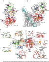
(A and B) Crystal structure of FI (generated using Pymol). Symbols indicate the following: purple circles = variant sites; yellow circles = serine protease site; light blue hexagons = N-glycosylation sites. Thick arrow-shaped structures in the various domains are beta pleated sheets. The dotted beige line in the linker represents a structure that is not visible in the crystal structure, and its location is therefore hypothetical. Serine protease, red; FIMAC, blue; linker, beige; LDLR1, yellow; LDLR2, orange; SRCR, green. C. Mapping of the FI variants on the surface of C3b in a complex with FI and FH (complement control repeats 1–4 and 19–20). Color coding is as follows: C3b, gray; FH, cyan. Refer to text for further explanation of the structural evaluation for each variant. R, arginine; Q, glutamine; S, serine; G, glycine; N, asparagine; K, lysine.
Figure 4
Figure 4
Functional evaluation of rare FI variant G362A: cofactor activity. The fluid-phase C3b cofactor activity of the variant FI with a cofactor protein (FH, MCP or CR1) was assessed by cleavage of purified C3b to iC3b and compared with WT FI derived from transfected cells. The percentage of alpha chain remaining and the percentage of generation of alpha 41 or alpha 43 indicates cleavage of C3b to iC3b at 0, 10, 20 and 30 min. Cleavage was quantitated by densitometric analysis of the remaining alpha chain and generation of alpha 43 or alpha 41. Data represent three separate experiments with bars corresponding to the SEM. Upon comparison to WT FI, the cofactor activity of variant G362A was significantly different with MCP and CR1 (P < 0.05) but not significant with FH as a cofactor (P 0.05). G, glycine; A, alanine. Bar charts demonstrating comparison of WT and variant cofactor activity at the 30 min time point. The numbers on the Y-axis in the bar charts reflect %.
Figure 5
Figure 5
Functional evaluation of rare FI variant N536K: cofactor activity. (A) The fluid-phase C3b cofactor activity of the variant FI with a cofactor protein (FH, MCP or CR1) was assessed by cleavage of purified C3b to iC3b and compared with WT FI derived from transfected cells. Data represent three separate experiments with bars corresponding to the SEM. There was no significant difference in the percentage of alpha chain remaining between WT and N536K variant for FH (P > 0.05) and CR1 (P > 0.05). The P-value for the difference in the percentage of alpha chain remaining between WT and N536K variant for MCP was not significant (P = 0.07) but reached significance for alpha 43 generation (P < 0.05). Bar charts demonstrate a comparison of WT and variant cofactor activity at the 30 min time point. The numbers on Y-axis reflect %. (B) Representative Western Blotting for MCP.
Figure 6
Figure 6
Functional evaluation of rare FI variants K441R, I492L and Q462H: cofactor activity. The fluid-phase C3b cofactor activity of the FI variants K441R, Q462H and I492L with a cofactor protein (FH, MCP or CR1) was assessed by cleavage of purified C3b to iC3b and compared with WT FI derived from transfected cells. Data represent three separate experiments with bars corresponding to the SEM. There was no significant difference (P > 0.05) in the percentage of alpha chain remaining between WT and variants for either of the cofactors, FH, MCP or CR1.

References

    1. Wong, W.L., Su, X., Li, X., Cheung, C.M., Klein, R., Cheng, C.Y. and Wong, T.Y. (2014) Global prevalence of age-related macular degeneration and disease burden projection for 2020 and 2040: a systematic review and meta-analysis. Lancet Glob. Health, 2, e106–e116. - PubMed
    1. Seddon, J.M. (2017) Macular degeneration epidemiology: nature-nurture, lifestyle factors, genetic risk, and gene-environment interactions—the Weisenfeld Award Lecture. Invest. Ophthalmol. Vis. Sci., 58, 6513–6528. - PMC - PubMed
    1. Schramm, E.C., Clark, S.J., Triebwasser, M.P., Raychaudhuri, S., Seddon, J.M. and Atkinson, J.P. (2014) Genetic variants in the complement system predisposing to age-related macular degeneration: a review. Mol. Immunol., 61, 118–125. - PMC - PubMed
    1. Seddon, J.M., Yu, Y., Miller, E.C., Reynolds, R., Tan, P.L., Gowrisankar, S., Goldstein, J.I., Triebwasser, M., Anderson, H.E., Zerbib, J. et al. (2013) Rare variants in CFI, C3 and C9 are associated with high risk of advanced age-related macular degeneration. Nat. Genet., 45, 1366–1370. - PMC - PubMed
    1. Kavanagh, D., Yu, Y., Schramm, E.C., Triebwasser, M., Wagner, E.K., Raychaudhuri, S., Daly, M.J., Atkinson, J.P. and Seddon, J.M. (2015) Rare genetic variants in the CFI gene are associated with advanced age-related macular degeneration and commonly result in reduced serum factor I levels. Hum. Mol. Genet., 24, 3861–3870. - PMC - PubMed

Publication types