. 2022 Jul;596(13):1639-1646.
doi: 10.1002/1873-3468.14373. Epub 2022 May 16.

Cadherins can dimerize via asymmetric interactions

Affiliations

Cadherins can dimerize via asymmetric interactions

Andrew Vae Priest et al. FEBS Lett. 2022 Jul.

Abstract

Cadherins are essential cell-cell adhesion proteins that interact in two distinct conformations: X-dimers and strand-swap dimers. Both X-dimers and strand-swap dimers are thought to exclusively rely on symmetric sets of interactions between key amino acids on both cadherin binding partners. Here, we use single-molecule atomic force microscopy and computer simulations to show that symmetry in cadherin binding is dispensable and that cadherins can also interact in a novel conformation that asymmetrically incorporates key elements of both strand-swap dimers and X-dimers. Our results clarify the biophysical rules for cadherin binding and demonstrate that cadherins interact in a more diverse range of conformations than previously understood.

Keywords: AFM force measurements; X-dimer; classical cadherins; molecular dynamics; strand-swap dimer; trans interactions.


Conflict of interest statement

Declaration of Interests: The authors declare no competing interests.

Figures

Figure 1.
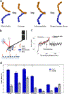
Figure 1.. Ecad dimer event rate measured with an Atomic Force Microscope.
(a) Ecad monomers (left) interact by first forming an X-dimer (middle-left) and transitioning to an intermediate conformation (middle-right) before fully converting to a strand-swap dimer (right) (strand-swap dimer corresponds to PDB ID: 3Q2V; X-dimer was visualized by alignment to PDB ID: 3LNH; the intermediate conformation was visualized by aligning to the structure found in ref (9)). (b) Biotinylated Ecad monomers were immobilized on AFM cantilevers and coverslips functionalized with polyethylene glycol (PEG) and streptavidin. Interactions between Ecad on the cantilever and the coverslip were measured. (c) A typical force-distance trace showing single rupture event. PEG stretching region of the force curve was fit to a worm like chain model (red). (d) Binding probabilities were measured using AFM in Ca2+ (blue) and EGTA (gray). Total number of Ca2+/EGTA measurements performed for each experiment were 12150/6534 measurements for WT-WT, 12147/6144 measurements for W2A-W2A, 11968/7350 measurements for K14E-K14E, 12150/6534 measurements for WT-Dblm, 12265/7350 measurements for W2A-Dblm, 12394/7350 measurements for K14E-Dblm, and 9654/7350 measurements for Dblm-Dblm. Error bars are the standard deviations of the bootstrapped event rates. Chi-squared tests show significant difference (p < 0.0001) between event rates for interacting dimers (WT-WT, K14E-K14E, W2A-W2A, WT-Dblm; in Ca2+) compared to nonspecific interactions (Dblm-Dblm; EGTA). WT-WT:Dblm-Dblm p = 1.0×10−40; K14E-K14E:Dblm-Dblm p = 1.1×10−43; W2A-W2A:Dblm-Dblm p = 1.5×10⁻²⁹; WT-Dblm:Dblm-Dblm p = 3.3×10¹⁷. Chi-squared tests also show that WT-WT, W2A-W2A, K14E-K14E, and WT-Dblm interactions are Ca2+ dependent (p < 0.0001) with Ca2+ vs. EGTA p-values of 8.6×10−44, 3.5×10⁻²⁰, 3.1×10⁻¹³, and 1.2×10⁻³⁴ respectively.
Figure 2.
Figure 2.. Dynamic force spectroscopy analysis of interacting Ecad dimers.
Loading rates measured in Ca2+ for a) WT-WT, b) K14E-K14E, c) W2A-W2A, and d) WT-Dblm were grouped together using k-means clustering. The Bell-Evans model (red line) was fit to the median unbinding forces and loading rates (black squares). Uncertainties in koff and xβ are median absolute deviations. Each circle represents a single unbinding event, and colors correspond to the data points from the same cluster.
Figure 3:
Figure 3:. Computational analysis of Ecad dimer structures.
(a) Structural alignment of the final frame of the MD simulations for WT-WT (orange), K14E-K14E (blue), and WT-Dblm (green). (b) Close-up of the W2 in the binding pocket of the opposing Ecad (from the black box in panel a). Structures were aligned to each other based on the binding pocket residues. (c) Violin plots of the distance between the center of mass of W2 and the respective binding pocket for strand-swap dimers. Distances for the mutated residues of WT-Dblm are also shown. The width of the violin signifies the density of data at that distance. The mean distance from each simulation repeat is overlayed on the violin plots (black circles). (d) Structural alignment of the final frame of the MD simulations for W2A-W2A (yellow) and WT-Dblm (green). (e) Close-up of the K14:D138 interaction (from the black box in panel d). (f) Violin plot of distances between K14 and D138 for X-dimers. The means from each simulation were used to determine the statistical significance between each pair in panels c and f; the distance determined from the mutated residues in WT-Dblm shows significant difference compared to the unmutated residues. Welch’s T-test of W2:pocket with W2:pocket had a p-value > 0.1 while W2:pocket with A2:pocket had a p-value ~0.0015. Welch’s T-test of K14:D138 with K14:D138 had a p-value > 0.1 while K14:D138 with E14:D138 had a p-value < 0.001. Table of p-values found in (Table S1).

References

    1. Mendonsa AM, Na T-Y, Gumbiner BM. E-cadherin in contact inhibition and cancer. Oncogene. 2018;37(35):4769–80. - PMC - PubMed
    1. Brasch J, Harrison OJ, Honig B, Shapiro L. Thinking outside the cell: how cadherins drive adhesion. Trends Cell Biol. 2012;22(6):299–310. - PMC - PubMed
    1. Priest AV, Koirala R, Sivasankar S. Single molecule studies of classical and desmosomal cadherin adhesion. Current Opinion in Biomedical Engineering. 2019;12:43–50. - PMC - PubMed
    1. Boggon TJ, Murray J, Chappuis-Flament S, Wong E, Gumbiner BM, Shapiro L. C-cadherin ectodomain structure and implications for cell adhesion mechanisms. Science. 2002;296(5571):1308–13. - PubMed
    1. Pertz O, Bozic D, Koch AW, Fauser C, Brancaccio A, Engel J. A new crystal structure, Ca2+ dependence and mutational analysis reveal molecular details of E-cadherin homoassociation. EMBO J. 1999;18(7):1738–47. - PMC - PubMed

Publication types