

Development of HEK-293 Cell Lines Constitutively Expressing Flaviviral Antigens for Use in Diagnostics
- PMID: 35532242
- PMCID: PMC9241944
- DOI: 10.1128/spectrum.00592-22
Development of HEK-293 Cell Lines Constitutively Expressing Flaviviral Antigens for Use in Diagnostics
Abstract
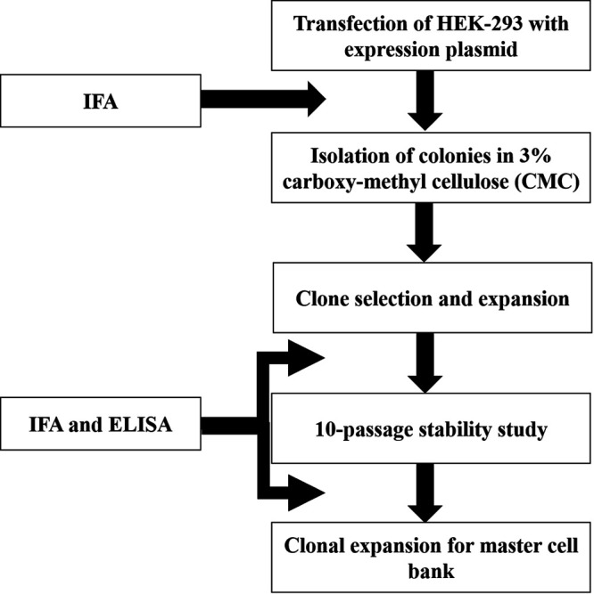
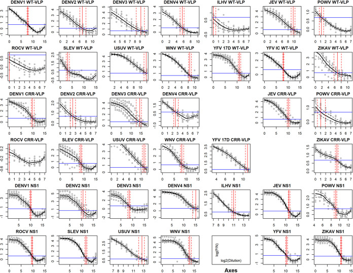
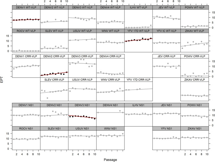
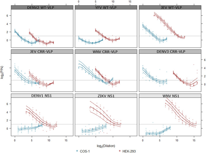
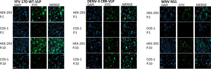
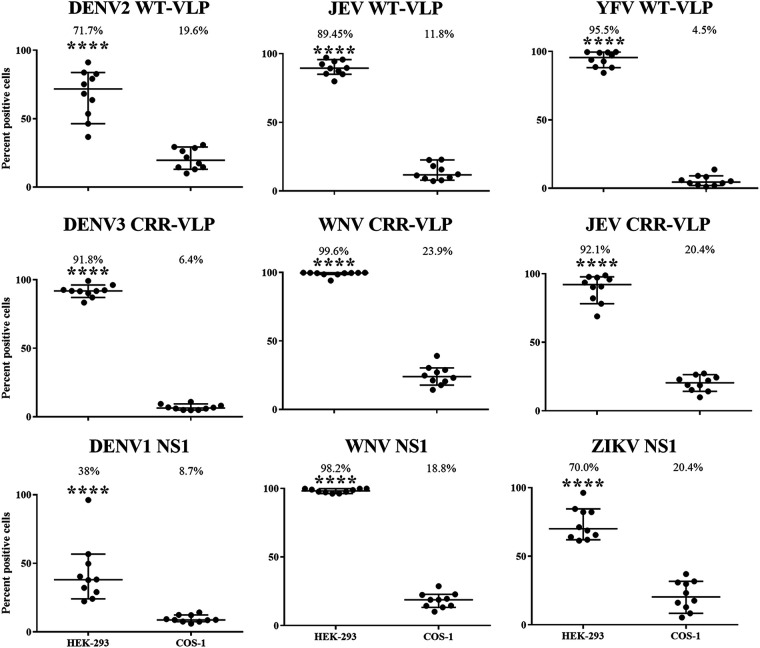
Flaviviruses are important human pathogens worldwide. Diagnostic testing for these viruses is difficult because many of the pathogens require specialized biocontainment. To address this issue, we generated 39 virus-like particle (VLP)- and nonstructural protein 1 (NS1)-secreting stable cell lines in HEK-293 cells of 13 different flaviviruses, including dengue, yellow fever, Japanese encephalitis, West Nile, St. Louis encephalitis, Zika, Rocio, Ilheus, Usutu, and Powassan viruses. Antigen secretion was stable for at least 10 cell passages, as measured by enzyme-linked immunosorbent assays and immunofluorescence assays. Thirty-five cell lines (90%) had stable antigen expression over 10 passages, with three of these cell lines (7%) increasing in antigen expression and one cell line (3%) decreasing in antigen expression. Antigen secretion in the HEK-293 cell lines was higher than in previously developed COS-1 cell line counterparts. These antigens can replace current antigens derived from live or inactivated virus for safer use in diagnostic testing. IMPORTANCE Serological diagnostic testing for flaviviral infections is hindered by the need for specialized biocontainment for preparation of reagents and assay implementation. The use of previously developed COS-1 cell lines secreting noninfectious recombinant viral antigen is limited due to diminished antigen secretion over time. Here, we describe the generation of 39 flaviviral virus-like particle (VLP)- and nonstructural protein 1 (NS1)-secreting stable cell lines in HEK-293 cells representing 13 medically important flaviviruses. Antigen production was more stable and statistically higher in these newly developed cell lines than in their COS-1 cell line counterparts. The use of these cell lines for production of flaviviral antigens will expand serological diagnostic testing of flaviviruses worldwide.
Keywords: arbovirus; diagnostics; flavivirus; recombinant protein production.
Conflict of interest statement
The authors declare a conflict of interest. CDC has filed a patent application describing these cell lines. Davis B.S. and Chang G.J. Stable human cell lines expressing flavivirus virus-like particles and uses thereof, 20 May 2020. US provisional patent application 63/021,852.
Figures
Similar articles
-
Comparative analysis of immunoglobulin M (IgM) capture enzyme-linked immunosorbent assay using virus-like particles or virus-infected mouse brain antigens to detect IgM antibody in sera from patients with evident flaviviral infections.J Clin Microbiol. 2005 Jul;43(7):3227-36. doi: 10.1128/JCM.43.7.3227-3236.2005. J Clin Microbiol. 2005. PMID: 16000440 Free PMC article.
-
Nonstructural protein 1-specific immunoglobulin M and G antibody capture enzyme-linked immunosorbent assays in diagnosis of flaviviral infections in humans.J Clin Microbiol. 2015 Feb;53(2):557-66. doi: 10.1128/JCM.02735-14. Epub 2014 Dec 10. J Clin Microbiol. 2015. PMID: 25502522 Free PMC article.
-
Noninfectious recombinant antigen for detection of St. Louis encephalitis virus-specific antibodies in serum by enzyme-linked immunosorbent assay.J Clin Microbiol. 2004 Oct;42(10):4709-17. doi: 10.1128/JCM.42.10.4709-4717.2004. J Clin Microbiol. 2004. PMID: 15472331 Free PMC article.
-
Secretion of flaviviral non-structural protein NS1: from diagnosis to pathogenesis.Novartis Found Symp. 2006;277:233-47; discussion 247-53. doi: 10.1002/0470058005.ch17. Novartis Found Symp. 2006. PMID: 17319166 Review.
-
The Role of NS1 Protein in the Diagnosis of Flavivirus Infections.Viruses. 2023 Feb 19;15(2):572. doi: 10.3390/v15020572. Viruses. 2023. PMID: 36851784 Free PMC article. Review.
Cited by
-
Development of a Diagnostic IgM Antibody Capture ELISA for Detection of Anti-Cache Valley Virus Human IgM.Am J Trop Med Hyg. 2024 Nov 19;112(2):386-395. doi: 10.4269/ajtmh.24-0360. Print 2025 Feb 5. Am J Trop Med Hyg. 2024. PMID: 39561397 Free PMC article.
References
-
- Kraemer MUG, Reiner RC Jr, Brady OJ, Messina JP, Gilbert M, Pigott DM, Yi D, Johnson K, Earl L, Marczak LB, Shirude S, Davis Weaver N, Bisanzio D, Perkins TA, Lai S, Lu X, Jones P, Coelho GE, Carvalho RG, Van Bortel W, Marsboom C, Hendrickx G, Schaffner F, Moore CG, Nax HH, Bengtsson L, Wetter E, Tatem AJ, Brownstein JS, Smith DL, Lambrechts L, Cauchemez S, Linard C, Faria NR, Pybus OG, Scott TW, Liu Q, Yu H, Wint GRW, Hay SI, Golding N. 2019. Past and future spread of the arbovirus vectors Aedes aegypti and Aedes albopictus. Nat Microbiol 4:854–863. doi:10.1038/s41564-019-0376-y. - DOI - PMC - PubMed
-
- Bhatt S, Gething PW, Brady OJ, Messina JP, Farlow AW, Moyes CL, Drake JM, Brownstein JS, Hoen AG, Sankoh O, Myers MF, George DB, Jaenisch T, Wint GR, Simmons CP, Scott TW, Farrar JJ, Hay SI. 2013. The global distribution and burden of dengue. Nature 496:504–507. doi:10.1038/nature12060. - DOI - PMC - PubMed
-
- Messina JP, Brady OJ, Golding N, Kraemer MUG, Wint GRW, Ray SE, Pigott DM, Shearer FM, Johnson K, Earl L, Marczak LB, Shirude S, Davis Weaver N, Gilbert M, Velayudhan R, Jones P, Jaenisch T, Scott TW, Reiner RC Jr, Hay SI. 2019. The current and future global distribution and population at risk of dengue. Nat Microbiol 4:1508–1515. doi:10.1038/s41564-019-0476-8. - DOI - PMC - PubMed
Publication types
MeSH terms
Substances
LinkOut - more resources
Full Text Sources
Medical
Research Materials
Miscellaneous
