Review
. 2022 Jul-Aug;11(4):252-274.
doi: 10.4103/EUS-D-21-00151.

Ultrasound elastography

Affiliations
Review

Ultrasound elastography

Xin-Wu Cui et al. Endosc Ultrasound. 2022 Jul-Aug.

Abstract

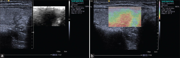
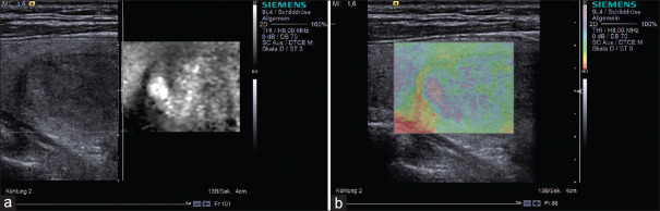
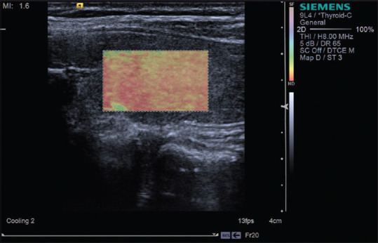
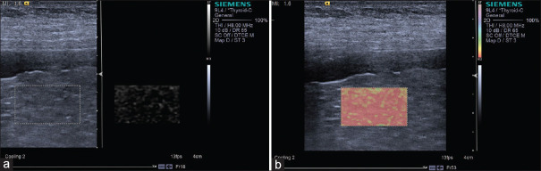
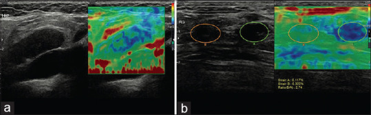
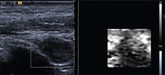
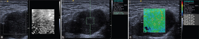
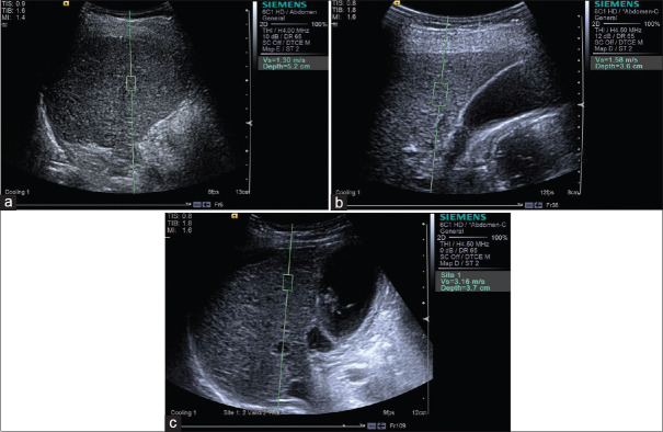
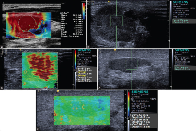
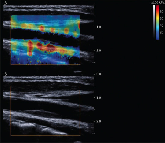
Physicians have used palpation as a diagnostic examination to understand the elastic properties of pathology for a long time since they realized that tissue stiffness is closely related to its biological characteristics. US elastography provided new diagnostic information about elasticity comparing with the morphological feathers of traditional US, and thus expanded the scope of the application in clinic. US elastography is now widely used in the field of diagnosis and differential diagnosis of abnormality, evaluating the degree of fibrosis and assessment of treatment response for a range of diseases. The World Federation of Ultrasound Medicine and Biology divided elastographic techniques into strain elastography (SE), transient elastography and acoustic radiation force impulse (ARFI). The ARFI techniques can be further classified into point shear wave elastography (SWE), 2D SWE, and 3D SWE techniques. The SE measures the strain, while the shear wave-based techniques (including TE and ARFI techniques) measure the speed of shear waves in tissues. In this review, we discuss the various techniques separately based on their basic principles, clinical applications in various organs, and advantages and limitations and which might be most appropriate given that the majority of doctors have access to only one kind of machine.

Keywords: acoustic radiation force impulse; elastography; shear wave; strain; ultrasound.


Conflict of interest statement

None

Figures

Figure 1
Figure 1
An example of strain image (breast cancer). The strain image is overlaid as a color scale with red being soft and blue being hard on the B-mode image
Figure 2
Figure 2
A breast mass of a 56-year-old woman which proved to be adenopathy with fibroadenomatous nodules (a and b). The Tsukuba score was 1 (a) and the strain ratio was 0.90 (b). A breast mass of a 72-year-old woman, which proved to be invasive carcinoma (c and d). The Tsukuba score was 4 (c) and the strain ratio was 3.04 (d)
Figure 3
Figure 3
An example of score 3 thyroid nodule, which is proven as papillary carcinoma; almost the whole lesion is displayed in hard blue (a), the strain ratio is 4.94 (b)
Figure 4
Figure 4
A 44-year-old female patient with a papillary microcarcinoma in the left thyroid gland. The strain ratio was 2.39
Figure 5
Figure 5
Strain elastography with Virtual Touch image showing a papillary carcinoma (a and b) is hard
Figure 6
Figure 6
Strain elastography with Virtual Touch image showing a nodular goiter (a and b) is soft
Figure 7
Figure 7
Strain elastography with Virtual Touch image showing Hashimoto thyroiditis is hard
Figure 8
Figure 8
Strain elastography with Virtual Touch image showing liver cirrhosis (F4) (a and b) is hard
Figure 9
Figure 9
Strain elastography revealed a metastatic lymph node in the neck with the Tsukuba score of 3 (a); inguinal metastatic lymph nodes with the Tsukuba score of 4 (b)
Figure 10
Figure 10
The neck lymph node metastasis appeared to be hard on strain elastography with Virtual Touch image
Figure 11
Figure 11
Non-Hodgkin's lymphoma is not so hard on strain elastography with Virtual Touch image (a) and shear wave elastography with Virtual Touch quantification (b) and Virtual Touch tissue quantification (c)
Figure 12
Figure 12
Acoustic radiation force impulse techniques with Virtual Touch quantification for diagnosis F2 (a), F3 (b), F4 (c) in hepatitis B patients
Figure 13
Figure 13
Acoustic radiation force impulse techniques with Virtual Touch quantification showing liver cirrhosis in a patient with alcoholic liver disease
Figure 14
Figure 14
SWE images of the liver in patients with hepatitis B: F1 – (a) E mean 7.1 kPa; F2 – (b) E mean 7.8 kPa; F3 – (c) E mean 9.5 kPa; F4 – (d) E mean 24.5 kPa
Figure 15
Figure 15
Acoustic radiation force impulse techniques with Virtual Touch quantification (a) and Virtual Touch image (b) showing hepatocellular carcinoma is hard in a patient with hepatocellular carcinoma
Figure 16
Figure 16
2D SWE showing the lesion is hard in patients with metastasis (a), hemangioma (b), and focal nodular hyperplasia (c)
Figure 17
Figure 17
Acoustic radiation force impulse with Virtual Touch quantification shows breast fibroadenoma is softer (a) and breast invasive ductal carcinoma is harder (b)
Figure 18
Figure 18
2D shear wave elastography shows fibroadenoma nodule is softer (a) and invasive ductal carcinoma is harder (b)
Figure 19
Figure 19
Acoustic radiation force impulse shows papillary carcinoma (a, red area) is harder and adenoma (b and c) is softer, while follicular carcinoma (d) is not so hard as papillary carcinoma
Figure 20
Figure 20
2D shear wave elastography shows papillary thyroid carcinoma is harder (a) and nodular goiter is softer (b)
Figure 21
Figure 21
Metastatic lymph node is shown to be hard on 2D shear wave elastography (a), acoustic radiation force impulse with Virtual Touch quantification (b) and Virtual Touch tissue quantification (c), while inflammatory lymph nodes appear to be soft, which is shown on acoustic radiation force impulse with Virtual Touch quantification (d) and Virtual Touch tissue quantification (e)
Figure 22
Figure 22
2D shear wave elastography image of a stable plaque in a 55-year-old man

References

    1. Ophir J, Céspedes I, Ponnekanti H, et al. Elastography: A quantitative method for imaging the elasticity of biological tissues. Ultrason Imaging. 1991;13:111–34. - PubMed
    1. Ferraioli G, Wong VW, Castera L, et al. Liver ultrasound elastography: An update to the World Federation for Ultrasound in Medicine and Biology guidelines and recommendations. Ultrasound Med Biol. 2018;44:2419–40. - PubMed
    1. Shiina T, Nightingale KR, Palmeri ML, et al. WFUMB guidelines and recommendations for clinical use of ultrasound elastography: Part 1: Basic principles and terminology. Ultrasound Med Biol. 2015;41:1126–47. - PubMed
    1. Bamber J, Cosgrove D, Dietrich CF, et al. EFSUMB guidelines and recommendations on the clinical use of ultrasound elastography. Part 1: Basic principles and technology. Ultraschall Med. 2013;34:169–84. - PubMed
    1. Cosgrove D, Piscaglia F, Bamber J, et al. EFSUMB guidelines and recommendations on the clinical use of ultrasound elastography. Part 2: Clinical applications. Ultraschall Med. 2013;34:238–53. - PubMed