. 2022 Aug;19(4):739-754.
doi: 10.1007/s13770-022-00447-3. Epub 2022 May 9.

Development of a Novel Perfusion Rotating Wall Vessel Bioreactor with Ultrasound Stimulation for Mass-Production of Mineralized Tissue Constructs

Affiliations

Development of a Novel Perfusion Rotating Wall Vessel Bioreactor with Ultrasound Stimulation for Mass-Production of Mineralized Tissue Constructs

Jae Min Cha et al. Tissue Eng Regen Med. 2022 Aug.

Abstract

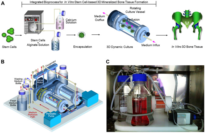
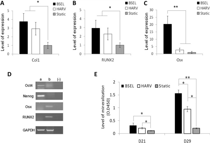
Background: As stem cells are considered a promising cell source for tissue engineering, many culture strategies have been extensively studied to generate in vitro stem cell-based tissue constructs. However, most approaches using conventional tissue culture plates are limited by the lack of biological relevance in stem cell microenvironments required for neotissue formation. In this study, a novel perfusion rotating wall vessel (RWV) bioreactor was developed for mass-production of stem cell-based 3D tissue constructs.

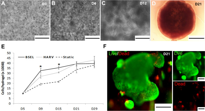
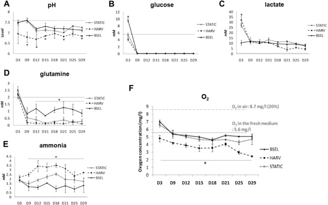
Methods: An automated RWV bioreactor was fabricated, which is capable of controlling continuous medium perfusion, highly efficient gas exchange with surrounding air, as well as low-intensity pulsed ultrasound (LIPUS) stimulation. Embryonic stem cells encapsulated in alginate/gelatin hydrogel were cultured in the osteogenic medium by using our bioreactor system. Cellular viability, growth kinetics, and osteogenesis/mineralization were thoroughly evaluated, and culture media were profiled at real time. The in vivo efficacy was examined by a rabbit cranial defect model.

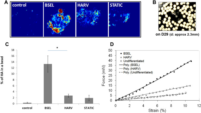
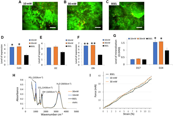
Results: Our bioreactor successfully maintained the optimal culture environments for stem cell proliferation, osteogenic differentiation, and mineralized tissue formation during the culture period. The mineralized tissue constructs produced by our bioreactor demonstrated higher void filling efficacy in the large bone defects compared to the group implanted with hydrogel beads only. In addition, the LIPUS modules mounted on our bioreactor successfully reached higher mineralization of the tissue constructs compared to the groups without LIPUS stimulation.

Conclusion: This study suggests an effective biomanufacturing strategy for mass-production of implantable mineralized tissue constructs from stem cells that could be applicable to future clinical practice.

Keywords: 3D mineralized tissue constructs; Low-intensity ultrasound; Perfusion; Rotating wall vessel bioreactor; Stem cells.


Conflict of interest statement

No competing financial interests exist.

Figures

Fig. 1
Fig. 1
Need for perfusion culture system. A, B Fed-batch bioreactors are problematic in the maintenance of a desirable culture environment due to the exhaustion of nutrients (A) and accumulation of toxic cellular metabolites (B). As cells grow over the culture time, cells in a fed-batch bioreactor can be more stressed by the abrupt exposure to a different culture environment caused by the exchange of medium. C, D Perfusion culture systems continuously supply the nutrients (C), prevent metabolic waste accumulation (D), and eliminate the potential stress caused by medium-exchange, facilitating the maintenance of a desirable environment
Fig. 2
Fig. 2
Integrated bioprocess for 3D stem cell culture and a perfusion RWV bioreactor (BSEL) system. (A) Approximately five-hundreds of stem cell-laden alginate beads were produced within 3 min by an automated hydrogel encapsulator (each bead contained about 20,000 cells). Large scale production of stem cell-based 3D-tissue constructs was achieved by using a novel perfusion RWV bioreactor system. (B) Two sets of the perfusion RWV system were mounted on a single base unit, which rotated together by a servo motor (a perfusion path driven by a peristaltic pump is indicated by the red arrows: fresh medium bottle → oxygenator → culture vessel → wasting medium bottle). A gas-permeable membrane covered the culture vessel to have a direct gas-exchange with the surrounding air. C The bioreactor system running in a CO2 incubator. Tissue constructs in the culture vessel were in a free-fall state while the culture vessel rotated
Fig. 3
Fig. 3
Growth kinetics of encapsulated ESCs. AD Microscopic observation of the ESC-colonies growing over the culture period. Scale bars are 500 μm on D3 (A), D6 (B), and D12 (C). The beads became visually opaque with fully packed ESC-colonies on D21 (D). The scale bar indicates 1000 μm. E Cellular growth kinetics of all 3D-culture conditions were examined by a DNA quantification method during the culture period. F Cell viability in the tissue constructs was examined on day 21 using a Live/Dead assay kit (live, green fluorescence; dead, red fluorescence). Scale bars are 200 μm
Fig. 4
Fig. 4
Profiling of culturing factors in the 3D-culture conditions. Culture media from all groups were collected during the culture period and the bioprofile information was analyzed by measuring A pH, B glucose, C lactate, D glutamine, and E ammonia, and F oxygen. The fresh culture medium was measured as a control and indicated in each plot by a dotted line. All the statistics were done between the BSEL and HARV groups by a student t test (n = 3). All error bars are standard deviation (SD). Asterisks indicate p < 0.05
Fig. 5
Fig. 5
Analyses of osteogenic differentiation and mineralization. Osteogenic constructs were collected at the end of culture period (D29) from all 3D-culture conditions. AC Comparisons of osteogenic gene expressions: A type I collagen (Col1), B Runx2 (RUNX2), and C Osterix (Osx). All quantitative comparisons were normalized by the correspondent gene expression level of the Static group based on the 2−ΔΔCT method. Statistical analysis was performed by one-way ANOVA (n = 5) at a level of significance of p < 0.05 or p < 0.001 (single and double asterisks, respectively). All error bars represent standard deviation (SD). D Verification of pluripotency marker gene expression of Oct4 and Nanog; a pluripotent samples 3D-cultured for 13 days in the BSEL bioreactor using the medium containing the leukemia inhibitory factor (LIF); b osteogenic samples cultured for 29 days in the BSEL bioreactor following the osteogenic differentiation protocol; (−) negative control (water blank). E Comparison of mineralization of the 3D-culture groups based on a quantitative Alizarin Red S (ARS) assay. Statistical analysis was performed by one-way ANOVA (n = 5) at a level of significance of p < 0.05 or p < 0.001 (single and double asterisks, respectively). All error bars represent standard deviation (SD)
Fig. 6
Fig. 6
Analyses of cumulated hydroxyapatite and mechanical strength of the mineralized tissue constructs collected on day 29 from all 3D-culture conditions. A Hydroxyapatite distribution throughout the mineralized tissue constructs was visualized by the ATR-FTIR imaging method. The infrared images were captured based on the spectral region of 1200–965 cm−1. The color gradient from yellow to red represents approaching to the peak in the spectra (PO4 at between 1026 and 1035 cm−1). B A photo of the mineralized tissue constructs (osteogenic beads) collected on day 29 from the BSEL bioreactor. C Quantitative comparison of hydroxyapatite composition in the mineralized tissue constructs. The relative amount of hydroxyapatite in each of the osteogenic beads was compared by calculating the percentage area that was registered as the presence of hydroxyapatite. Statistical analysis was performed by one-way ANOVA (n = 5) at a level of significance of p < 0.05. All errors bars represent standard deviation (SD). D Mechanical strength of the mineralized tissue constructs were examined by a micro-compression testing. The force–displacement characteristics were plotted and fitted by linear regression. Corresponding Young’s moduli were calculated based on this plot (Table 3)
Fig. 7
Fig. 7
Results of the mineralized tissue constructs produced by using the low-intensity pulsed ultrasound (LIPUS)-mounted BSEL bioreactor (10 mW and 30 mW). AC Cell viability in the tissue constructs was examined on day 24 using a Live/Dead assay kit (live, green fluorescence; dead, red fluorescence). Scale bars indicate 200 μm. DF Comparisons of osteogenic gene expressions: type I collagen (Col1; D), E Osteocalcin (OC; E), and Osteonectin (ON; F). All quantitative comparisons were normalized by the correspondent gene expression level of the Static group based on the 2−ΔΔCT method. All the statistics were done by one-way ANOVA (n = 3). All error bars represent standard deviation (SD). Asterisks indicate p < 0.05. G Comparison of mineralization based on a quantitative ARS assay. Statistical analysis was performed by one-way ANOVA (n = 5) at a level of significance of p < 0.05. All error bars represent standard deviation (SD). H FTIR spectra indicating the molecular composition of hydroxyapatite of mineralized tissue constructs. I Mechanical strength analysis of the mineralized tissue constructs. The force–displacement characteristics were plotted and fitted by linear regression (dotted lines). Corresponding Young’s moduli were calculated based on this plot (Table 4)

References

    1. Polak JM, Mantalaris S. Stem cells bioprocessing: An important milestone to move regenerative medicine research into the clinical arena. Pediatr Res. 2008;63:461–466. doi: 10.1203/PDR.0b013e31816a8c1c. - DOI - PubMed
    1. Peroglio M, Gaspar D, Zeugolis DI, Alini M. Relevance of bioreactors and whole tissue cultures for the translation of new therapies to humans. J Orthop Res. 2018;36:10–21. - PubMed
    1. Cha JM, Shin EK, Sung JH, Moon GJ, Kim EH, Cho YH, et al. Efficient scalable production of therapeutic microvesicles derived from human mesenchymal stem cells. Sci Rep. 2018;8:1171. doi: 10.1038/s41598-018-19211-6. - DOI - PMC - PubMed
    1. Gieseck Iii RL, Hannan NR, Bort R, Hanley NA, Drake RA, Cameron GW, et al. Maturation of induced pluripotent stem cell derived hepatocytes by 3D-culture. PloS One. 2014;9:e86372. doi: 10.1371/journal.pone.0086372. - DOI - PMC - PubMed
    1. Xie Y, Hardouin P, Zhu Z, Tang T, Dai K, Lu J. Three-dimensional flow perfusion culture system for stem cell proliferation inside the critical-size beta-tricalcium phosphate scaffold. Tissue Eng. 2006;12:3535–3543. doi: 10.1089/ten.2006.12.3535. - DOI - PubMed

Publication types

LinkOut - more resources