Review
. 2022 May 9;79(6):285.
doi: 10.1007/s00018-022-04321-2.

The role of NSD1, NSD2, and NSD3 histone methyltransferases in solid tumors

Affiliations
Review

The role of NSD1, NSD2, and NSD3 histone methyltransferases in solid tumors

Iuliia Topchu et al. Cell Mol Life Sci. .

Abstract

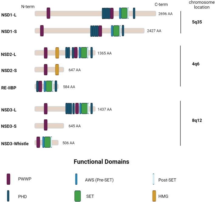
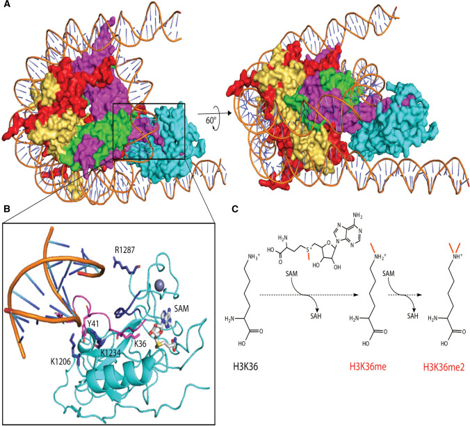
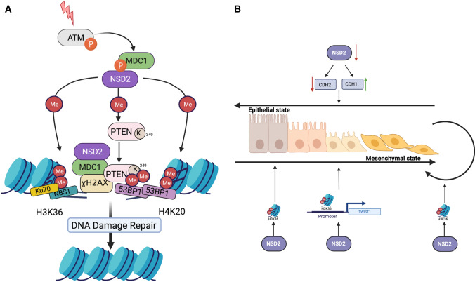
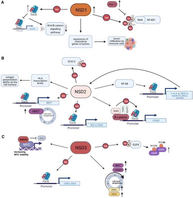
NSD1, NSD2, and NSD3 constitute the nuclear receptor-binding SET Domain (NSD) family of histone 3 lysine 36 (H3K36) methyltransferases. These structurally similar enzymes mono- and di-methylate H3K36, which contribute to the maintenance of chromatin integrity and regulate the expression of genes that control cell division, apoptosis, DNA repair, and epithelial-mesenchymal transition (EMT). Aberrant expression or mutation of members of the NSD family is associated with developmental defects and the occurrence of some types of cancer. In this review, we discuss the effect of alterations in NSDs on cancer patient's prognosis and response to treatment. We summarize the current understanding of the biological functions of NSD proteins, focusing on their activities and the role in the formation and progression in solid tumors biology, as well as how it depends on tumor etiologies. This review also discusses ongoing efforts to develop NSD inhibitors as a promising new class of cancer therapeutic agents.

Keywords: Cancer; H3K36 methyltransferases; Histone methylation; NSD1/KMT3B; NSD2/MMSET/WHSC1; NSD3/WHSC1L1; Solid tumors.


Conflict of interest statement

The author declares he has no conflict of interest.

Figures

Fig. 1
Fig. 1
The schematic structure of NSD proteins. Abbreviations of domain names: PWWP proline–tryptophan–tryptophan–proline, PHD plant homeo domain, SET suppressor of variegation, enhancer of zeste, and trithorax, HMG high-mobility group
Fig. 2
Fig. 2
H3K36 methylation by NSD family proteins. A Nucleosome-bound NSD3 (cyan) in a catalytically active state (PDB ID 7CRR). Included in this complex are H2A (red), H2B (yellow), H4 (green), and H3 (magenta). B Allosteric regulation of nucleosome binding leads to a 2.4 Å movement of the SET domain loop (dark blue) which allows access of the H3 tail (magenta) and proximity of K36 to SAM (gray). Salt bridge contacts between Lys1206 and Lys1234 of NSD3, further stabilized by π-stacking with Tyr41 of H3, enforce tight binding in this complex. Arg1287 on the post-SET loop of NSD3 makes additional phosphate backbone contacts to the anti-parallel DNA strand. C The cofactor SAM serves as a methyl donor for single or double H3K36 methylation events. H3K36me2, in turn, regulates gene expression and DNA damage repair, and interplays with other histone modifications
Fig. 3
Fig. 3
Role of NSD2 in DNA damage response and epithelial–mesenchymal transition. A NSD2 participates in DNA damage response and DNA repair. H3K36me2 mediated by NSD2 accumulates at DNA double-strands breaks (DSBs) and recruits early DNA repair components NBS1 and Ku70 at DSBs. Also, NSD2 is involved in γH2AX-MDC1 pathway: MDC1 interacts with phosphorylated Ser 102 of NSD2, in turn NSD2 methylates H4K20 and recruits 53BP1 at the sites of DNA damage. NSD2 also dimethylates PTEN protein, then dimethylated PTEN is recognized by the 53BP1 and is recruited to DNA damage sites. B H3K36me2 plays role in promoting EMT and to maintain cells in mesenchymal state. In the cells in epithelial state, NSD2 mediates H3K36me2 and promotes EMT to change the cells from epithelial state to mesenchymal state. NSD2 mediates H3K36me2 at the promoter of TWIST1 gene that maintains its expression, which supports mesenchymal state of cells. Similarly, inhibition of NSD2 downregulates N-cadherin and upregulates E-cadherin promoting epithelial states of cells. ATM Ataxia-Telangiectasia Mutated kinase, MDC1 mediator of DNA damage Checkpoint protein 1, PTEN phosphatase and tensin homolog, NBS1 Nijmegen breakage syndrome 1, 53BP1 P53-binding protein 1, CDH1 Cadherin-1 or E-Cadherin, CDH2 Cadherin-2 or N-Cadherin, TWIST1 Twist family bHLH transcription factor 1
Fig. 4
Fig. 4
The scheme of signaling cancer pathways involving the NSD family proteins. A NSD1 methylates lysines 218 and 221 of RelA, resulting in NF-kB activation, while FBXL11 demethylates lysines 218 and 221 of RelA, which results in an inactivation of NF-kB in colon cancer cells. NSD1 also activates Wnt/β-catenin-signaling pathway, regulating expression of Wnt10b in HCC. Moreover, NSD1 is involved in regulation of chemokine genes expression and thus influences T-cell infiltration in HNSCC tumors. B NSD2 directly activates NF-kB and additionally increases expression of NF-kB target genes by dimethylation of H3K36 on their promoters in CRPC. Also, by dimethylating H3K36 on gene specific promoters, NSD2 increases expression of CCND1, BCL-2 in CRPC, NEK-7 in HNSCC, SOX2 in osteosarcoma; NSD2 methylates STAT3 at K163, thus promoting the activation of STAT3 pathway in colorectal adenocarcinoma; besides, NSD2 plays the role in the antigen presentation ability on the cell surface in prostate cancer. C NSD3 methylates EGFR at K721, which subsequently leads to increasing of phospho-Y1148 and phospho-Y1173 EGFR levels in HNSCC. NSD3 regulates expression of cell cycle-related genes as CDK2, CDC6 in HNSCC, NEK7, and CCNG1 in bladder cancer lung cancer. NSD3 also stabilizes and supports transcriptional activity of MYC oncoprotein. Wnt10b Wingless Type MMTV Integration Site Family, Member 10B, NF-kB Nuclear Factor-kappa B, FBXL11 F-box and leucine-rich repeat protein 11, STAT3 Signal transducer and activator of transcription 3, HLA human leukocyte antigen, NEK7 NIMA-related Kinase-7, BCL-2 B-Cell Lymphoma 2, SOX2 SRY-box transcription factor 2, TIAM1 TIAM Rac1 Associated GEF 1, IQGAP1 IQ Motif-containing GTPase-activating protein 1, CCND1 Cyclin D1, IL-6,8 Interleukin-6,-8, VEGF-A Vascular endothelial growth factor A, FBXW7 F-box and WD repeat domain-containing 7, CDK2 Cyclin-Dependent Kinase 2, CDC6 Cell Division Cycle 6, CCNG1 Cyclin G1, EGFR Epidermal Growth Factor Receptor
Fig. 5
Fig. 5
Chemical diversity of compounds reported to show inhibition of NSD family proteins. A Numerous diverse chemical scaffolds have been reported to show broad spectrum activity against NSD family members. B Covalent inhibitors BT3 and BT5; a crystal structure of BT3 (green) was solved in complex with NSD1 (gray) and SAM (purple) (PDB ID 6KQQ). C Inhibitor BI-9321 (cyan) was solved in complex with NSD3-PWWP1 (PDB ID 6G2O). D Inhibitor UNC6934 (yellow) was solved in complex with NSD2-PWWP1 (PDB ID 6XCG)

References

    1. Martin C, Zhang Y. The diverse functions of histone lysine methylation. Nat Rev Mol Cell Biol. 2005;6(11):838–849. doi: 10.1038/nrm1761. - DOI - PubMed
    1. Strahl BD, Allis CD. The language of covalent histone modifications. Nature. 2000;403(6765):41–45. doi: 10.1038/47412. - DOI - PubMed
    1. Husmann D, Gozani O. Histone lysine methyltransferases in biology and disease. Nat Struct Mol Biol. 2019;26(10):880–889. doi: 10.1038/s41594-019-0298-7. - DOI - PMC - PubMed
    1. Zaghi M, Broccoli V, Sessa A. H3K36 methylation in neural development and associated diseases. Front Genet. 2019;10:1291. doi: 10.3389/fgene.2019.01291. - DOI - PMC - PubMed
    1. Kim A, Kiefer CM, Dean A. Distinctive signatures of histone methylation in transcribed coding and noncoding human beta-globin sequences. Mol Cell Biol. 2007;27(4):1271–1279. doi: 10.1128/MCB.01684-06. - DOI - PMC - PubMed

MeSH terms