. 2023 Nov;17(6):1563-1572.
doi: 10.1177/19322968221093362. Epub 2022 May 9.

In Vivo Study of the Inflammatory Tissue Response Surrounding a Novel Extended-Wear Kink-Resistant Insulin Infusion Set Prototype Compared With a Commercial Control Over Two Weeks of Wear Time

Affiliations

In Vivo Study of the Inflammatory Tissue Response Surrounding a Novel Extended-Wear Kink-Resistant Insulin Infusion Set Prototype Compared With a Commercial Control Over Two Weeks of Wear Time

Jasmin R Kastner et al. J Diabetes Sci Technol. 2023 Nov.

Abstract

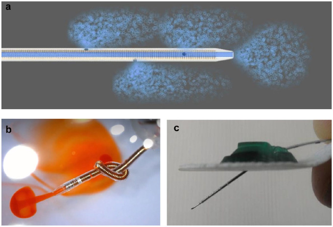
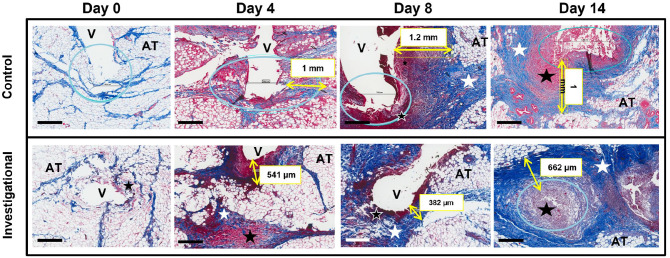
Background: Infusion set function remains the limiting factor of insulin pump therapy due to nonmetabolic complications. Here, we tested an investigational extended-wear infusion set prototype with a soft, angled, wire-reinforced cannula with three additional side holes, and compared failure mechanisms and tissue response with a commercial Teflon control.

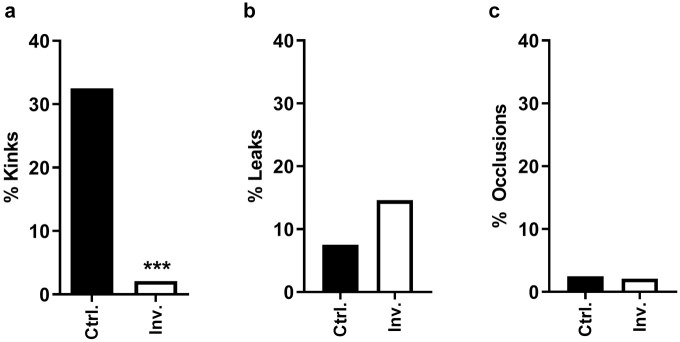
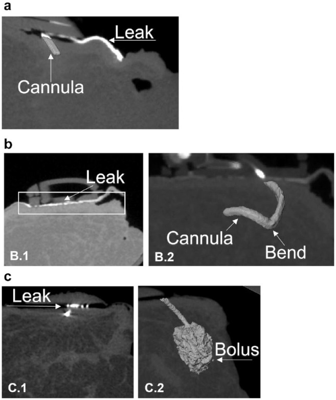
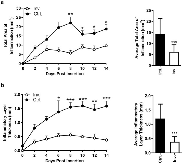
Methods: A total of 48 Teflon and 48 prototype infusion sets were inserted subcutaneously every other day for 14 days in 12 swine and infused with dilute insulin. After two weeks, tissue around cannulas was excised, and occlusions, leaks, and kinks were determined. Tissue was processed and stained to assess the total area of inflammation (TAI) and the inflammatory layer thickness (ILT) around the cannulas. Data were analyzed using Fisher's exact, analysis of variance-general linear model, Kruskal-Wallis, and post hoc tests.

Results: On average, the TAI surrounding the investigational cannula was 52.6% smaller than around the commercial control. The ILT was 66.3% smaller around investigational cannulas. Kinks occurred in 2.1% (investigational) vs 32.4% (commercial) cannulas (P < .001). There was no difference in occlusion alarms and leaks onto skin.

Conclusions: The data suggest that the infusion set prototype elicits less inflammation over an extended wear time and is resistant to kinking, compared with a commercial Teflon device. This is consistent with previously published data on the impact of cannula material/angle on the inflammatory tissue response. We highlight the following important aspects of infusion set design: (1) secure skin adhesion, (2) reliable cannula insertion, (3) automatic removal of the stylet, (4) cannula material/design that resists kinking, and (5) minimization of local tissue inflammation.

Keywords: CSII therapy; extended-wear infusion set; inflammatory tissue response; insulin infusion set; insulin pump therapy; swine model.


Conflict of interest statement

Declaration of Conflicting InterestsThe author(s) declared the following potential conflicts of interest with respect to the research, authorship, and/or publication of this article: JIJ is a founder, equity owner, and consultant to Capillary Biomedical and has received research supports. PJS is a founder, equity owner, and employee of Capillary Biomedical. MCT is an equity owner and member of Capillary Biomedical’s Scientific Advisory Board. JRK is a consultant to and equity owner of Capillary Biomedical.

Figures

Figure 1.
Figure 1.
(a) Schematic representation of insulin flow (blue) through the investigational prototype extended-wear cannula and out through the four holes. (b) The coil-reinforced wall guarantees flexibility and kink resistance of the cannula. This demonstration shows orange dye flowing through the four holes, even when the 24-gauge cannula is tied in a knot. (c) The investigational IIS prototype with a soft Nylon-derivative cannula and stiff stainless-steel stylet with a sharp needle distal tip. Markings facilitated manual insertion of the 13.5-mm-long cannula into the subcutaneous tissue at an angle of approximately 35°. The stylet was removed after insertion. Abbreviation: IIS, insulin infusion set.
Figure 2.
Figure 2.
Insulin infusion set failure mechanisms. (a) Percentage of cannula kinks/bends. (b) Percentage of leaks onto the skin surface (all types included). (c) Percentage of cannula occlusions as evidenced by insulin pump alarm. Abbreviations: Inv., investigational; Ctrl., control.
Figure 3.
Figure 3.
Types of leaks using micro-CT imaging. (a) “Type 1 leak”: Incomplete and superficial cannula insertion caused the most proximal hole to remain above the skin surface, and insulin/X-ray contrast (white) leaked on the IIS adhesive and skin surface. No insulin/contrast was delivered into the SC tissue. (b) “Type 2 leak”: The first-generation spring-loaded inserter caused incomplete and superficial cannula insertion. Subsequent manual advancement of the cannula into the SC tissue using forceps led to both a bend in the cannula (b2) and a misalignment between the IIS tubing pin and the septum within the IIS hub, causing insulin/contrast to leak into the hub. No insulin/contrast was delivered into the SC tissue (b1). (c) “Type 3 leak”: High resistance to flow around a Teflon control IIS cannula caused insulin/contrast to reflux from the SC tissue onto the skin surface, along the path of least resistance (c1). In general, the volume of insulin/contrast that refluxed from the SC tissue onto the skin surface was minimal, as evidenced by the large SC bolus (c2). Note, that because of the angled cannula, it is only possible to view the leak and the bolus in two separate 2D images—c1 and c2 show the same cannula from different angles/depths in the SC tissue. Abbreviations: CT, computerized tomography; IIS, insulin infusion set; SC, subcutaneous.
Figure 4.
Figure 4.
Subcutaneous inflammatory tissue response to the insulin infusion cannula. (a) TAI (tissue disruption, immune cells, bleeding, etc) around the cannula insertion channel. The TAI around the investigational IIS cannula (Inv.) stabilizes after four days of wear time at a low level, while the TAI around the control Teflon cannula (Ctrl.) continues to increase, and peaks on day 8 of wear time. (b) ILT (cellular debris, immune cells, collagen deposition, etc) around the inserted IIS cannula. The ILT around the investigational IIS cannula stabilizes at a low level at four to six days of wear time, while the ILT around the control Teflon cannula continues to increase in thickness and peaks around day 10 of wear time. Abbreviations: TAI, total area of inflammation; IIS, insulin infusion set; Inv., investigational; Ctrl., control; ILT, inflammatory layer thickness. *P < .05. **P < .005. ***P < .001.
Figure 5.
Figure 5.
Progression of the acute inflammatory response in the subcutaneous tissue around the insulin infusion set cannula over 14 days of wear time. The black bar in the left lower corner is equal to 500 µm. After two hours of cannula insertion (day 0), the SC tissue is merely disrupted by stylet/cannula insertion with no cellular infiltration. On day 4 of wear time, there is infiltration of inflammatory cells and fibrin (dense red areas) and collagen deposition (light blue). The area around the cannula contains red blood cells, platelets, fibrin clot, immune cells, damaged adipose cells, and connective tissue. By day 8 and eventually day 14, there is a dense layer of inflammatory tissue and collagen deposited around the cannula. Abbreviations: blue circle, cannula tip; yellow arrow and box, inflammatory layer thickness; white star, collagen deposition; black star, inflamed abnormal tissue; AT, healthy adipose tissue; V, void caused by cannula insertion channel; SC, subcutaneous.

Similar articles

Cited by

References

    1. Summers JC, Briganti EM, Fitzgerald ZA, Lambers LNJ, Cohen ND. Long-term effectiveness of continuous subcutaneous insulin infusion in the prevention of hypoglycemia in adults with type 1 diabetes. Diabetes Technol Ther. 2019;21:423-429. - PubMed
    1. Senn JD, Fischli S, Slahor L, Schelbert S, Henzen C. Long-term effects of initiating Continuous Subcutaneous Insulin Infusion (CSII) and Continuous Glucose Monitoring (CGM) in people with type 1 diabetes and unsatisfactory diabetes control. J Clin Med. 2019;8:E394. - PMC - PubMed
    1. Tubili C, Folco UD, Nardone MR, Clementi A. A single-center long-term Continuous Subcutaneous Insulin Infusion (CSII) experience: higher fractional use is associated with less diabetes complications. J Diabetes Sci Technol. 2017;11:1057-1058. - PMC - PubMed
    1. Heinemann L. Insulin infusion sets: a critical reappraisal. Diabetes Technol Ther. 2016;18:327-333. - PubMed
    1. Pickup JC, Yemane N, Brackenridge A, Pender S. Nonmetabolic complications of continuous subcutaneous insulin infusion: a patient survey. Diabetes Technol Ther. 2014;16:145-149. - PMC - PubMed

Publication types