. 2022 May 9;18(5):e1010532.
doi: 10.1371/journal.ppat.1010532. eCollection 2022 May.

Ubiquitination of Ebola virus VP35 at lysine 309 regulates viral transcription and assembly

Affiliations

Ubiquitination of Ebola virus VP35 at lysine 309 regulates viral transcription and assembly

Sarah van Tol et al. PLoS Pathog. .

Abstract

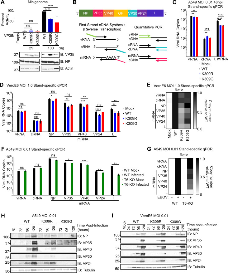
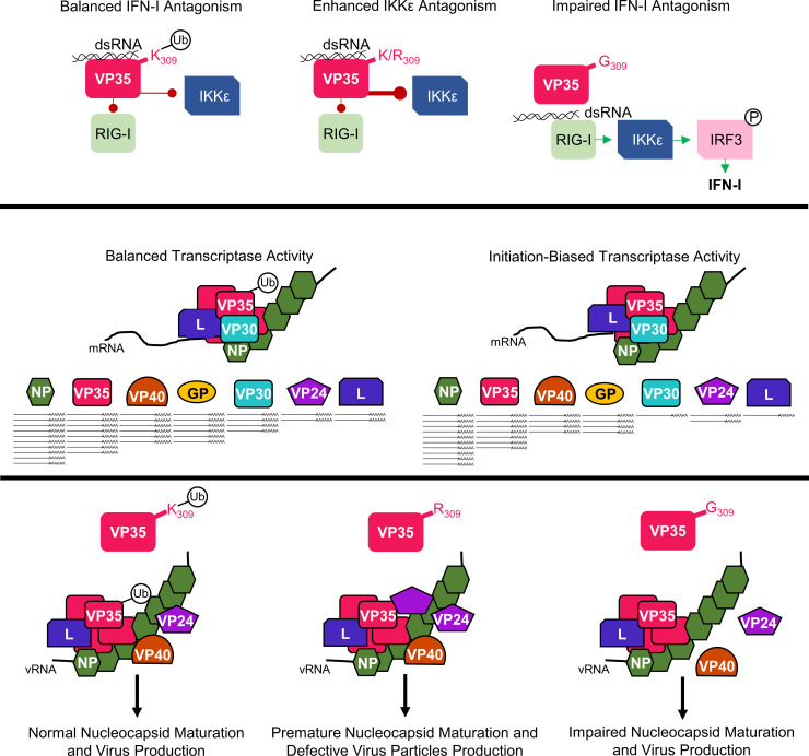
Ebola virus (EBOV) VP35 is a polyfunctional protein involved in viral genome packaging, viral polymerase function, and host immune antagonism. The mechanisms regulating VP35's engagement in different functions are not well-understood. We previously showed that the host E3 ubiquitin ligase TRIM6 ubiquitinates VP35 at lysine 309 (K309) to facilitate virus replication. However, how K309 ubiquitination regulates the function of VP35 as the viral polymerase co-factor and the precise stage(s) of the EBOV replication cycle that require VP35 ubiquitination are not known. Here, we generated recombinant EBOVs encoding glycine (G) or arginine (R) mutations at VP35/K309 (rEBOV-VP35/K309G/-R) and show that both mutations prohibit VP35/K309 ubiquitination. The K309R mutant retains dsRNA binding and efficient type-I Interferon (IFN-I) antagonism due to the basic residue conservation. The rEBOV-VP35/K309G mutant loses the ability to efficiently antagonize the IFN-I response, while the rEBOV-VP35/K309R mutant's suppression is enhanced. The replication of both mutants was significantly attenuated in both IFN-competent and -deficient cells due to impaired interactions with the viral polymerase. The lack of ubiquitination on VP35/K309 or TRIM6 deficiency disrupts viral transcription with increasing severity along the transcriptional gradient. This disruption of the transcriptional gradient results in unbalanced viral protein production, including reduced synthesis of the viral transcription factor VP30. In addition, lack of ubiquitination on K309 results in enhanced interactions with the viral nucleoprotein and premature nucleocapsid packaging, leading to dysregulation of virus assembly. Overall, we identified a novel role of VP35 ubiquitination in coordinating viral transcription and assembly.


Conflict of interest statement

The authors have declared that no competing interests exist.

Figures

Fig 1
Fig 1. A basic residue and lack of ubiquitination at VP35/309 is required for most efficient IFN-I antagonism.
(A) VP35 K309 is located in the IFN-inhibitory domain and is involved in binding double-stranded RNA (dsRNA) to prevent the activation of the host’s cytoplasmic RNA sensor RIG-I and is ubiquitinated (white circle with ‘Ub’) at this position. Mutation of K309 to an arginine (R) is predicted to prevent ubiquitination at this site without disrupting dsRNA binding due to the conservation of a basic residue. The glycine (G) mutant is predicted to lose both ubiquitination and dsRNA binding, allowing enhanced activation of RIG-I, IRF3 phosphorylation (white circle with ‘P’), and downstream IFN-I induction. (B) Peptide purified FLAG-VP35 WT and mutants were mixed with 500 ng biotin-poly(I:C), followed by biotin pulldown. The quantification (ImageJ) represents data from three independent experiments. The percent binding was calculated as follows: the ratio of VP35 bound to poly(I:C) (IP) to the VP35 input levels for each VP35 construct was divided by the wt VP35 ratio. (C) Whole cell extracts (WCE) from 293T cells co-expressing HA-Ub and untagged VP35 (wt, K309R, or K309G) were used for immunoprecipitation (IP) with anti-HA beads. The presented western blot is representative of three independent experiments. (D) Lysates (WCE) from mock or rEBOV-VP35/wt, -K309R, or -K309G infected VeroE6 cells were used for IP with IgG (control) or anti-VP35 antibody, followed by immunoblot. The presented western blot is representative of two independent blots. (E) 293T cells were transfected with IFNβ luciferase reporter and Renilla luciferase plasmid and transfected 24 hours later with 3.125 ug/mL high molecular weight (HMW) poly(I:C). The ratio of firefly luciferase (IFNβ promoter activity) to renilla luciferase (transfection efficiency normalization) luminance was measured for each VP35 construct in the presence and absence of poly(I:C) stimulation. The percent activity relative to empty vector is presented. The quantification is from three independent experiments conducted in biological triplicate, and the IB is representative of the corresponding lysates. (F) As in E, but 2 ng IKKε was transfected along with the luciferase plasmids. The ratio of firefly luciferase (IFNβ promoter activity) to renilla luciferase (transfection efficiency normalization) luminance was measured for each VP35 construct in the presence and absence of IKKε over-expression. The percent activity relative to empty vector is presented. The quantification is from two independent experiments conducted in biological triplicate, and the IB is representative of the corresponding lysates. (G) Untagged VP35 constructs were incubated with FLAG-IKKε K38A, and lysates were immunoprecipitated with anti-FLAG-beads. The quantification (ImageJ) represents three independent experiments. The binding ratio ((IP: VP35/FLAG-IKKε K38A)/(WCE: (VP35/FLAG-IKKε K38A)/Tubulin)) for each VP35 construct was divided by wt VP35’s ratio to determine percent binding. Analysis was done using a one-way ANOVA with Tukey’s post-test for comparison between groups. P-value: *<0.05, ***<0.001, ****<0.0001.
Fig 2
Fig 2. The replication of rEBOV-VP35/K309R and -G mutants is attenuated in IFN-competent cells.
A549 cells were mock infected (grey) or infected in triplicate wells with rEBOV-eGFP-VP35/wt (black), -K309R (blue), or -K309G (red) at an MOI of 0.01 (A-C) or 2.5 PFU/cell (D-F; J-K). At different time points, supernatants were collected for virus titration (A, D) or for IFNβ ELISA (I, 48 hpi). The limit of detection (LOD) for the titrations (10 PFU/mL) (A and D) and IFNβ (50 pg/mL) (I) is indicated (black line). Cells were lysed in either TRIzol for RNA analysis (B, E, H, J) or in Laemmli buffer for immunoblot analysis (K). qPCR for EBOV RNA (B and D), IFNβ mRNA (H) or ISG mRNA (J) is shown. The fluorescence microscopy images (GFP) are representative of the three images taken (C and F). The difference in titer (log10) between the mutant and wt viruses at the time point corresponding to the wt peak titer is summarized (G). The area under the curve (AUC) for each protein was calculated using ImageJ to determine the relative activation of the interferon pathway regulators TBK1, IRF3, and STAT1 (phosphorylated protein/(respective total protein/tubulin)) was normalized to the activation levels in wt-infected cells. The western blots are representative of two independent experiments run in duplicate or triplicate (K). The titration (A and D), qRT-PCR (B, D, H and J), and ELISA (I) were done in biological triplicate and are representative of two independent experiments. The data analysis was done using a two-way ANOVA (A, B, D, E, H, and J) or one-way ANOVA (I) with Bonferroni’s or Tukey’s post-test for comparison between groups, respectively. P-value: *<0.05, **< 0.01, ***<0.001, ****<0.0001. For two-way ANOVA with Bonferroni’s post-test statistical analysis, the non-significant differences (P> 0.05) are not indicated on the graph to prevent cluttering. Red and blue stars represent K309G and K309R comparison to wt, respectively.
Fig 3
Fig 3. TRIM6-mediated VP35/K309 ubiquitination is required for efficient replication.
Wild-type (WT) or TRIM6 knockout (T6-KO) A549 cells were infected with rEBOV-eGFP/VP35-wt, -K309R, or -K309G at a multiplicity of infection (MOI) of 0.01 PFU/cell for 120 hours (A and B) or 2.5 PFU/cell for 72 hours (C and D) corresponding to the peak titer for wt virus. The limit of detection (LOD), 10 PFU/mL, is indicated. Fluorescence images representative of three independent wells corresponding to an MOI of 0.01 PFU/cell (B) or 2.5 PFU/cell (D). (E) Cell-sorted CD11b+CD11c+ bone marrow-derived dendritic cells (BMDCs) from WT or Trim6-/- mice were infected with rEBOV-eGFP/VP35-wt, -K309R, or -K309G for 24 hours and RNA was collected for strand-specific qPCR for viral genomic RNA (vRNA). (F) Titer from WT or Trim6-/- BMDCs infected with rEBOV-eGFP/VP35-wt or -K309R at an MOI of 0.5 PFU/cell for 96 hours (n = 3). The data analysis was done using a one-way ANOVA (A, C, E, and F) with Tukey’s post-test for comparison between groups. P-value: *<0.05, **<0.01, ****<0.0001, and ns, not significant (p> 0.05).
Fig 4
Fig 4. The replication of rEBOV-VP35/K309R and -G mutants is attenuated in IFN-incompetent cells.
VeroE6 cells were mock infected (grey) or infected with rEBOV-eGFP-VP35/wt (black), -K309R (blue), or -K309G (red) viruses at an MOI of 0.01 (A-B) or 1.0 PFU/cell (C-D). The fluorescence microscopy images (GFP) are representative of the three images taken (B and D). The limit of detection (LOD), 10 PFU/mL, is indicted (A and C). (E) The difference in titer (log10) between the mutant and wt viruses at the time point corresponding to the wt peak titer is summarized. The titrations were collected in biological triplicate (A, C). The data analysis was done using a two-way ANOVA (A and B) with Bonferonni’s post-test for comparison between groups. P-value: *<0.05, ***<0.001, ****<0.0001. Red and blue stars represent K309G and K309R comparison to wt, respectively. Non-significant differences, P-value >0.05, are not indicated to prevent cluttering on the image.
Fig 5
Fig 5. Ubiquitination of VP35/309 enhances viral transcriptase function.
(A) Minigenome components (renilla, VP30, NP, L, T7 polymerase, and EBOV minigenome luciferase plasmid) were co-expressed with 25 or 100 ng of empty vector (pCAGGS) or VP35/wt, -K309R, or K309G in 293T cells. At 50 hours post-transfection, the cells were lysed to measure luciferase and evaluate protein expression. Quantification is from two independent experiments conducted in biological triplicate. (B) Graphical representation of the EBOV genome and the strand-specific qPCR approach. (C) A549 cells were infected with rEBOV-eGFP-VP35/wt (black), -K309R (blue), or -K309G (red) viruses at an MOI of 0.01 PFU/cell for 48 hours and RNA was collected for strand-specific qRT-PCR (triplicates). (D) VeroE6 cells were mock (grey) treated or infected with rEBOV-eGFP-VP35/wt, -K309R, or -K309G viruses at an MOI of 1.0 PFU/cell for 24 hours and RNA was collected for strand-specific qRT-PCR (three biological replicates from two independent experiments with qRT-PCR run in triplicate). (E) Heat map representing the ratio of copy number relative to wt for each viral RNA species corresponding to the data presented in panel D. (F) WT (black) or TRIM6-knockout (T6-KO) (green) A549 cells were infected with rEBOV-eGFP-VP35/wt at an MOI of 2.5 PFU/cell for 24 hours and RNA was collected for strand-specific qRT-PCR (triplicates). (G) Heat map representing the ratio of copy number relative to wt for each viral RNA species corresponding to the data presented in panel D. (H-I) Protein lysates from A549 (H) or VeroE6 cells (I) infected with rEBOV-eGFP-VP35/wt, -K309R, or -K309G at an MOI of 0.01 PFU/cell, or mock-infected, were analyzed for the time-course expression of viral proteins. The data analysis was done using a one-way ANOVA with Tukey’s post-test for comparison between groups (A, C, and D) or a student’s t-test (F). P-value: *<0.05, **<0.01, ***<0.001, ****<0.0001; ns, not significant (p>0.05).
Fig 6
Fig 6. Mutation of VP35 at K309 dysregulates VP35’s interaction with the EBOV polymerase but not TRIM6 or itself.
(A) Lysates (WCE) and HA-immunoprecipitation (IP) from 293T cells co-transfected with untagged VP35 (wt, K309R, or K309G) with HA-TRIM6 or pCAGGS (empty vector). The quantification is based on immunoblot densitometry (area under the curve) determined using ImageJ from three independent experiments. The binding ratio ((IP: VP35/HA-TRIM6)/(WCE: (VP35/HA-TRIM6)/Tubulin)) for each VP35 construct was divided by wt VP35’s ratio to determine relative binding. (B) 293T cells were co-transfected with His- or FLAG-tagged VP35 and FLAG IPs were performed. The quantification is based on AUC determined using ImageJ from three independent experiments. The binding ratio ((IP: His-VP35/FLAG-VP35)/(WCE: (His-VP35/FLAG-VP35)/Tubulin)) for each VP35 construct was divided by wt VP35’s ratio to determine relative binding. (C) WCE and HA-IP from 293T cells co-transfected with untagged VP35 (wt, K309R, or K309G) with HA-L1-505 or empty vector. Immunoblot quantification from two independent experiments. The binding ratio ((IP: VP35/HA-L1-505)/(WCE: (VP35/HA-L1-505)/Tubulin)) for each VP35 construct was divided by wt VP35’s ratio to determine relative binding. (D) HA-L1-505 and untagged wt VP35 were co-transfected into wt or TRIM6 knockout (T6-KO) A549 cells, and WCE were immunoprecipitated with anti-HA-tagged beads. The quantification is from data collected from three independent experiments. The binding ratio ((IP: VP35/HA-L1-505)/(WCE: (VP35/HA-L1-505)/Tubulin)) for lysates from wt and T6-KO cells was divided by the wt’s ratio to determine relative binding. (E) HA-L1-505 and untagged wt VP35 were co-transfected with FLAG-tagged TRIM6 wt or -C15A or empty vector into T6-KO A549 cells, and WCE were immunoprecipitated with anti-HA-tagged beads. The quantification is from data collected from two independent experiments. The binding ratio ((IP: VP35/HA-L1-505)/(WCE: (VP35/HA-L1-505)/Tubulin)) for lysates from T6-KO cells transfected with empty vector, HA-TRIM6-wt, or -C15A was divided by the ratio of empty vector transfected cells to determine relative binding. The data analysis was done using a one-way ANOVA with Tukey’s post-test for comparison between groups (A-E). P-value: *<0.05, **<0.01, ***<0.001, ****<0.0001; ns, not significant (p>0.05).
Fig 7
Fig 7. Ubiquitination of VP35 at K309 impedes interaction with EBOV nucleoprotein.
(A) HA-NP (input) was added to beads bound with lysates from empty vector or FLAG-VP35 (wt, K309R, or K309G) transfected 293T cells, washed, and FLAG-eluted. Immunoblot quantification from two independent experiments. The binding ratio (HA-NP/FLAG-VP35) for each VP35 construct was divided by the ratio of wt VP35 to determine relative binding. (B) HA-NP and untagged wt VP35 were co-transfected into wt or T6-KO A549 cells and the WCE were immunoprecipitated with anti-HA beads. The blot quantifications are representative of two independent experiments. The binding ratio ((IP: VP35/HA-NP)/(WCE: (VP35/HA-NP)/Tubulin)) for lysates from wt and T6-KO cells was divided by the wt’s ratio to determine relative binding. (C) Diagram depicting the experiment set-up for the deubiquitinase experiment. When wild-type VP35 (pink rectangle) is expressed, several ubiquitin molecules will be ubiquitinated (white circle with ‘Ub’) at lysine 309. When co-expressed with catalytically active, wt ovarian tumor (OTU) deubiquitinase, the covalently linked ubiquitin will be cleaved from VP35. The catalytically inactive mutant, OTU-2A, has two key cysteine residues mutated to alanine and is not able to cleave ubiquitin from substrates. Lysates cells co-expressing VP35 and FLAG-OTU were added onto beads coated with either HA-NP (antibody molecule with green hexagon) or VP35-specific antibody (antibody with pink rectangle). (D) 293T cells were co-transfected with untagged VP35 (wt, K309R, or K-all-R) and empty vector or FLAG-OTU (wt or -2A). The WCE from VP35 FLAG-OTU co-transfected cells were incubated with the anti-HA (IP:HA), IgG-protein A, or anti-VP35-protein (IP: VP35) coated beads, bound with lysates from HA-NP or empty vector transfected cells to pulldown VP35 (IP:HA) or ubiquitin (IP: VP35). The western blot is representative of two independent experiments run in duplicate. (E) The area under the curve (AUC) for each protein from the western blots in panel D were calculated using ImageJ. The relative binding ratio (VP35 IP: (Ub/VP35)/WCE: (Ub/VP35)/tubulin) for VP35-associated ubiquitin was determined for each condition and divided by the ratio for wt VP35 without OTU treatment. (F) The area under the curve (AUC) for each protein from the western blots in panel D were calculated using ImageJ. The relative binding ratio (HA-NP IP: (VP35/HA-NP)/WCE: (VP35/tubulin) for VP35-NP binding was determined for each condition and divided by the ratio for wt VP35 without OTU treatment. The data analysis was done using a one-way ANOVA with Tukey’s post-test (A and B) or two-way ANOVA with Bonferroni’s post-test (E and F) for comparison between groups. P-value: *<0.05, **<0.01, ***<0.001, ****<0.0001; ns, not significant (p>0.05).
Fig 8
Fig 8. Lack of ubiquitination and a basic residue at VP35/309 dysregulates virus assembly.
(A) Lysates from mock or rEBOV-VP35/wt/-K309R or -K309G infected (MOI = 0.01 PFU/cell for 144 hours) VeroE6 cells were immunoprecipitated (IP) with IgG or anti-VP35 antibody with protein A beads in RIPA complete and used for western blot to assess interaction with viral proteins VP40, NP, VP24 and VP30. Lysates used for this experiment were also used for Fig 1C. The area under the curve (AUC) for each protein was calculated using ImageJ from western blots run in triplicate. The relative binding ratio (IP: (viral protein/VP35)/WCE: (viral protein/VP35)) was for all VP35 constructs and divided by wt VP35’s ratio. (B) Protein lysates (WCE) from VeroE6 cells infected cells (MOI = 0.01 PFU/cell, 144 hours) with rEBOV-eGFP-VP35/wt (WT), -K309R (R), or -K309G (G) and corresponding sucrose-gradient purified virus. The area under the curve (AUC) for each antibody were calculated using ImageJ from western blots run in triplicate. The packaging ratio (purified virus: (viral protein/VP35)/WCE (panel B): (viral protein/VP35)) was for all VP35 constructs and divided by wt VP35’s ratio. (C) The number of viral genome copies was determined using strand-specific qPCR of sucrose-gradient-purified virus. (D) The ratio of packaged to intracellular RNA copies was determined using strand-specific qPCR for genomic RNA on lysates from cells and the purified virus, and the ratio was normalized to the value for wt virus. (E) The ratio of infectious virus to packaged genome copies was determined by titrating the sucrose-gradient purified virus (PFU/mL) and strand-specific PCR to calculate the vRNA copies in the corresponding sample, and the ratio was normalized to the value for the wt virus. (F) The ratio of infectious virus to intracellular genome copies was determined using the supernatant titer and intracellular genome copy number, and the ratio was normalized to the value for the wt virus. This experiment was performed in triplicate (C-F). The data analysis was done using a one-way ANOVA with Tukey’s post-test for comparison between groups (C-F). P-value: *<0.05, **<0.01, ***<0.001, ****<0.0001; ns, not significant (p>0.05).
Fig 9
Fig 9. A basic residue and the ubiquitination capacity of VP35/309 coordinates VP35’s functions.
Both ubiquitinated (white circle with Ub) and non-ubiquitinated (K/R309) VP35 bind double-stranded RNA (dsRNA) to antagonize RIG-I. Loss of a basic residue (G309) impairs dsRNA binding which allows RIG-I activation and downstream IRF3 phosphorylation (white circle with P) leading to type I interferon (IFN-I) induction. In the absence of ubiquitination, VP35 (K/R309) impedes IKKε activation more efficiently. In the context of the viral transcriptase, comprised of the viral polymerase (L), VP35, and the transcription factor (VP30), the capacity for VP35/K309 to be ubiquitinated enables balanced transcriptional activity. Under this balanced transcriptase function, a 3’-to-5’ transcriptional gradient is generated and viral proteins are produced in an optimal ratio. When VP35/309 is unable to receive ubiquitination, the transcriptase is biased toward transcriptional initiation and the transcriptional gradient is dysregulated resulting in unbalanced intracellular viral protein ratios. When VP35/309 has the capacity for ubiquitination, the recruitment of VP24 and VP40 is regulated and progeny virions are assembled normally. In the absence of ubiquitin and when a basic residue is present at VP35/309, VP24 is more efficiently recruited to VP35 prematurely and some immature nucleocapsids are incorporated into progeny virions resulting in the defective viruses. When the basic residue is lost (K309G), interaction with VP24 and VP40 is impaired which reduces virus production.

Similar articles

Cited by

References

    1. Kuhn JH, Amarasinghe GK, Basler CF, Bavari S, Bukreyev A, Chandran K, et al.. ICTV Virus Taxonomy Profile: Filoviridae. The Journal of general virology. 2019;100(6). doi: 10.1099/jgv.0.001252 - DOI - PMC - PubMed
    1. Jacob ST, Crozier I, Fischer WA 2nd, Hewlett A, Kraft CS, Vega MA, et al.. Ebola virus disease. Nat Rev Dis Primers. 2020;6(1):13. doi: 10.1038/s41572-020-0147-3 - DOI - PMC - PubMed
    1. CDC. Ebola (Ebola Virus Disease) History of Ebola Outbreaks: CDC; 2021. [updated 2021. Available from: https://www.cdc.gov/vhf/ebola/history/chronology.html#anchor_1526565058132.
    1. Feldmann H, Sprecher A, Geisbert TW. Ebola. The New England journal of medicine. 2020;382(19). - PubMed
    1. Iversen PL, Kane CD, Zeng X, Panchal RG, Warren TK, Radoshitzky SR, et al.. Recent successes in therapeutics for Ebola virus disease: no time for complacency. The Lancet Infectious diseases. 2020;20(9). doi: 10.1016/S1473-3099(20)30282-6 - DOI - PMC - PubMed

Publication types

MeSH terms

Substances