

MRI-based prostate and dominant lesion segmentation using cascaded scoring convolutional neural network
- PMID: 35533237
- PMCID: PMC9388615
- DOI: 10.1002/mp.15687
MRI-based prostate and dominant lesion segmentation using cascaded scoring convolutional neural network
Abstract
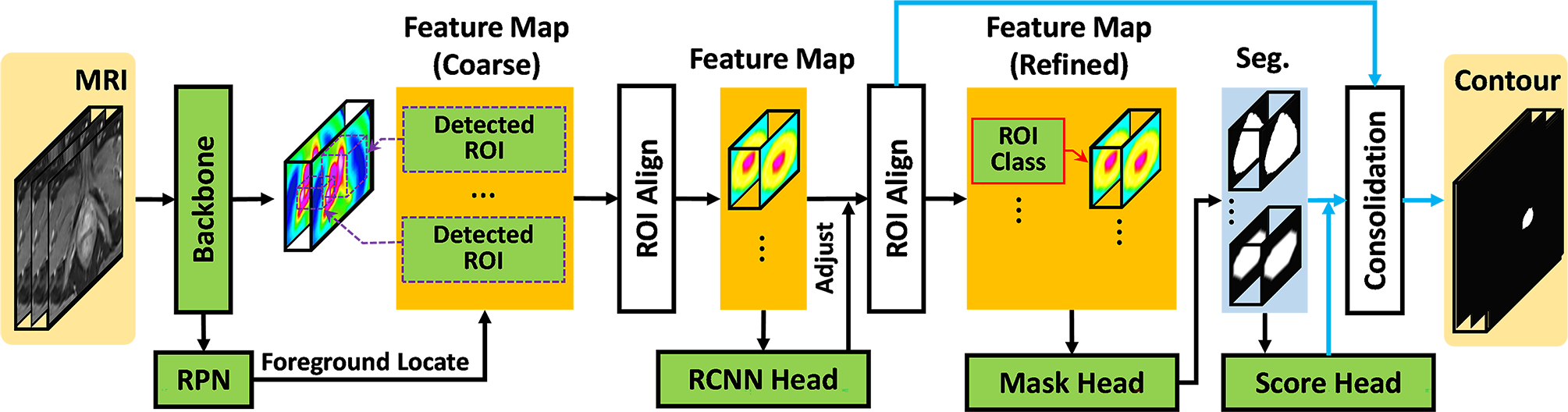
Purpose: Dose escalation to dominant intraprostatic lesions (DILs) is a novel treatment strategy to improve the treatment outcome of prostate radiation therapy. Treatment planning requires accurate and fast delineation of the prostate and DILs. In this study, a 3D cascaded scoring convolutional neural network is proposed to automatically segment the prostate and DILs from MRI.
Methods and materials: The proposed cascaded scoring convolutional neural network performs end-to-end segmentation by locating a region-of-interest (ROI), identifying the object within the ROI, and defining the target. A scoring strategy, which is learned to judge the segmentation quality of DIL, is integrated into cascaded convolutional neural network to solve the challenge of segmenting the irregular shapes of the DIL. To evaluate the proposed method, 77 patients who underwent MRI and PET/CT were retrospectively investigated. The prostate and DIL ground truth contours were delineated by experienced radiologists. The proposed method was evaluated with fivefold cross-validation and holdout testing.
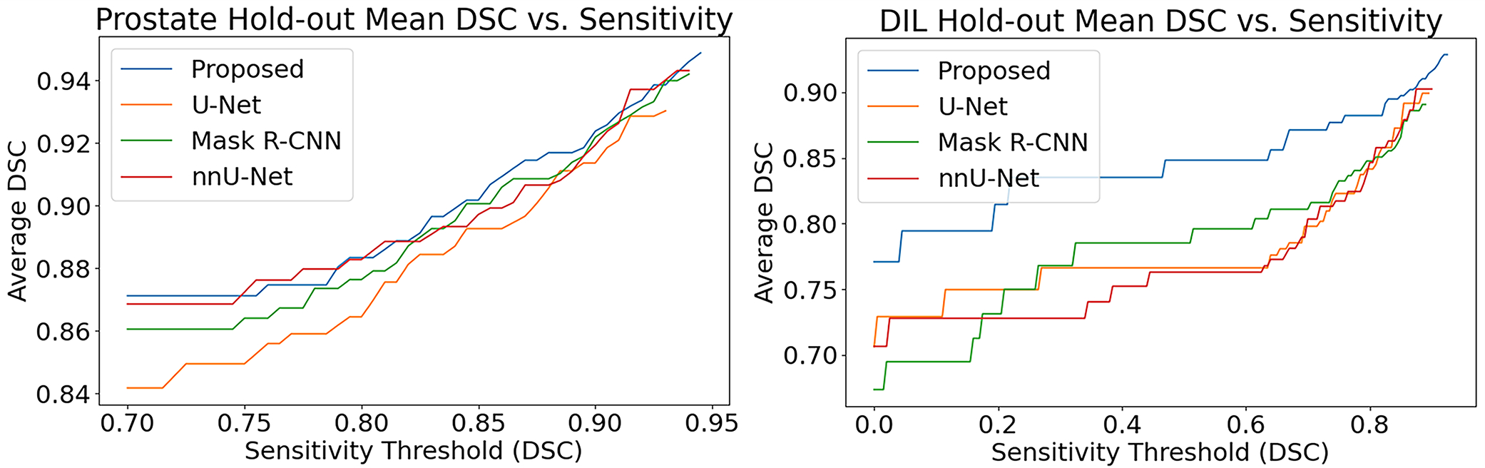
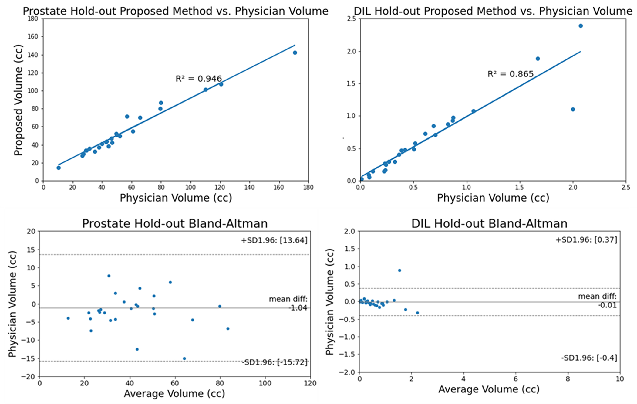
Results: The average centroid distance, volume difference, and Dice similarity coefficient (DSC) value for prostate/DIL are 4.3 ± 7.5/3.73 ± 3.78 mm, 4.5 ± 7.9/0.41 ± 0.59 cc, and 89.6 ± 8.9/84.3 ± 11.9%, respectively. Comparable results were obtained in the holdout test. Similar or superior segmentation outcomes were seen when compared the results of the proposed method to those of competing segmentation approaches.
Conclusions: The proposed automatic segmentation method can accurately and simultaneously segment both the prostate and DILs. The intended future use for this algorithm is focal boost prostate radiation therapy.
Keywords: MRI; deep learning; prostate and dominant lesion segmentation.
© 2022 American Association of Physicists in Medicine.
Conflict of interest statement
Disclosures
The author declares no conflicts of interest.
Figures


References
-
- Arrayeh E, Westphalen AC, Kurhanewicz J, et al. Does local recurrence of prostate cancer after radiation therapy occur at the site of primary tumor? Results of a longitudinal MRI and MRSI study [published online ahead of print 2012/02/15]. Int J Radiat Oncol Biol Phys. 2012;82(5):e787–793. - PMC - PubMed
MeSH terms
Grants and funding
LinkOut - more resources
Full Text Sources
