. 2022 Jul:146:105540.
doi: 10.1016/j.compbiomed.2022.105540. Epub 2022 Apr 30.

ECG-iCOVIDNet: Interpretable AI model to identify changes in the ECG signals of post-COVID subjects

Affiliations

ECG-iCOVIDNet: Interpretable AI model to identify changes in the ECG signals of post-COVID subjects

Amulya Agrawal et al. Comput Biol Med. 2022 Jul.

Abstract

Objective: Studies showed that many COVID-19 survivors develop sub-clinical to clinical heart damage, even if subjects did not have underlying heart disease before COVID. Since Electrocardiogram (ECG) is a reliable technique for cardiovascular disease diagnosis, this study analyzes the 12-lead ECG recordings of healthy and post-COVID (COVID-recovered) subjects to ascertain ECG changes after suffering from COVID-19.

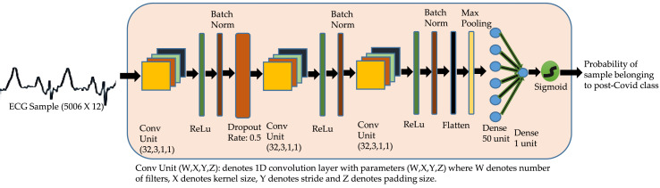
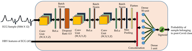
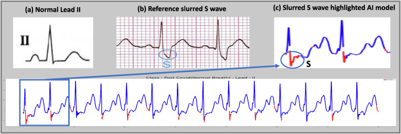
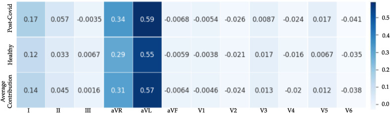
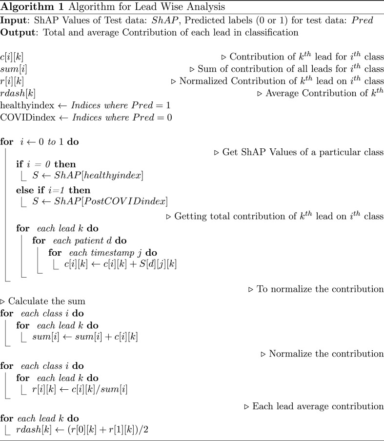
Method: We propose a shallow 1-D convolutional neural network (CNN) deep learning architecture, namely ECG-iCOVIDNet, to distinguish ECG data of post-COVID subjects and healthy subjects. Further, we employed ShAP technique to interpret ECG segments that are highlighted by the CNN model for the classification of ECG recordings into healthy and post-COVID subjects.

Results: ECG data of 427 healthy and 105 post-COVID subjects were analyzed. Results show that the proposed ECG-iCOVIDNet model could classify the ECG recordings of healthy and post-COVID subjects better than the state-of-the-art deep learning models. The proposed model yields an F1-score of 100%.

Conclusion: So far, we have not come across any other study with an in-depth ECG signal analysis of the COVID-recovered subjects. In this study, it is shown that the shallow ECG-iCOVIDNet CNN model performed good for distinguishing ECG signals of COVID-recovered subjects from those of healthy subjects. In line with the literature, this study confirms changes in the ECG signals of COVID-recovered patients that could be captured by the proposed CNN model. Successful deployment of such systems can help the doctors identify the changes in the ECG of the post-COVID subjects on time that can save many lives.

Keywords: AI in ECG; CNN; COVID; Electrocardiogram (ECG); Interpretability; Post-COVID; Shapley additive exPlanations (ShAP).


Conflict of interest statement

The authors declare that they have no conflict of interest.

Figures

Fig. 1
Fig. 1
Block Diagram of ECG-iCOVIDNet Architecture: The model comprises of three convolutional blocks followed by a global average pool layer. The output of global average pool layer is flattened and passed through a fully connected layer, which is connected to a single neuron with sigmoid activation function to obtain the probability of the sample belonging to each class.
Fig. 2
Fig. 2
Block Diagram of ECG-HiCOVIDNet Architecture: The model comprises of three convolutional blocks followed by a global average pool layer. The output is flattened and concatenated with the HRV features and then passed through fully connected dense layers. The output is passed through the sigmoid activation function to obtain the probability. For each split of the data, one classifier of ECG-HiCOVIDNet is trained. The details of the CNN blocks in these classifiers is as follows. Classifier-1: (W = 32,X = 3,Y = 1,Z = 1); Classifier-2: (W = 32,X = 5,Y = 1,Z = 1); Classifier-3: (W = 32,X = 5,Y = 1,Z = 1); Classifier-4: (W = 64,X = 5,Y = 1,Z = 1); Classifier-5: (W = 96,X = 3,Y = 1,Z = 1).
Fig. 3
Fig. 3
t-SNE plots of ECG data being separated layer after layer when features are learnt gradually by the ECG-iCOVIDNet model.
Fig. 4
Fig. 4
t-SNE plots of data being separated layer after layer when features are learnt gradually by the ECG-HiCOVIDNet model. Here, HRV features are concatenated with the latent space embedding of the convolutional blocks.
Fig. 5
Fig. 5
Wider P wave and/or notching of P wave highlighted in lead I of ECG, in subjects having ejection fraction less than 45%: (a) Showing healthy lead I ECG, (b) wide/notching P wave as reported in the literature, and (c) segments of ECG highlighted in red color by our ECG-iCOVIDNet model correspond to wide/notching P wave regions.).
Fig. 6
Fig. 6
Slurred S wave highlighted in lead II of ECG, in subjects having left ventricular dysfunction: (a) Showing healthy lead II ECG, (b) slurred S wave reported in the literature, and (c) segments of ECG highlighted in red color by our ECG-iCOVIDNet model correspond to slurred S wave.
Fig. 7
Fig. 7
Lead wise Importance: showing the impact of all the 12 leads on the two classes. The average contribution of lead-aVL is the highest followed by lead aVR.
Image 1

Similar articles

Cited by

References

    1. Yamayoshi S., Sakai-Tagawa Y., Koga M., Akasaka O., Nakachi I., Koh H., Maeda K., Adachi E., Saito M., Nagai H., et al. Comparison of rapid antigen tests for COVID-19. Viruses. 2020;12:1420. - PMC - PubMed
    1. Singh P., Gupta A. Generalized SIR (GSIR) epidemic model: an improved framework for the predictive monitoring of COVID-19 pandemic. ISA Trans. 2021 doi: 10.1016/j.isatra.2021.02.016. In press. - DOI - PMC - PubMed
    1. Singh P., Singhal A., Fatimah B., Gupta A. ICASSP 2021-2021 IEEE International Conference on Acoustics, Speech and Signal Processing (ICASSP) IEEE; 2021. An improved data driven dynamic SIRD model for predictive monitoring of COVID-19; pp. 8158–8162.
    1. Majumder J., Minko T. Recent developments on therapeutic and diagnostic approaches for COVID-19. AAPS J. 2021;23:1–22. - PMC - PubMed
    1. Gasecka A., Pruc M., Kukula K., Gilis-Malinowska N., Filipiak K.J., Jaguszewski M.J., Szarpak L. Post-COVID-19 heart syndrome. Cardiol. J. 2021;28:353–354. - PMC - PubMed

Publication types