

Myoferlin targeting triggers mitophagy and primes ferroptosis in pancreatic cancer cells
- PMID: 35533575
- PMCID: PMC9096673
- DOI: 10.1016/j.redox.2022.102324
Myoferlin targeting triggers mitophagy and primes ferroptosis in pancreatic cancer cells
Abstract
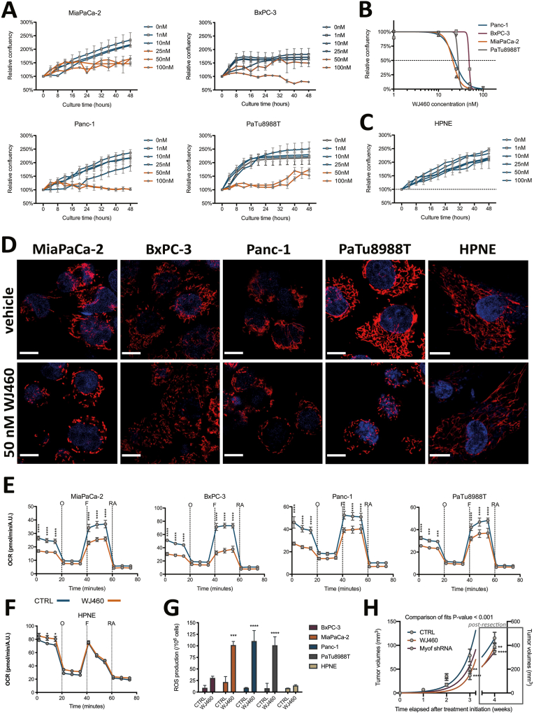
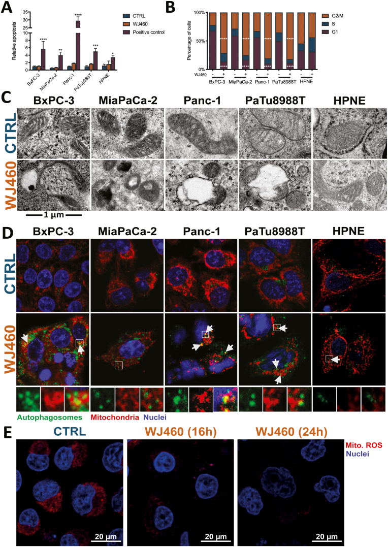
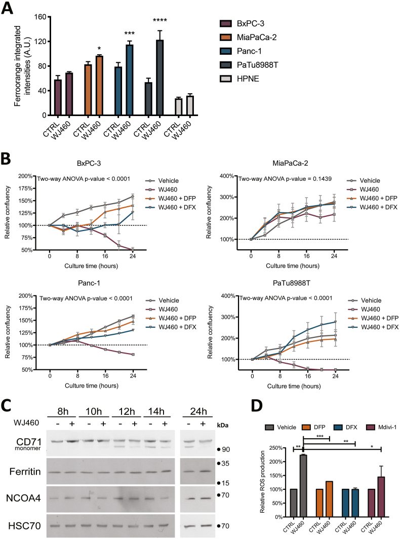
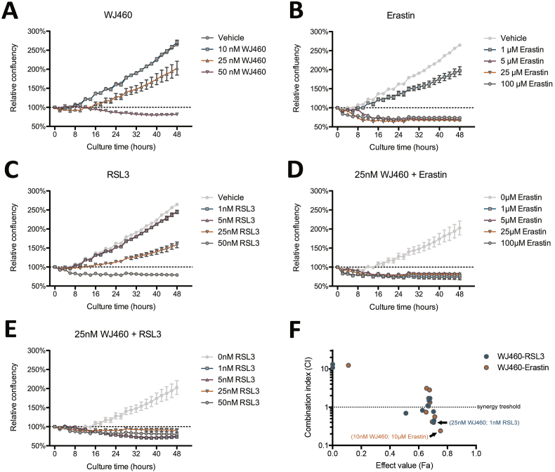
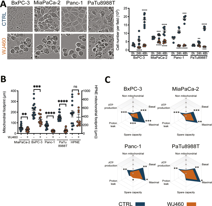
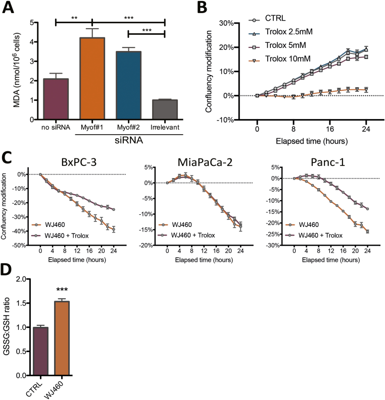
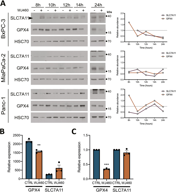
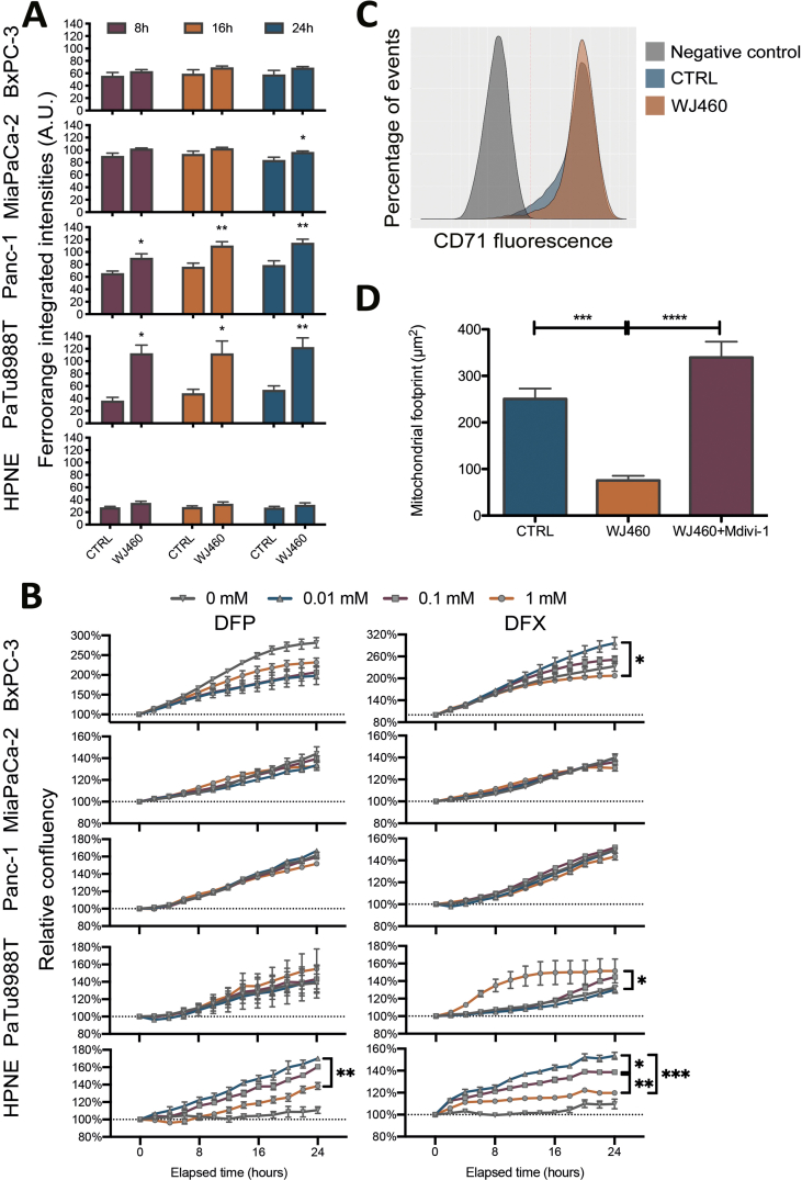
Myoferlin, an emerging oncoprotein, has been associated with a low survival in several cancer types including pancreas ductal adenocarcinoma where it controls mitochondria structure and respiratory functions. Owing to the high susceptibility of KRAS-mutated cancer cells to iron-dependent cell death, ferroptosis, and to the high iron content in mitochondria, we investigated the relation existing between mitochondrial integrity and iron-dependent cell death. We discovered that myoferlin targeting with WJ460 pharmacological compound triggered mitophagy and ROS accumulation culminating with lipid peroxidation and apoptosis-independent cell death. WJ460 caused a reduction of the abundance of ferroptosis core regulators xc- cystine/glutamate transporter and GPX-4. Mitophagy inhibitor Mdivi1 and iron chelators inhibited the myoferlin-related ROS production and restored cell growth. Additionally, we reported a synergic effect between ferroptosis inducers, erastin and RSL3, and WJ460.
Keywords: Ferroptosis; Mitochondria; Mitophagy; Myoferlin; Pancreas cancer.
Copyright © 2022 The Authors. Published by Elsevier B.V. All rights reserved.
Conflict of interest statement
The authors declare that they have no known competing financial interests or personal relationships that could have appeared to influence the work reported in this paper.
Figures


Similar articles
-
RSL3 and Erastin differentially regulate redox signaling to promote Smac mimetic-induced cell death.Oncotarget. 2016 Sep 27;7(39):63779-63792. doi: 10.18632/oncotarget.11687. Oncotarget. 2016. PMID: 27588473 Free PMC article.
-
Lipid Peroxidation-Dependent Cell Death Regulated by GPx4 and Ferroptosis.Curr Top Microbiol Immunol. 2017;403:143-170. doi: 10.1007/82_2016_508. Curr Top Microbiol Immunol. 2017. PMID: 28204974 Review.
-
Susceptibility of Mitophagy-Deficient Tumors to Ferroptosis Induction by Relieving the Suppression of Lipid Peroxidation.Adv Sci (Weinh). 2025 Feb;12(6):e2412593. doi: 10.1002/advs.202412593. Epub 2024 Dec 16. Adv Sci (Weinh). 2025. PMID: 39679775 Free PMC article.
-
A Shortage of FTH Induces ROS and Sensitizes RAS-Proficient Neuroblastoma N2A Cells to Ferroptosis.Int J Mol Sci. 2021 Aug 18;22(16):8898. doi: 10.3390/ijms22168898. Int J Mol Sci. 2021. PMID: 34445601 Free PMC article.
-
Ferroptosis: process and function.Cell Death Differ. 2016 Mar;23(3):369-79. doi: 10.1038/cdd.2015.158. Epub 2016 Jan 22. Cell Death Differ. 2016. PMID: 26794443 Free PMC article. Review.
Cited by
-
Mitophagy alleviates cisplatin-induced renal tubular epithelial cell ferroptosis through ROS/HO-1/GPX4 axis.Int J Biol Sci. 2023 Feb 13;19(4):1192-1210. doi: 10.7150/ijbs.80775. eCollection 2023. Int J Biol Sci. 2023. PMID: 36923942 Free PMC article.
-
Ferroptosis in cancer: From molecular mechanisms to therapeutic strategies.Signal Transduct Target Ther. 2024 Mar 8;9(1):55. doi: 10.1038/s41392-024-01769-5. Signal Transduct Target Ther. 2024. PMID: 38453898 Free PMC article. Review.
-
Iron-Based Nanovehicle Delivering Fin56 for Hyperthermia-Boosted Ferroptosis Therapy Against Osteosarcoma.Int J Nanomedicine. 2024 Jan 4;19:91-107. doi: 10.2147/IJN.S441112. eCollection 2024. Int J Nanomedicine. 2024. PMID: 38192634 Free PMC article.
-
Advances in mitophagy and mitochondrial apoptosis pathway-related drugs in glioblastoma treatment.Front Pharmacol. 2023 Jun 30;14:1211719. doi: 10.3389/fphar.2023.1211719. eCollection 2023. Front Pharmacol. 2023. PMID: 37456742 Free PMC article. Review.
-
International consensus guidelines for the definition, detection, and interpretation of autophagy-dependent ferroptosis.Autophagy. 2024 Jun;20(6):1213-1246. doi: 10.1080/15548627.2024.2319901. Epub 2024 Mar 24. Autophagy. 2024. PMID: 38442890 Free PMC article. Review.
References
-
- Turtoi A., Musmeci D., Wang Y., Dumont B., Somja J., Bevilacqua G., Pauw E.D., Delvenne P., Castronovo V. Identification of novel accessible proteins bearing diagnostic and therapeutic potential in human pancreatic ductal adenocarcinoma. J. Proteome Res. 2011;10:4302–4313. doi: 10.1021/pr200527z. - DOI - PubMed
-
- Rademaker G., Hennequière V., Brohée L., Nokin M.-J., Lovinfosse P., Durieux F., Gofflot S., Bellier J., Costanza B., Herfs M., Peiffer R., Bettendorff L., Deroanne C., Thiry M., Delvenne P., Hustinx R., Bellahcène A., Castronovo V., Peulen O. Myoferlin controls mitochondrial structure and activity in pancreatic ductal adenocarcinoma, and affects tumor aggressiveness. Oncogene. 2018;66:1–15. doi: 10.1038/s41388-018-0287-z. - DOI - PMC - PubMed
-
- Fahmy K., Gonzalez A., Arafa M., Peixoto P., Bellahcène A., Turtoi A., Delvenne P., Thiry M., Castronovo V., Peulen O. Myoferlin plays a key role in VEGFA secretion and impacts tumor-associated angiogenesis in human pancreas cancer. Int. J. Cancer. 2016;138:652–663. doi: 10.1002/ijc.29820. - DOI - PubMed
-
- Blomme A., Fahmy K., Peulen O., Costanza B., Fontaine M., Struman I., Baiwir D., Pauw E.D., Thiry M., Bellahcène A., Castronovo V., Turtoi A. Myoferlin is a novel exosomal protein and functional regulator of cancer-derived exosomes. Oncotarget. 2016;7:83669–83683. doi: 10.18632/oncotarget.13276. - DOI - PMC - PubMed
Publication types
MeSH terms
Substances
LinkOut - more resources
Full Text Sources
Medical
Miscellaneous
