Review
. 2022 Aug;1867(8):159179.
doi: 10.1016/j.bbalip.2022.159179. Epub 2022 May 6.

Retinol-binding protein 2 (RBP2): More than just dietary retinoid uptake

Affiliations
Review

Retinol-binding protein 2 (RBP2): More than just dietary retinoid uptake

Jacqueline Plau et al. Biochim Biophys Acta Mol Cell Biol Lipids. 2022 Aug.

Abstract

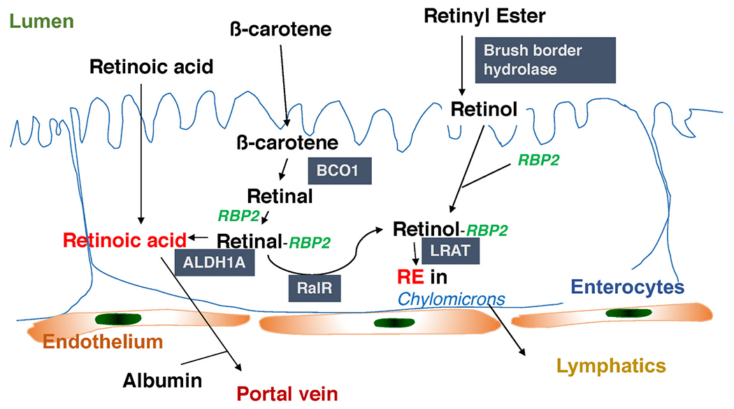
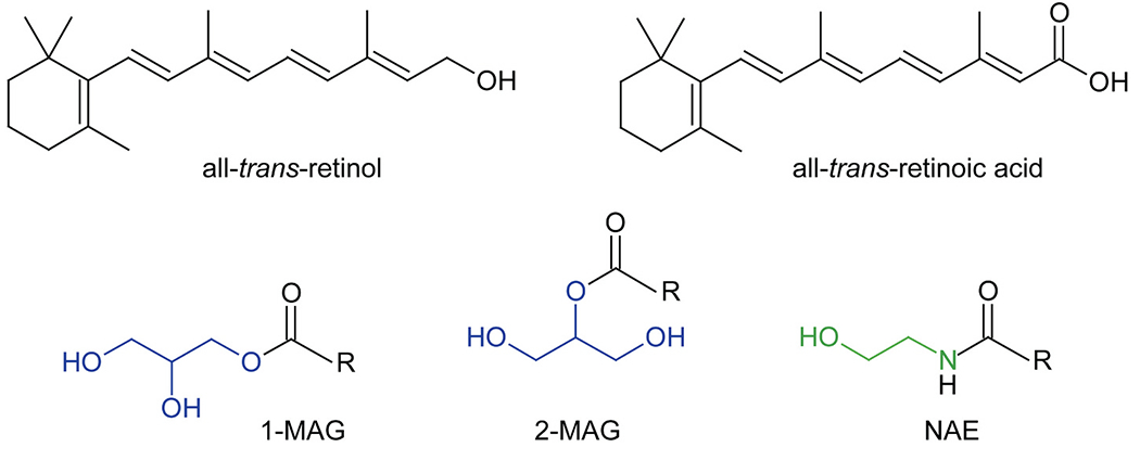
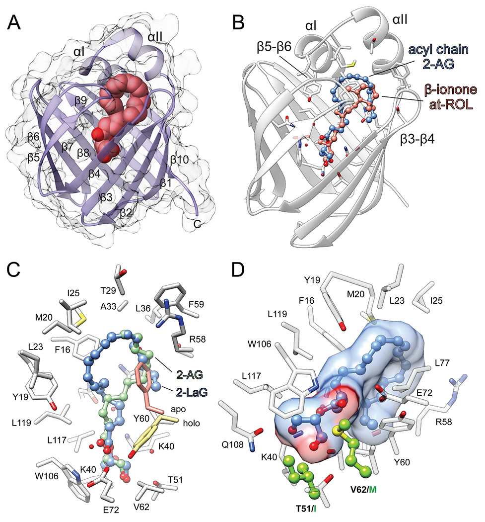
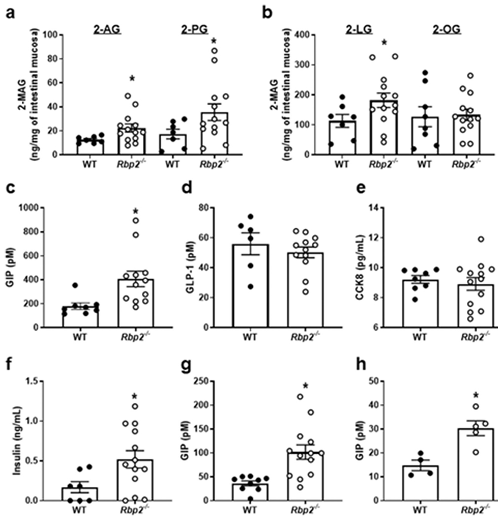
Retinol-binding protein 2 (RBP2, also known as cellular retinol-binding protein 2 (CRBP2)) is a member of the fatty acid-binding protein family and has been extensively studied for its role in facilitating dietary vitamin A (retinol) uptake and metabolism within enterocytes of the small intestine. RBP2 is present in highest concentrations in the proximal small intestine where it constitutes approximately 0.1-0.5% of soluble protein. Recent reports have established that RBP2 binds monoacylglycerols (MAGs) with high affinity, including the canonical endocannabinoid 2-arachidonoylglycerol (2-AG). Crystallographic studies reveal that retinol, 2-AG, or other long-chain MAGs alternatively can bind in the retinol-binding pocket of RBP2. It also has been demonstrated recently that Rbp2-deficient mice are more susceptible to developing obesity and associated metabolic phenotypes when exposed to a high fat diet, or as they age when fed a conventional chow diet. When subjected to an oral fat challenge, the Rbp2-deficient mice release into the circulation significantly more, compared to littermate controls, of the intestinal hormone glucose-dependent insulinotropic polypeptide (GIP). These new findings regarding RBP2 structure and actions within the intestine are the focus of this review.

Keywords: 2-monoacylglycerol; All-trans-retinoic acid; CRBP2; Cellular retinol-binding protein 2; Glucose-dependent insulinotropic polypeptide (GIP); Incretin; Obesity; RBP2; Retinoids; Retinol-binding proteins; Vitamin A.


Conflict of interest statement

Declaration of competing interest

None of the authors of this review article have any conflicts of interest that have influenced the writing of the review.

Figures

Fig. 1.
Fig. 1.
RBP2 acts to facilitate retinoid metabolism within enterocytes. Dietary preformed vitamin A (retinol) or provitamin A carotenoids (like β-carotene) are taken up by enterocytes. Dietary retinyl esters must first be hydrolyzed to retinol before uptake. Retinol binds RBP2, which channels it to lecithin:retinol acyltransferase (LRAT), for retinyl ester formation and packaging into nascent chylomicrons. Provitamin A carotenoids are absorbed as such and cleaved within the enterocyte by β-carotene monooxygenase 1 (BCO1) to retinal (retinaldehyde) which then binds RBP2 which channels it to retinal reductase (RalR) for reduction to retinol, allowing for its esterification and packaging into chylomicrons. It also has been reported that RBP2-bound retinol can be channeled to retinol dehydrogenases (RDHs) that catalyze its oxidation to retinal (not shown). RBP2-bound retinal can then be oxidized to retinoic acid by aldehyde dehydrogenases, ALDH1A enzymes. Retinoic acid is needed and used locally to regulate retinoic acid receptor (RAR)-mediated gene transcription.
Fig. 2.
Fig. 2.
Chemical structures of lipids which interact with selected intracellular retinoid-binding proteins. 1-MAG and 2-MAG stands for 1- and 2-monoacylglicerides. The glycerol backbone in these compounds is colored blue. NAE corresponds to N-acylethanolamides. The ethanolamine moiety is shown in green. R – indicate a fatty acid moiety, including C14 – C22 long acyl chains that can be saturated, mono- or polyunsaturated. (For interpretation of the references to colour in this figure legend, the reader is referred to the web version of this article.)
Fig. 3.
Fig. 3.
Interaction of human RBP2 with its lipid ligands. (A) Representation of the overall RBP2 fold (PDB: 6BTH). Location of the ligand binding site is marked by a molecule of 2-AG depicted as spheres. (B) Comparison of the spatial positions of all-trans-retinol (at-ROL) and 2-AG in the binding cavity of RBP2 (PDBs: 4QZT and 6BTH, respectively). Geometric configuration of arachidonoyl chain of 2-AG mimics the structure of β-ionone ring of at-ROL, interacting with the portal region of the protein. (C) The opposite orientation of the acyl chains of 2-AG (blue) and 2-lauroylglycerol (2-LaG) (green) (PDB: 7K3I) in the binding site of RBP2. Regardless the differences in the binding modes both ligands cause repositioning of the Y60 side chain as compared with the apo-form of the protein. (D) The key difference in the amino acid composition of the binding pocket of RBP1 that prevents its high-affinity interaction with MAGs. Residues I51 and M62 present in RBP1 are colored green. 2-AG molecule within the binding site is represented in ball and stick style. (For interpretation of the references to colour in this figure legend, the reader is referred to the web version of this article.)
Fig. 4.
Fig. 4.
Following an oral fat challenge to chow-fed Rbp2−/− and WT mice of approximately 7-months-of-age, levels of 2-AG, 2-PG, and 2-LG but not 2-OG were significantly elevated in mucosal scrapings from Rbp2−/− mice (A and B). Plasma levels of GIP (C) and insulin (F) but not GLP-1 (D) or CCK8 (E) were significantly elevated for Rbp2−/− mice receiving the corn oil challenge. GIP levels in plasma of both fed (G) and fasted (H) Rbp2−/− mice were also significantly elevated compared to those of matched WT mice. Data are provided as means ± SEM with N = 6 or more for each group, *, p < 0.05. Taken directly from Lee et al. [12].
Fig. 5.
Fig. 5.
Potential involvement of RBP2 in ATRA synthesis in intestine. (A)Three equal length sections of small intestine (SI 1–3, proximal to distal) were taken to prepare mucosal scrapings. For these scrapings, all-trans-retinoic acid (ATRA) synthesis activity was determined upon incubation with 1 μM all-trans-retinaldehyde and 15 μg protein and is expressed as ATRA synthesis rate (pmol ATRA/μg protein/min). (B) Relative mRNA expression levels of ATRA (Aldh1a1 and Aldh1a2) or retinyl ester (Lrat) synthesizing enzymes, and Rbp2 in colon. (C) ATRA synthesis capacity of colon for Aldh1a1-KO and WT mice.

Similar articles

Cited by

References

    1. Ong DE, A novel retinol-binding protein from rat. Purification and partial characterization, J. Biol. Chem 259 (1984) 1476–1482. - PubMed
    1. Ong DE, Cellular transport and metabolism of vitamin a: roles of the cellular retinoid-binding proteins, Nutr. Rev 52 (1994) S24–S31. - PubMed
    1. Napoli JL, Functions of intracellular retinoid binding-proteins, Subcell Biochem. 81 (2016) 21–76. - PMC - PubMed
    1. Blaner WS, Brun PJ, Calderon RM, Golczak M, Retinol-binding protein 2 (RBP2): biology and pathobiology, Crit. Rev. Biochem. Mol. Biol 55 (2020) 197–218. - PMC - PubMed
    1. Napoli JL, Cellular retinoid binding-proteins CRBP, CRABP, FABP5: effects on retinoid metabolism, function and related diseases, Pharmacol. Ther 173 (2017) 19–33. - PMC - PubMed

Publication types

MeSH terms