Review
. 2022 May;60(3):359-369.
doi: 10.1016/j.rcl.2022.01.004. Epub 2022 Jan 11.

Review of Thoracic Imaging Manifestations of COVID-19 and Other Pathologic Coronaviruses

Affiliations
Review

Review of Thoracic Imaging Manifestations of COVID-19 and Other Pathologic Coronaviruses

Ayushi P Singh et al. Radiol Clin North Am. 2022 May.

Abstract

Severe acute respiratory syndrome coronavirus 2 (SARS-CoV-2) is an easily transmissible coronavirus that emerged in late 2019 and has caused a global pandemic characterized by acute respiratory disease named coronavirus disease 2019 (COVID-19). Diagnostic imaging can be helpful as a complementary tool in supporting the diagnosis of COVID-19 and identifying alternative pathology. This article presents an overview of acute and postacute imaging findings in COVID-19.

Keywords: Computed tomography; Coronavirus disease 2019 (COVID-19); Middle East respiratory syndrome coronavirus (MERS-CoV); Severe acute respiratory syndrome coronavirus (SARS-CoV); Severe acute respiratory syndrome coronavirus 2 (SARS-CoV-2).


Figures

Fig. 1
Fig. 1
Spectrum severity of COVID-19 on chest radiography. (A) No acute consolidation or other radiographic findings. (B) Mild bilateral ill-defined hazy opacities (arrow) in a peripheral and lower lobe distribution. (C) Moderate multifocal patchy opacities (arrows) in a predominately peripheral distribution. (D) Severe bilateral diffuse patchy opacities (arrows).
Fig. 2
Fig. 2
A 75-year-old man with fever and cough. (A) CXR on admission shows mild multifocal hazy and patchy opacities in a peripheral distribution (arrows). (B) Follow-up CXR performed 5 days later shows worsening now moderate multifocal peripheral opacities (arrow). (C) Repeat CXR 14 days later shows interval worsening large consolidations in the upper to mid-lungs (arrows).
Fig. 3
Fig. 3
Severity of COVID-19 on CT. (A) No acute pulmonary findings. (B) Mild bilateral ground-glass and reticular densities in a peripheral distribution. (C) Moderate bilateral peripheral consolidative and ground-glass opacities. (D) Severe diffuse peripheral ground-glass opacities.
Fig. 4
Fig. 4
A 60-year-old positive man with COVID-19 with focal ground-glass exhibiting a “crazy paving” pattern (arrow).
Fig. 5
Fig. 5
A 70-year-old positive patient with COVID-19 with bilateral ground-glass opacities in a peripheral distribution.
Fig. 6
Fig. 6
A 74-year-old positive man with COVID-19 with large bilateral pleural effusions (arrows).
Fig. 7
Fig. 7
A 79-year-old man with diffuse ground-glass opacification and mild bibasilar predominant bronchial dilatation (arrows).
Fig. 8
Fig. 8
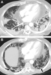
Necrotizing pneumonia in a 69-year-old woman with RT-PCR-test–proven COVID-19. (A) Admission CT shows a large dense right lower lobe consolidation. (B) Follow-up CT performed 10 days later due to worsening respiratory symptoms shows new numerous air-filled cystic lucencies (arrow) within the consolidation, which suggests necrotizing pneumonia.
Fig. 9
Fig. 9
Halo sign in a 31-year-old man with RT-PCR-test–proven COVID-19. Axial chest CT images show rounded dense consolidations surrounded by ground-glass opacities (arrows) in the left lower lobe, findings consistent with the halo sign.
Fig. 10
Fig. 10
Cardiac MR imaging performed in a 38-year-old woman with new-onset cardiomyopathy in the setting of COVID-19. (A) Axial T2-weighted sequence demonstrates multifocal areas of increased signal intensity (arrows) in a peripheral distribution. (B) Short-axis STIR sequence shows increased STIR signal in the corresponding areas of abnormality (arrows). STIR, short tau inversion recovery.
Fig. 11
Fig. 11
A 19-year-old man with metastatic alveolar rhabdomyosarcoma presenting for follow-up PET/CT after radiation therapy. Incidentally found to be COVID positive. (A, B) CT demonstrates mild multifocal bibasilar ground-glass opacities (arrowheads) with corresponding FDG uptake (arrows) on the fused PET image. (C, D) Follow-up PET/CT performed 4 months later shows resolution of ground-glass opacity and FDG uptake.
Fig. 12
Fig. 12
Examples of COVID-19 categorization endorsed by the RSNA. (A, B) Axial and coronal views of a patient exhibiting commonly reported imaging features of COVID-19 pneumonia including peripheral, bilateral, ground-glass opacities. (C) Indeterminate appearance showing bilateral reticular and ground-glass opacities lacking typical COVID features. (D) Atypical appearance showing uncommonly or not reported features of COVID-19 pneumonia such as cavitation (arrow).
Fig. 13
Fig. 13
CTA chest radiography in a 98-year-old man with dyspnea in the setting of COVID-19. (A) Large saddle pulmonary embolism (arrow) extending to the lobar branches. (B) There is mild right ventricular enlargement with flattening of the interventricular septum (arrow), which suggests right heart strain. (C) Brain MR imaging performed concurrently shows a small area of infarct (arrow) in the right middle cerebral artery (MCA) territory.
Fig. 14
Fig. 14
A 50-year-old man presenting 1 month after recovering from COVID-19. (A) Chest CT shows moderate multifocal peripheral reticular opacities (arrows) with scattered areas of traction bronchiectasis. (B) DECT images obtained concurrently show perfusion defects (arrows) in the corresponding areas of lung involvement. DECT, dual-energy CT.
Fig. 15
Fig. 15
A 64-year-old COVID-19 positive male. (A) Portable CXR on admission shows severe bilateral patchy opacities (arrows). (B) Follow-up CXR 1 year later shows complete resolution of pulmonary findings.
Fig. 16
Fig. 16
A 71-year-old COVID-19 positive patient showing evolution of pathology over the span of 3 months. (A) CT in the acute phase shows severe bilateral dense peripheral consolidations. (B) Follow-up scan 3 months later shows resolution of ground-glass and consolidative densities with evolution into moderate mostly peripheral subpleural reticulation (arrows) and scarring with regions of bronchiectasis, findings consistent with fibrosis.
Fig. 17
Fig. 17
A 38-year-old man with shortness of breath found to be COVID-19 positive on PCR. (A) Scan performed on admission demonstrates severe bilateral multifocal dense and ground-glass opacities in a peripheral distribution. (B) Follow-up CT scan performed 1 year later demonstrates mild fibrotic-like changes including mild reticular opacities (arrow) with traction bronchiectasis.
Fig. 18
Fig. 18
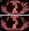
PET/CT in a patient with a history of squamous cell carcinoma of the pharynx. (A) PET/CT performed 1 week following administration of the COVID vaccine in the left arm shows multiple enlarged and hypermetabolic left axillary lymph nodes (arrow). (B) Follow-up PET performed 2 months later shows decrease in size and FDG avidity within these lymph nodes.

Similar articles

References

    1. Hu B., Guo H., Zhou P., et al. Characteristics of SARS-CoV-2 and COVID-19. Nat Rev Microbiol. 2021;19(3):141–154. - PMC - PubMed
    1. Stawicki S.P., Jeanmonod R., Miller A.C., et al. The 2019-2020 Novel Coronavirus (Severe Acute Respiratory Syndrome Coronavirus 2) Pandemic: A Joint American College of Academic International Medicine-World Academic Council of Emergency Medicine Multidisciplinary COVID-19 Working Group Consensus Paper. J Glob Infect Dis. 2020;12(2):47–93. - PMC - PubMed
    1. Johns Hopkins Coronavirus Resource Center. https://coronavirus.jhu.edu/map.html Available at: Accessed October 6, 2021.
    1. Centers for Disease Control CDC Coronavirus Disease 2019 (COVID 2019) Basics. 2020. www.cdc.gov Available at: Accessed September 10, 2021.
    1. Chen B., Tian E.K., He B., et al. Overview of lethal human coronaviruses. Signal Transduct Target Ther. 2020;5(1):89. - PMC - PubMed