Review
. 2022 Dec 1;12(12):a041178.
doi: 10.1101/cshperspect.a041178.

Buttons and Zippers: Endothelial Junctions in Lymphatic Vessels

Affiliations
Review

Buttons and Zippers: Endothelial Junctions in Lymphatic Vessels

Peter Baluk et al. Cold Spring Harb Perspect Med. .

Abstract

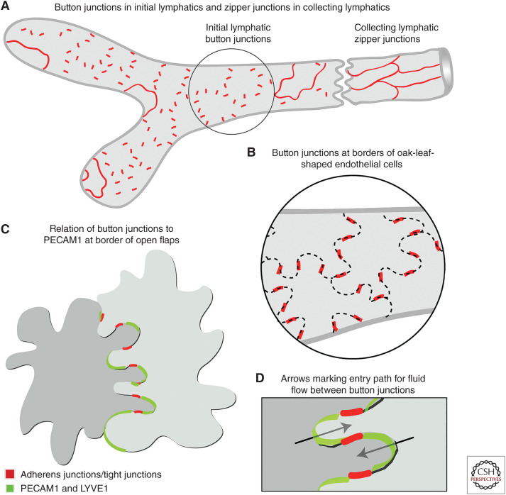
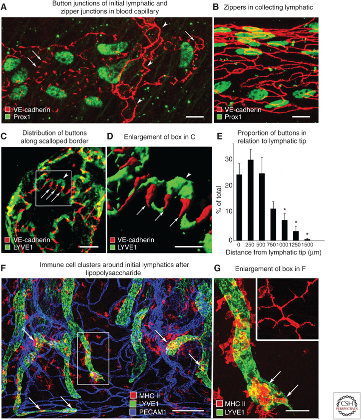
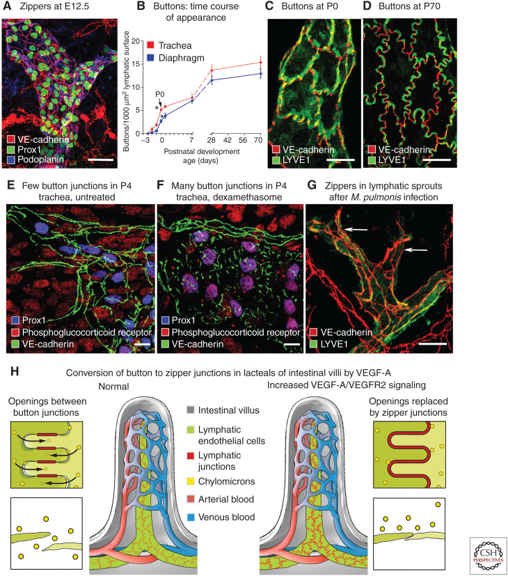
Button-like junctions are discontinuous contacts at the border of oak-leaf-shaped endothelial cells of initial lymphatic vessels. These junctions are distinctively different from continuous zipper-like junctions that create the endothelial barrier in collecting lymphatics and blood vessels. Button junctions are point contacts, spaced about 3 µm apart, that border valve-like openings where fluid and immune cells enter lymphatics. In intestinal villi, openings between button junctions in lacteals also serve as entry routes for chylomicrons. Like zipper junctions that join endothelial cells, buttons consist of adherens junction proteins (VE-cadherin) and tight junction proteins (claudin-5, occludin, and others). Buttons in lymphatics form from zipper junctions during embryonic development, can convert into zippers in disease or after experimental genetic or pharmacological manipulation, and can revert back to buttons with treatment. Multiple signaling pathways and local microenvironmental factors have been found to contribute to button junction plasticity and could serve as therapeutic targets in pathological conditions ranging from pulmonary edema to obesity.


Figures

Figure 1.
Figure 1.
Early studies of lymphatics. (A) The first illustrations of intraluminal (secondary) valves in lymphatics. (Left panel) Dog lymphatics drawn by Jan Swammerdam in June 1664 but only published in 1666 in a commentary on a contemporary textbook of anatomy. (Panel A reprinted from Table 24 in Blasius 1666 without restriction because figure is in the public domain.) (Right panel) Drawings by Frederik Ruysch published 1 year earlier (1665). The lymphatic labeled A has been dissected longitudinally to show the valves (labeled a). (Lymphatic labeled A, right side reprinted from Figure 1 in Ruysch 1665 without restriction because figure is in the public domain.) (B,C) Early drawings of the shape of lymphatic endothelial cells stained by silver nitrate. (B) Faint endothelial cell borders in lymphatics of guinea-pig diaphragm drawn in 1862. (Panel B reprinted from Table 2, Figure 2 in von Recklinghausen (1862) and reprinted without restriction because table and figure are in the public domain.) (C) Endothelial cells in lymphatic, where the cells are oak-leaf shaped, and in artery and vein of cat omentum drawn in 1918. The transverse lines over the artery and veins are outlines of smooth muscle cells. (Panel C is reprinted from Figure 1 in Casparis 1918 without restriction because figure is in the public domain.)
Figure 2.
Figure 2.
Evolving concepts of open junctions between lymphatic endothelial cells. (A) Three-dimensional rendering of initial lymphatic reconstructed from transmission electron micrographs. Anchoring filaments link endothelial cells to the surrounding connective tissue. (Panel A from Figure 25 in Leak and Burke 1968b; reprinted, with permission, from Rockefeller University Press © 1968.) (B) Concept of changes in endothelial cell junctions in initial lymphatic. (Top) Normal condition, junctions are closed. (Middle) Moderate edema, slits form between endothelial cells. (Bottom) Severe edema, endothelial cells are fully detached and widely separated from one another. (Panel B from Figure 12.14 in Majno and Joris 1996; reprinted, with permission, from John Wiley © 1996.) (C) Three-dimensional renderings of lymphatic endothelial cell borders based on scanning electron microscopy (SEM) images of initial lymphatics with interstitial pressure at normal level (left), moderately increased (middle), and greatly increased (right). Focal regions of intercellular junction detachment are shown as shaded openings that enlarge as lymphatics dilate. (Panel C from Figure 16 in Castenholz 1987; reprinted, with permission, from the International Society of Lymphology © 1987.) (D) Concept of “expansion” phase, when primary valves are open, lymph enters the initial lymphatic along the hydrostatic pressure gradient and secondary valves are closed to prevent backflow, and “compression” phase, when primary valves are closed, secondary valves are open, and lymph is pushed through the collecting lymphatic. (Panel D is from Figure 7 in Mendoza and Schmid-Schonbein 2003; reprinted, with permission, from the American Society of Mechanical Engineers © 2003.)
Figure 3.
Figure 3.
Abluminal surface of lymphatic endothelial cell borders. Scanning electron microscopy view of abluminal surface of lymphatics after removal of surrounding connective tissue. (A) Initial lymphatic: Loosely apposed, overlapping, scallop-shaped cell borders of endothelial cells. (B) Collecting lymphatic: Tightly apposed linear borders of adjacent endothelial cells. Scale bars, 1 µm. (Panels A and B from Figure 1 and Supplemental Figure 1 in Baluk et al. 2007; reprinted courtesy of Creative Commons License (Attribution–Noncommercial–Share Alike 4.0 Unported license).)
Figure 4.
Figure 4.
Diagrams of button and zipper junctions in lymphatics. (A) Drawing showing discontinuous button junctions (red line segments) in endothelium of initial lymphatics and continuous zipper junctions (continuous red lines) in collecting lymphatics. Both types of junctions consist of proteins typical of adherens junctions and tight junctions. (B) More detailed view showing the oak-leaf shape of endothelial cells (dashed lines) of an initial lymphatic. Buttons (red) appear to be oriented perpendicular to the cell border but are in fact parallel to the sides of flaps. (C,D) Enlarged views of buttons along the sides of flaps of adjacent oak-leaf-shaped endothelial cells with overlapping edges. PECAM1 and LYVE1 are located at the tip of flaps. Button-like adherens junctions and tight junctions at the sides of flaps direct fluid entry (arrows) through the junction-free region at the tip. Fluid traverses the basement membrane (not shown) and enters initial lymphatics through these openings without disruption of junctions. (Panels AD from Figure 7 in Baluk et al. 2007; reprinted courtesy of Creative Commons License (Attribution–Noncommercial–Share Alike 4.0 Unported license).)
Figure 5.
Figure 5.
Buttons and zippers and cell entry into initial lymphatics. Confocal microscopic images of mouse tracheal lymphatics. (A) Staining for VE-cadherin marks discontinuous buttons (arrows) in an initial lymphatic. Arrowheads point to zipper junctions in a blood capillary. (B) Continuous zipper junctions in a collecting lymphatic. (C,D) Confocal images showing VE-cadherin at buttons (arrows) and LYVE1 between buttons (arrowhead) at the border of oak-leaf-shaped endothelial cells of initial lymphatic. (D) Imaris isosurface rendering of confocal image stack of enlarged boxed region in panel C. (E) Gradient in abundance of buttons shown as a function of distance from the tip of initial lymphatics. Mean ± SEM; *P < 0.05 compared to proportion at the tip. (Panels AE from Figure 1 in Baluk et al. 2007; reprinted courtesy of Creative Commons License (Attribution–Noncommercial–Share Alike 4.0 Unported license).) (F) Lymphatics (LYVE1, green) and MHC II–positive immune cells (arrows, red) in whole mount of mouse trachea 24 h after intratracheal instillation of lipopolysaccharide. (G) Enlargement of boxed region in F of an initial lymphatic containing MHC II–positive cells (arrows) that are rounded and have fewer processes than a corresponding cell in a pathogen-free mouse (inset). Scale bars, 10 µm (AC); 5 µm (D); 200 µm (F); 50 µm (G, inset). (Panels F and G from Figure 6 in Baluk et al. 2007; reprinted courtesy of Creative Commons License (Attribution–Noncommercial–Share Alike 4.0 Unported license).)
Figure 6.
Figure 6.
Plasticity of button junctions in initial lymphatics. (A) Endothelial cells joined by zipper junctions (arrows) in embryonic lymphatics at E12.5. (B) Time course of development of button junctions in lymphatics of trachea (red) and diaphragm (blue) from E16.5 to P70. Buttons are expressed as number of VE-cadherin-stained junctional segments. Buttons that appear before birth represent only about 35% of the eventual number in adult initial lymphatics in the trachea and 20% in the diaphragm at P70. *P < 0.05, values at P0 (arrow) are significantly different from those at E18.5. (C,D) Confocal microscopic images that illustrate the increasing proportion of buttons in initial lymphatics from P0 to P70. (E,F) Confocal images of tracheal lymphatics of P4 pups, either untreated (E) or treated with dexamethasone from P0 to P4 (F). Without treatment (E), lymphatic endothelial cells (Prox1, blue nuclei) have zipper junctions (VE-cadherin, green) and faint staining for phosphoglucocorticoid receptor (Ser211, red). After dexamethasone (F), lymphatics have button junctions and strong phosphoglucocorticoid receptor staining that colocalizes with Prox1 in lymphatic endothelial cell nuclei (purple, arrows). (G) Zippers in lymphatic endothelium after Mycoplasma pulmonis infection for 14 d. (Panels AG from Figures 1, 2, 4, and 7 in Yao et al. 2012; reprinted, with permission, from the American Society for Investigative Pathology published by Elsevier © 2012.) (H) Entry of chylomicrons through openings between button junctions in normal intestinal lacteal (left) and lack of entry after conversion of buttons to zippers by activation of VEGF-A/VEGFR2 signaling in lacteal endothelial cells (right). Scale bars, 20 µm (A, EG); 10 µm (C,D). (Panel H from McDonald 2018; adapted, with permission, from the author.)

Similar articles

Cited by

References

    1. Arasa J, Collado-Diaz V, Halin C. 2021a. Structure and immune function of afferent lymphatics and their mechanistic contribution to dendritic cell and T cell trafficking. Cells 10: 1269. 10.3390/cells10051269 - DOI - PMC - PubMed
    1. Arasa J, Collado-Diaz V, Kritikos I, Medina-Sanchez JD, Friess MC, Sigmund EC, Schineis P, Hunter MC, Tacconi C, Paterson N, et al. 2021b. Upregulation of VCAM-1 in lymphatic collectors supports dendritic cell entry and rapid migration to lymph nodes in inflammation. J Exp Med 218: 1–18. 10.1084/jem.20201413 - DOI - PMC - PubMed
    1. Arif N, Zinnhardt M, Nyamay'Antu A, Teber D, Brückner R, Schaefer K, Li YT, Trappmann B, Grashoff C, Vestweber D. 2021. PECAM-1 supports leukocyte diapedesis by tension-dependent dephosphorylation of VE-cadherin. EMBO J 40: e106113. 10.15252/embj.2020106113 - DOI - PMC - PubMed
    1. Aselli G. 1627. De lactibus sive lacteis venis, quarto vasorum mesarai corum genere novo invento. G.B. Bidelli, Milan, Italy.
    1. Azzali G. 2006. On the transendothelial passage of tumor cell from extravasal matrix into the lumen of absorbing lymphatic vessel. Microvasc Res 72: 74–85. 10.1016/j.mvr.2006.03.005 - DOI - PubMed

Publication types