. 2022 Jun 8;42(23):4755-4765.
doi: 10.1523/JNEUROSCI.0140-22.2022. Epub 2022 May 9.

Motor Impairments and Dopaminergic Defects Caused by Loss of Leucine-Rich Repeat Kinase Function in Mice

Affiliations

Motor Impairments and Dopaminergic Defects Caused by Loss of Leucine-Rich Repeat Kinase Function in Mice

Guodong Huang et al. J Neurosci. .

Abstract

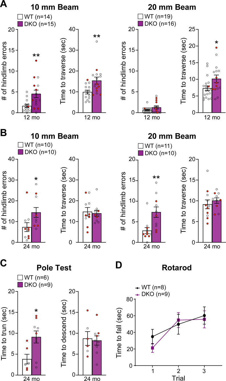
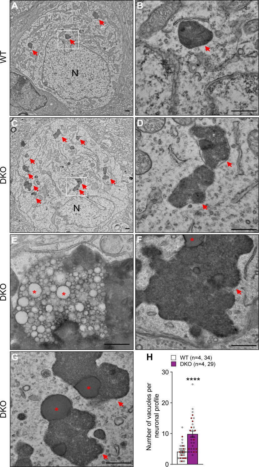
Mutations in leucine-rich repeat kinase 2 (LRRK2) are the most common genetic cause of Parkinson's disease (PD), but the pathogenic mechanism underlying LRRK2 mutations remains unresolved. In this study, we investigate the consequence of inactivation of LRRK2 and its functional homolog LRRK1 in male and female mice up to 25 months of age using behavioral, neurochemical, neuropathological, and ultrastructural analyses. We report that LRRK1 and LRRK2 double knock-out (LRRK DKO) mice exhibit impaired motor coordination at 12 months of age before the onset of dopaminergic neuron loss in the substantia nigra (SNpc). Moreover, LRRK DKO mice develop age-dependent, progressive loss of dopaminergic terminals in the striatum. Evoked dopamine (DA) release measured by fast-scan cyclic voltammetry in the dorsal striatum is also reduced in the absence of LRRK. Furthermore, LRRK DKO mice at 20-25 months of age show substantial loss of dopaminergic neurons in the SNpc. The surviving SNpc neurons in LRRK DKO mice at 25 months of age accumulate large numbers of autophagic and autolysosomal vacuoles and are accompanied with microgliosis. Surprisingly, the cerebral cortex is unaffected, as shown by normal cortical volume and neuron number as well as unchanged number of apoptotic cells and microglia in LRRK DKO mice at 25 months. These findings show that loss of LRRK function causes impairments in motor coordination, degeneration of dopaminergic terminals, reduction of evoked DA release, and selective loss of dopaminergic neurons in the SNpc, indicating that LRRK DKO mice are unique models for better understanding dopaminergic neurodegeneration in PD.SIGNIFICANCE STATEMENT Our current study employs a genetic approach to uncover the normal function of the LRRK family in the brain during mouse life span. Our multidisciplinary analysis demonstrates a critical normal physiological role of LRRK in maintaining the integrity and function of dopaminergic terminals and neurons in the aging brain, and show that LRRK DKO mice recapitulate several key features of PD and provide unique mouse models for elucidating molecular mechanisms underlying dopaminergic neurodegeneration in PD.

Keywords: LRRK2; Parkinson's disease; SNpc; dopamine release; knock-out mice; striatum.


Figures

Figure 1.
Figure 1.
Impairment of motor coordination in LRRK DKO mice. A, In the 10 mm beam walk test, compared to WT mice, LRRK DKO mice at 12 months of age show significantly more hindlimb slips (WT, 1.6 ± 0.4; DKO, 4.5 ± 0.9; p = 0.0074, unpaired two-tailed Student's t test) and longer traversal time (WT, 9.9 ± 0.9 s; DKO, 15.4 ± 1.7 s; p = 0.0093). In the less challenging 20 mm beam walk test, LRRK DKO mice display few hindlimb slips (WT, 0.6 ± 0.1; DKO, 1.3 ± 0.3; p = 0.0835) but longer traversal time (WT, 7.3 ± 0.7 s; DKO, 10.3 ± 1.1 s; p = 0.0268). B, At 24 months of age, relative to WT mice, LRRK DKO mice exhibit markedly more hindlimb errors while traversing the 10 mm beam (WT, 7.4 ± 2.1; DKO, 14.4 ± 2.4; p = 0.0404; unpaired two-tailed Student's t test), but there is no difference in the time to traverse the beam (WT, 14.7 ± 2.0 s; DKO, 14.0 ± 1.5; p = 0.7678). In the 20 mm beam walk test, LRRK DKO mice (7.4 ± 1.3) show more hindlimb slips compared with WT controls (2.9 ± 0.7, p = 0.0056), but there is no difference in traversal time (WT, 9.2 ± 1.1 s; DKO, 10.1 ± 0.7 s; p = 0.4929). A, B, Within the WT or DKO group, there is no significant difference between male and female mice in their performance (p > 0.05). Mice that stalled >120 s on the beam or fell off the beam during both trials were excluded. C, In the pole test, LRRK DKO mice (9.1 ± 1.4 s) at 24 months of age exhibit significantly longer turning time than WT mice (3.8 ± 1.2 s, p = 0.0204, unpaired two-tailed Student's t test), but there is no sex-specific difference within the genotypic group (p > 0.05; specific p values can be found in in Extended Data Figure 1-1). The descending time is similar between WT (8.8 ± 1.7 s) and DKO (8.3 ± 1.1 s, p = 0.8053) mice, but the descending time of male DKO mice is significantly longer than that of female DKO mice (p = 0.0466). D, In the rotarod test, LRRK DKO (Trial 1, 21.2 ± 5.4 s; Trial 2, 54.8 ± 9.0 s; Trial 3, 55.7 ± 10.2 s) and WT mice (Trial 1, 34.9 ± 8.8 s; Trial 2, 49.8 ± 12.0 s; Trial 3, 60.3 ± 10.4 s) show similar latencies to fall off an accelerating rotating rod (F(1,15) = 0.1351, p = 0.7183; Trial 1, p = 0.9334; Trial 2, p > 0.9999; Trial 3, p > 0.9999, two-way ANOVA with Bonferroni's post hoc multiple comparisons). The average time before falling off the rod is shown for each of the three consecutive trials. All data are expressed as mean ± SEM. Red filled and open circles represent data obtained from individual male and female mice, respectively; *p < 0.05, **p < 0.01.
Figure 2.
Figure 2.
Age-dependent impairment of evoked DA release in the striatum of LRRK DKO mice. A, Representative 3D voltammograms from five-pulse stimulation in striatal slices of WT and LRRK DKO mice at 15–16 months of age; x-axis, recording time; y-axis, applied potential; color map, recorded current. Arrows represent time of stimulation. B, Two-dimensional current versus voltage plot showing oxidation and reduction peaks of DA (cross section at the dashed line in A). C, Summary of FSCV recordings measured at the peak oxidative voltage evoked by five-pulses stimulations in slices from LRRK DKO and WT mice at 15–16 months of age. D, Quantification of peak DA release electrically evoked by 5 pulses (25 Hz) in the dorsal striatum in LRRK DKO and WT mice at 2 months of age (WT, 1.33 ± 0.31 μm; DKO, 1.43 ± 0.30 μm; p = 0.8225, unpaired two-tailed Student's t test), 8–10 months (WT, 1.07 ± 0.18 μm; DKO, 1.29 ± 0.14 μm; p = 0.3314), and 15–16 months (WT, 1.67 ± 0.16 μm; DKO, 1.16 ± 0.11 μm; p = 0.0385) show a significant reduction of DA concentration in striatal slices from LRRK DKO mice at 15–16 months. E, Quantification of paired-pulse ratios (PPR) determined by FSCV shows normal PPR at the ages of 2 months (p = 0.8898, unpaired two-tailed Student's t test), 8–10 months (p = 0.7619), and 15–16 months (p = 0.7560). The paired-pulse ratio values were calculated by measuring the peak oxidative voltage evoked by each pulse and computing the ratio of the second pulse divided by the first pulse. The value in the column indicates the number of the mice used in each experiment. The number in the parentheses indicates the number of mice and the number of evoked recordings. All data are expressed as mean ± SEM. Red filled and open circles represent data obtained from individual male and female mice, respectively, and there is no sex-specific difference within the genotypic group (p > 0.05; specific p values can be found in Extended Data Figure 2-1); *p < 0.05.
Figure 3.
Figure 3.
Age-dependent loss of TH+ DA terminals in the striatum of LRRK DKO mice. A, Representative TH immunostaining images in the striatum of WT and LRRK DKO mice between the ages of 2 and 25 months. B, Quantification of TH immunoreactivity in the striatum of LRRK DKO mice and WT controls shows age-dependent decreases of TH immunoreactivity in LRRK DKO mice beginning at the age of 12 months (∼16%, p = 0.0781, unpaired two-tailed Student's t test). TH immunoreactivity is further diminished at 15 months (∼21%, p = 0.0378), 20 months (p = 0.0088), and 25 months (p < 0.0001). Average TH immunoreactivity in WT controls was defined as 100%. The value in the column indicates the number of the mice used in each experiment. All data are expressed as mean ± SEM. Red filled and open circles represent data obtained from individual male and female mice, respectively, and there is no sex-specific difference within the genotypic group (p > 0.05; specific p values can be found in Extended Data Figure 3-1); *p < 0.05, **p < 0.01, ****p < 0.0001. Scale bar, 100 μm.
Figure 4.
Figure 4.
LRRK DKO mice at 20–25 months of age develop substantial loss of dopaminergic neurons in the SNpc but exhibit no cortical neurodegeneration. A, Representative coronal sections show TH-immunoreactive dopaminergic neurons in the SNpc of LRRK DKO and WT mice at the ages of 20 and 25 months. B, Stereological neuron count reveals that the number of TH+ dopaminergic neurons in the SNpc of LRRK DKO mice at 20 months (10,356.0 ± 852.1) is markedly reduced, compared with WT mice (14,514.0 ± 549.4; F(1,19)= 13.29, p = 0.0017; p = 0.0005, two-way ANOVA with Bonferroni's post hoc multiple comparisons). By 25 months of age, the reduction of dopaminergic neurons in the SNpc of LRRK DKO mice (8342.9 ± 306.7) is even more severe, compared with WT mice (12,013.3 ± 742.9, p = 0.0005). C, Nissl staining of coronal sections of LRRK DKO and WT brains at 25 months of age shows normal gross brain morphology in LRRK DKO mice. D, Quantification indicates that the volume of the neocortex in LRRK DKO mice (36.79 ± 0.69 mm3) and WT controls (35.57 ± 1.32 mm3) is similar (p = 0.4090, unpaired two-tailed Student's t test). E, NeuN staining of coronal sections of LRRK DKO brains and WT controls at 25 months of age. F, Stereological quantification of the number of NeuN+ neurons in the neocortex shows similar numbers of cortical neurons in LRRK DKO (4.93 ± 0.10 × 10⁶) and WT brains (5.09 ± 0.31 × 10⁶, p = 0.6073, unpaired two-tailed Student's t test) at 25 months of age. The value in the column indicates the number of the mice used in each experiment. All data are expressed as mean ± SEM. Red filled and open circles represent data obtained from individual male and female mice, respectively, and no sex-specific difference was found within the genotypic group (p > 0.05; specific p values can be found in Extended Data Figure 4-1); ***p < 0.001. Scale bar, 100 μm.
Figure 5.
Figure 5.
Increases of apoptosis in the SNpc and striatum of LRRK DKO mice at 25 months of age. A, Representative images of active Caspase-3 immunostaining show active Caspase-3-immunoreactive apoptotic cells (black arrowheads). B, Quantification of active Caspase-3+ cells reveals significant increases of apoptotic cells in the SNpc (WT, 40.0 ± 6.8; DKO, 102.9 ± 15.4; p = 0.0048, unpaired two-tailed Student's t test) and the striatum (WT, 200.0 ± 113.7; DKO, 753.3 ± 24.0, p = 0.0089) of LRRK DKO mice, relative to WT mice, whereas the number of apoptotic cells is not significantly different in the neocortex (NC) between LRRK DKO mice (167.1 ± 17.0) and WT controls (269.2 ± 59.0, p = 0.1023). The value in the column indicates the number of the mice used in each experiment. All data are expressed as mean ± SEM. Red filled and open circles represent data obtained from individual male and female mice, respectively, and no sex-specific difference was found within the genotypic group (p > 0.05; specific p values can be found in Extended Data Figure 5-1); *p < 0.05, **p < 0.01. Scale bar, 100 μm.
Figure 6.
Figure 6.
Accumulation of large autophagy vacuoles in the SNpc of LRRK DKO mice at 25 months of age. A–D, Representative EM images showing electron-dense vacuoles (arrowheads) in the SNpc neurons of WT and LRRK DKO mice (A, C). Higher power views of the boxed areas in A and C showing large autolysosome (arrowheads) vacuoles (B, D). E–G, Higher power views showing large vacuoles including lipofuscin inclusions (asterisks). H, The average number of electron-dense vacuoles (>0.5 μm in diameter) in the SNpc of LRRK DKO mice (9.8 ± 1.0) is significantly increased compared with WT controls (4.0 ± 0.4, p < 0.0001, unpaired two-tailed Student's t test). The value in parentheses indicates the number of mice (left) and neuron profiles (right) used in each experiment. All data are expressed as mean ± SEM. Red filled and open circles represent data obtained from individual male and female mice, respectively, and no sex-specific difference was found within the WT or DKO group (p > 0.05; specific p values can be found in Extended Data Figure 6-1); *p < 0.05, ****p < 0.0001. Scale bar, 0.5 μm.
Figure 7.
Figure 7.
Elevated microgliosis in the SNpc of LRRK DKO mice. A, Representative images of Iba1+ microglia (red, marked by yellow arrowheads) and TH immunoreactivity (green) in the SNpc of LRRK DKO mice and WT controls at 25 months of age. B, Quantification of Iba1+ microglia shows a significant increase in the number of Iba1+ microglia in the SNpc of LRRK DKO mice (5097.1 ± 127.0), compared with WT mice (3186.7 ± 72.0; p < 0.0001, unpaired two-tailed Student's t test). C, Representative images of Iba1+ microglia in the neocortex of LRRK DKO mice and WT controls at 25 months of age. D, Stereological quantification shows that the number of Iba1+ cells in the neocortex of LRRK DKO mice (8.7 ± 0.3 × 10⁵) and WT controls (8.1 ± 0.3 × 10⁵) is similar (p = 0.1263, unpaired two-tailed Student's t test). The value in the column indicates the number of mice used in each experiment. Red filled and open circles represent data obtained from individual male and female mice, respectively. No sex-specific difference in microgliosis was found within the WT or DKO group (p > 0.05; specific p values can be found in Extended Data Figure 7-1). All data are expressed as mean ± SEM; ****p < 0.0001. Scale bar, 100 µm.

Similar articles

Cited by

References

    1. Bosgraaf L, Van Haastert PJ (2003) Roc, a Ras/GTPase domain in complex proteins. Biochim Biophys Acta 1643:5–10. 10.1016/j.bbamcr.2003.08.008 - DOI - PubMed
    1. Cookson MR (2016) Cellular functions of LRRK2 implicate vesicular trafficking pathways in Parkinson's disease. Biochem Soc Trans 44:1603–1610. 10.1042/BST20160228 - DOI - PubMed
    1. Di Fonzo A, Rohé CF, Ferreira J, Chien HF, Vacca L, Stocchi F, Guedes L, Fabrizio E, Manfredi M, Vanacore N, Goldwurm S, Breedveld G, Sampaio C, Meco G, Barbosa E, Oostra BA, Bonifati V (2005) A frequent LRRK2 gene mutation associated with autosomal dominant Parkinson's disease. Lancet 365:412–415. 10.1016/S0140-6736(05)17829-5 - DOI - PubMed
    1. Erb M, Moore DJ (2020) LRRK2 and the endolysosomal system in Parkinson's disease. J Parkinsons Dis 10:1271–1291. 10.3233/JPD-202138 - DOI - PMC - PubMed
    1. Giaime E, Tong Y, Wagner LK, Yuan Y, Huang G, Shen J (2017) Age-dependent dopaminergic neurodegeneration and impairment of the autophagy-lysosomal pathway in LRRK-deficient mice. Neuron 96:796–807.e6. 10.1016/j.neuron.2017.09.036 - DOI - PMC - PubMed

Publication types

Substances