Review
. 2022 Nov:131:134-145.
doi: 10.1016/j.semcdb.2022.04.021. Epub 2022 May 6.

Hydrostatic pressure as a driver of cell and tissue morphogenesis

Affiliations
Review

Hydrostatic pressure as a driver of cell and tissue morphogenesis

Mayank Chugh et al. Semin Cell Dev Biol. 2022 Nov.

Abstract

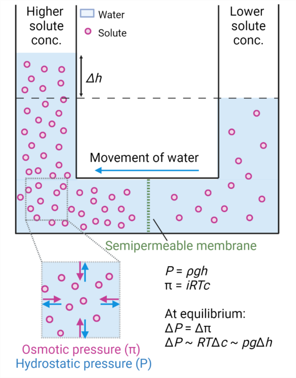
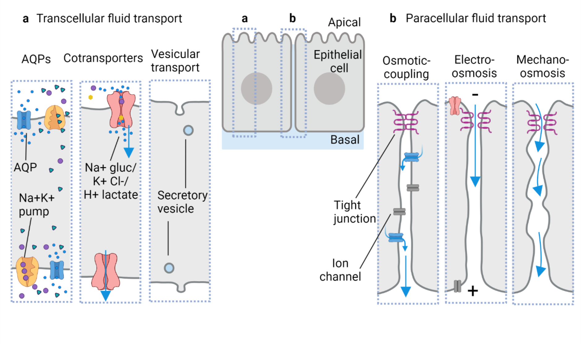
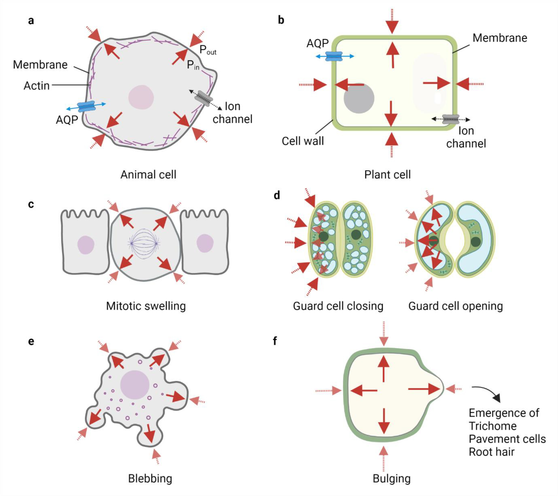
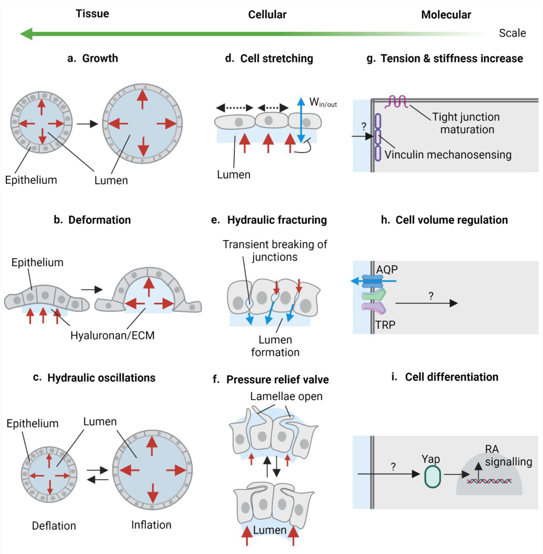
Morphogenesis, the process by which tissues develop into functional shapes, requires coordinated mechanical forces. Most current literature ascribes contractile forces derived from actomyosin networks as the major driver of tissue morphogenesis. Recent works from diverse species have shown that pressure derived from fluids can generate deformations necessary for tissue morphogenesis. In this review, we discuss how hydrostatic pressure is generated at the cellular and tissue level and how the pressure can cause deformations. We highlight and review findings demonstrating the mechanical roles of pressures from fluid-filled lumens and viscous gel-like components of the extracellular matrix. We also emphasise the interactions and mechanochemical feedbacks between extracellular pressures and tissue behaviour in driving tissue remodelling. Lastly, we offer perspectives on the open questions in the field that will further our understanding to uncover new principles of tissue organisation during development.

Keywords: Epithelial fluid transport; Epithelial lumen; Hyaluronan; Hydrostatic pressure; Mechanochemical feedback; Tissue morphogenesis.


Conflict of interest statement

Declaration of Competing Interest No conflict of interest. Conflict of interest The authors declare no conflict of interest.

Figures

Fig. 1:
Fig. 1:. Schematic representation for generation of osmotic and hydrostatic pressure.
Imbalance in the solute concentration across the semipermeable membrane generates osmotic pressure π leading to the flow of water from lower solute concentration to higher solute concentration. π = iRTc, where i is the van’t Hoff factor, R is the gas constant, T is the absolute temperature and c is the solute concentration. The counteractive pressure to osmotic pressure is hydrostatic pressure P = ρgh, where ρ is the density of the liquid, g is gravity, and h is the height of the liquid. At chemical equilibrium, osmotic pressure is equal to hydrostatic pressure.
Fig. 2:
Fig. 2:. Proposed routes of water movement across epithelial cell and tissue barriers.
a) Transcellular transport of water through the cells can be achieved via multiple modes: (left to right) solute imbalances within the cell for example due to the Na+/K+ pump can lead to a flow of water through aquaporins (AQPs) and membrane diffusion; water can also be transported via cotransporters such as Na+/glucose, H+/lactate, and K+/Cl− and vesicular transport such as pinocytosis and exocytosis can also enable water transport. b) Paracellular transport of water has been proposed across diverse epithelia: (left to right) the osmotic coupling model suggests water transport by following the ionic flux (osmosis) in the lateral intercellular spaces; electro-osmosis shown in the rabbit corneal endothelium suggests electric potential generation across the epithelial cells via differential ion pumping that eventually leads to flow of counterion current with water; mechano-osmosis model suggests an active role of cellular junctions and lateral membranes in transporting water flow by generating micro-peristalsis movements. Blue arrows represent the flow of water.
Fig. 3:
Fig. 3:. Comparative role of mechano-hydraulic coupling in animal and plant cells.
a-b) Animal and plant cell shape and volume are regulated by steady maintenance of ion and water flux through the cells, i.e. there is always an osmotic and hydrostatic pressure difference across the cell. The hydrostatic pressure gradient is balanced by the mechanical tension in the membrane, cell surface proteins, and cytoskeletal (actomyosin) cortex in animal cells as depicted by the red arrows. Solid red arrows represent Pin and dotted red arrows represent Pout. Blue arrows represent water flux. Plant cells have a high turgor pressure which is primarily balanced by the compressive forces from the stiff cell walls. c-f) Changes in mechano-hydraulic coupling can lead to alteration in hydrostatic pressure, cell shape, and cell volume and are important for cellular function. c) Animal cells undergo mitotic swelling due to increased hydrostatic pressure which is potentially useful for faithful segregation of chromosomes. d) Plant (guard) cells utilise swelling to open and close the stomatal apparatus for transpiration. e) Localised increases in the hydrostatic pressure in areas of the weaker cytoskeleton can lead to an inflation of animal membranes called blebbing, which is seen in various cellular processes such as cell division or cell death. f) Localised increases in the hydrostatic pressure due to weaker or thinner cell wall areas in plant cells can also lead to bulges which are important in the emergence of plant structures such as trichomes or root hairs
Fig. 4:
Fig. 4:. Hydrostatic pressure drives multiscale remodelling.
a-c) Hydrostatic pressure in dynamic tissue changes. a) Increase in the hydrostatic pressure in the epithelial lumen or lumen expansion as shown in mammalian blastocysts, zebrafish inner ear, and organoids regulate growth and size of the organ. b) Hydrostatic pressure arising due to hyaluronan hydrogel (water-dependent swelling of hyaluronan) in the ECM causes tissue deformation as shown during semicircular canal morphogenesis in the zebrafish inner ear. c) Inflation-deflation cycles of luminal pressure called hydraulic oscillations can regulate pressure in the lumen and control the size of the organ as demonstrated in mammalian blastocysts and organoids. d-f) Effects of hydrostatic pressure at the cellular scale. d) Hydrostatic pressure can cause epithelial cell stretching, which in zebrafish inner ear has been shown to negatively regulate water influx into the lumen. e) Pressurised microlumens in the lateral intercellular space can cause transient breakage of cellular junctions. This process called hydraulic fracturing has been demonstrated in mouse blastocoel formation. f) Oscillations in the luminal pressure can lead to quick release or build-up of pressure in the epithelial lumens. Epithelial cells can respond to pressure oscillations via membrane protrusions such as lamellae in the pressure relief valve in the zebrafish endolymphatic sac. g-i) Molecular perception and response to hydrostatic pressure. g) Hydrostatic pressure can elicit increased cell tension and stiffness and can allow pressure mechanosensing via vinculin and allow maturation of tight junctions as demonstrated in the mouse blastocyst. h) Cells can perceive hydrostatic pressure via stretch sensitive TRP family of proteins and/or Piezo proteins. TRP can physically interact with AQPs which can modulate water flux in the cell. This is a possible mechanism with extracellular hyaluronan during cell volume regulation during zebrafish heart valve morphogenesis. i) Hydrostatic pressure can also elicit a transcriptional response in the responding cells such as RA signalling activation via Yap nuclear localisation in the lung organoids. Red arrows represent hydrostatic pressure exerted by the lumen (a-f) and hyaluronan-rich ECM (b). Blue arrows represent water flux through the cells (d) and through the paracellular spaces via fracturing (e).

Similar articles

Cited by

References

    1. Lecuit T, Lenne P-F, Munro E, Force generation, transmission, and integration during cell and tissue morphogenesis, Annu. Rev. Cell Dev. Biol 27 (2011) 157–184. 10.1146/annurev-cellbio-100109-104027. - DOI - PubMed
    1. Matis M, The Mechanical Role of Microtubules in Tissue Remodeling, Bioessays. 42 (2020) e1900244. 10.1002/bies.201900244. - DOI - PubMed
    1. Mathur J, Hülskamp M, Microtubules and microfilaments in cell morphogenesis in higher plants, Curr. Biol 12 (2002) R669–76. 10.1016/s0960-9822(02)01164-8. - DOI - PubMed
    1. LeGoff L, Lecuit T, Mechanical Forces and Growth in Animal Tissues, Cold Spring Harb. Perspect. Biol 8 (2015) a019232. 10.1101/cshperspect.a019232. - DOI - PMC - PubMed
    1. Heisenberg C-P, Bellaïche Y, Forces in tissue morphogenesis and patterning, Cell. 153 (2013) 948–962. 10.1016/j.cell.2013.05.008. - DOI - PubMed

Publication types

LinkOut - more resources