

Cisplatin exposure acutely disrupts mitochondrial bioenergetics in the zebrafish lateral-line organ
- PMID: 35534350
- PMCID: PMC9745743
- DOI: 10.1016/j.heares.2022.108513
Cisplatin exposure acutely disrupts mitochondrial bioenergetics in the zebrafish lateral-line organ
Abstract
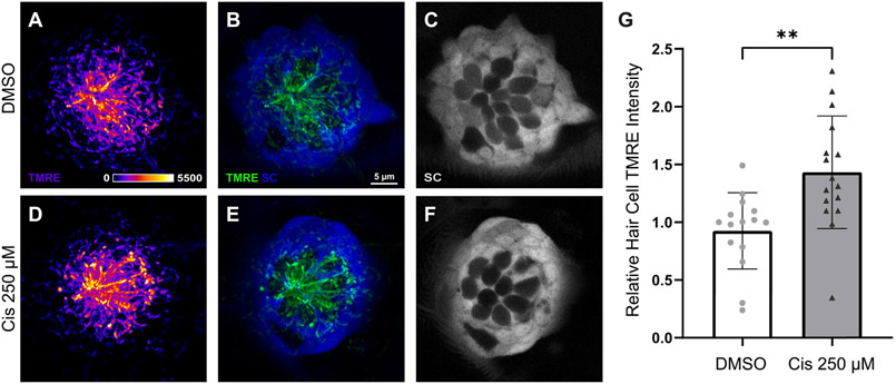
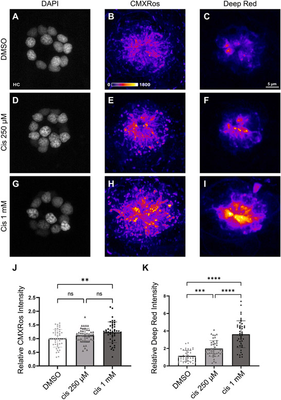
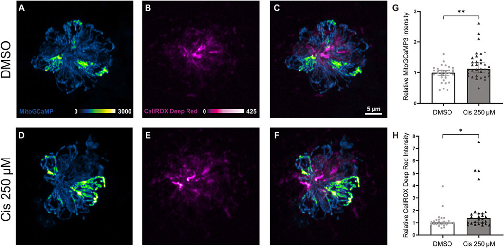
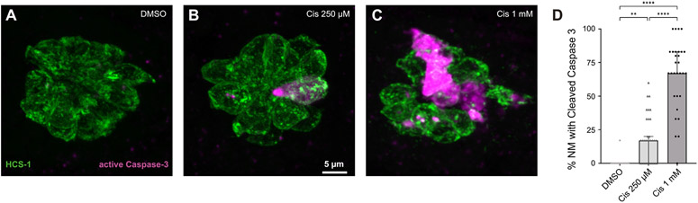
Cisplatin is a commonly used chemotherapeutic agent that causes debilitating high-frequency hearing loss. No targeted therapies currently exist to treat cisplatin ototoxicity, partly because the underlying mechanisms of cisplatin-induced hair cell damage are not completely defined. Zebrafish may offer key insights to cisplatin ototoxicity because their lateral-line organ contains hair cells that are remarkably similar to those within the cochlea but are optically accessible, permitting observation of cisplatin injury in live intact hair cells. In this study, we used a combination of genetically encoded biosensors in zebrafish larvae and fluorescent indicators to characterize changes in mitochondrial bioenergetics in response to cisplatin. Following exposure to cisplatin, confocal imaging of live intact neuromasts demonstrated increased mitochondrial activity. Staining with fixable fluorescent dyes that accumulate in active mitochondria similarly showed hyperpolarized mitochondrial membrane potential. Zebrafish expressing a calcium indicator within their hair cells revealed elevated levels of mitochondrial calcium immediately following completion of cisplatin treatment. A fluorescent ROS indicator demonstrated that these changes in mitochondrial function were associated with increased oxidative stress. After a period of recovery, cisplatin-exposed zebrafish demonstrated caspase-3-mediated apoptosis. Altogether, these findings suggest that cisplatin acutely disrupts mitochondrial bioenergetics and may play a key role in initiating cisplatin ototoxicity.
Keywords: Cisplatin ototoxicity; Mitochondrial bioenergetics; Mitochondrial dysfunction; Zebrafish.
Copyright © 2022 The Author(s). Published by Elsevier B.V. All rights reserved.
Conflict of interest statement
Declaration of Competing Interest None.
Figures
Similar articles
-
Protective effects of Y-27632 against cisplatin-induced ototoxicity: A zebrafish model Y-27632 and cisplatin-induced ototoxicity.Food Chem Toxicol. 2024 Aug;190:114792. doi: 10.1016/j.fct.2024.114792. Epub 2024 Jun 6. Food Chem Toxicol. 2024. PMID: 38849049
-
Apigenin attenuates cisplatin-induced hair cell damage in the zebrafish lateral line.Food Chem Toxicol. 2024 Dec;194:115099. doi: 10.1016/j.fct.2024.115099. Epub 2024 Nov 7. Food Chem Toxicol. 2024. PMID: 39521239
-
Novel synthetic protective compound, KR-22335, against cisplatin-induced auditory cell death.J Appl Toxicol. 2014 Feb;34(2):191-204. doi: 10.1002/jat.2852. Epub 2013 Jan 8. J Appl Toxicol. 2014. PMID: 23297007
-
Cisplatin-induced ototoxicity: From signaling network to therapeutic targets.Biomed Pharmacother. 2023 Jan;157:114045. doi: 10.1016/j.biopha.2022.114045. Epub 2022 Nov 28. Biomed Pharmacother. 2023. PMID: 36455457 Review.
-
Role of mitochondrial dysfunction and oxidative stress in sensorineural hearing loss.Hear Res. 2023 Jul;434:108783. doi: 10.1016/j.heares.2023.108783. Epub 2023 Apr 29. Hear Res. 2023. PMID: 37167889 Review.
Cited by
-
Mitochondrial form and function in hair cells.Hear Res. 2023 Feb;428:108660. doi: 10.1016/j.heares.2022.108660. Epub 2022 Nov 25. Hear Res. 2023. PMID: 36525891 Free PMC article. Review.
-
Evaluation of Cisplatin-Induced Pathology in the Larval Zebrafish Lateral Line.Int J Mol Sci. 2022 Nov 18;23(22):14302. doi: 10.3390/ijms232214302. Int J Mol Sci. 2022. PMID: 36430778 Free PMC article.
-
Protective Effects of Fasudil Against Cisplatin-Induced Ototoxicity in Zebrafish: An In Vivo Study.Int J Mol Sci. 2024 Dec 13;25(24):13363. doi: 10.3390/ijms252413363. Int J Mol Sci. 2024. PMID: 39769128 Free PMC article.
-
Putative COVID-19 therapies imatinib, lopinavir, ritonavir, and ivermectin cause hair cell damage: A targeted screen in the zebrafish lateral line.Front Cell Neurosci. 2022 Aug 24;16:941031. doi: 10.3389/fncel.2022.941031. eCollection 2022. Front Cell Neurosci. 2022. PMID: 36090793 Free PMC article.
-
Molecular Characteristics of Cisplatin-Induced Ototoxicity and Therapeutic Interventions.Int J Mol Sci. 2023 Nov 20;24(22):16545. doi: 10.3390/ijms242216545. Int J Mol Sci. 2023. PMID: 38003734 Free PMC article. Review.
References
-
- Behra M, Gallardo VE, Bradsher J, Torrado A, Elkahloun A, Idol J, Sheehy J, Zonies S, Xu L, Shaw KM, Satou C, Higashijima S-I, Weinstein BM, Burgess SM, 2012. Transcriptional signature of accessory cells in the lateral line, using the Tnk1bp1:EGFP transgenic zebrafish line. BMC Dev. Biol 12 (1), 6. doi:10.1186/1471-213x-12-6. - DOI - PMC - PubMed
-
- Bilan DS, Pase L, Joosen L, Gorokhovatsky AY, Ermakova YG, Gadella TW, Grabher C, Schultz C, Lukyanov S, Belousov VV, 2013. HyPer-3: a genetically encoded H(2)O(2) probe with improved performance for ratiometric and fluorescence lifetime imaging. ACS Chem. Biol 8 (3), 535–542. doi:10.1021/cb300625g. - DOI - PubMed
-
- Borse V, Al Aameri RFH, Sheehan K, Sheth S, Kaur T, Mukherjea D, Tupal S, Lowy M, Ghosh S, Dhukhwa A, Bhatta P, Rybak LP, Ramkumar V, 2017. Epigallocatechin-3-gallate, a prototypic chemopreventative agent for protection against cisplatin-based ototoxicity. Cell Death Dis. 8 (7), e2921. doi:10.1038/cddis.2017.314, - DOI - PMC - PubMed
Publication types
MeSH terms
Substances
Grants and funding
LinkOut - more resources
Full Text Sources
Research Materials
