. 2022 May 9;12(1):7568.
doi: 10.1038/s41598-022-11484-2.

Ultrastructure of setae of a planktonic diatom, Chaetoceros coarctatus

Affiliations

Ultrastructure of setae of a planktonic diatom, Chaetoceros coarctatus

Yuka Owari et al. Sci Rep. .

Abstract

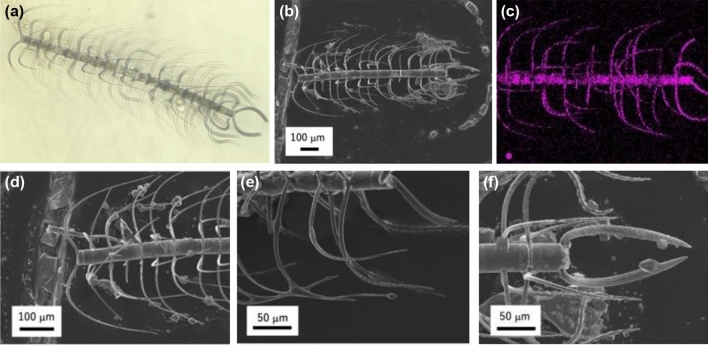
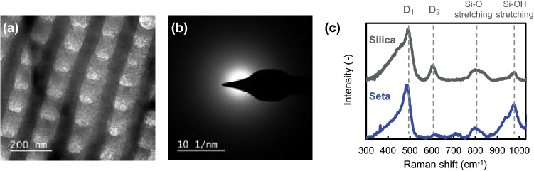
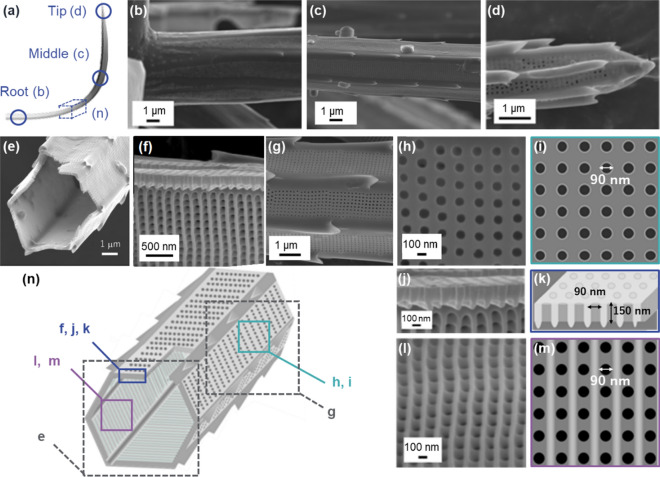
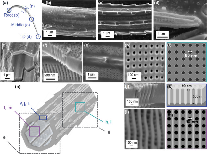
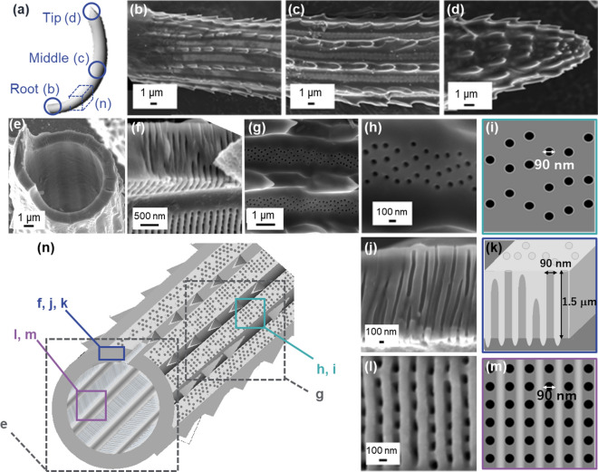
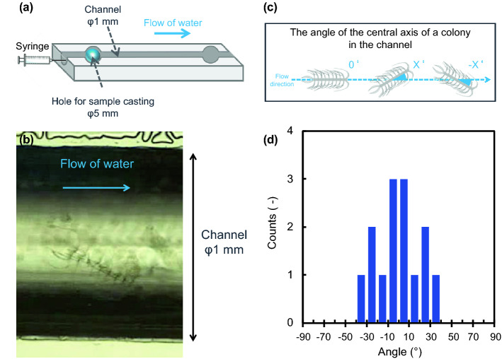
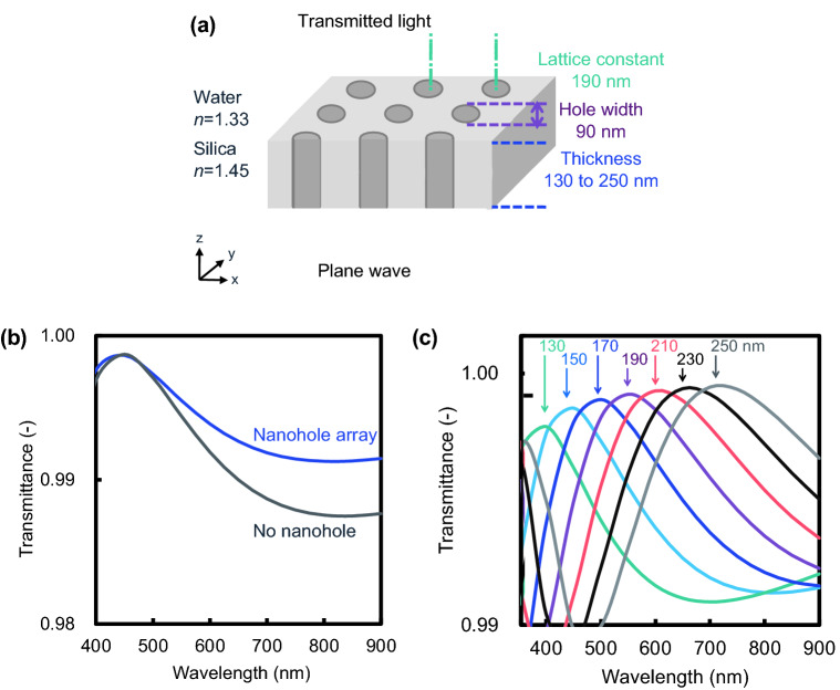
Silica frustules of most planktonic diatoms have many shallow holes in which the length (L) is smaller than the width (W). The present study focuses on a silica ultrastructure of setae of a planktonic diatom having deep (L/W > 1) holes. Here, we characterized microscopically patterned nanoholes on the silica walls of thick, robust, and hollow setae of a colony of Chaetoceros coarctatus. Basically, tetragonal poroid arrangements with and without a costa pattern are observed on the inner and outer surfaces, respectively, for three kinds of curving hollow setae attached to the anterior, intercalary, and posterior parts of the colony. The seta structures including specific poroid arrangements and continuity of deep nanoholes depend on the location. The deep nanoholes ∼90 nm wide are elongated from 150 to 1500 nm (L/W ∼17) with an increase in the wall thickness of the polygonal tubes of the setae. The inside poroid array, with a period of 190 nm in the extension direction of setae, is lined by parallel plates of the costae. However, the poroid arrangement on the outer surface is disordered, with several holes obstructed with increasing wall thickness of the posterior terminal setae. According to the movement of a colony in a fluid microchannel, the thick curving terminal setae is suggested to involve attitude control and mechanical protection. Using an optical simulation, the patterned deep through-holes on the intercalary setae were suggested to contribute anti-reflection of blue light in the wavelength range of 400 to 500 nm for the promotion of photosynthesis in seawater.


Conflict of interest statement

The authors declare no competing interests.

Figures

Figure 1
Figure 1
Overall view and enlarged images of a colony of C. coarctatus. Optical microscope image (a), SEM image (b), and elemental mapping of Si by energy-dispersive spectroscopy (EDS) (c) of a colony. Enlarged SEM images of M-shaped anterior terminal setae (d), intercalary setae (e), and U-shaped posterior terminal setae (f).
Figure 2
Figure 2
Structural analysis of setae. TEM (a) and SAED (b) images of a seta wall. Raman spectra of a seta and commercial amorphous silica particles (Reolosil, Tokuyama) (c).
Figure 3
Figure 3
Detailed observation of intercalary setae with SEM images (b-h, j, l) and schematic illustrations (a, i, k, m, n). The appearance of the root (b), middle (c, g), and apex (d), the fractured cross section of the middle (e, f), the poroid arrays on the outer and inner surfaces, and the cross-sectional view of the poroid array (h, j, l).
Figure 4
Figure 4
Detailed observation of anterior setae with SEM images (bh, j, l) and schematic illustrations (a, i, k, m, n). The appearance of the root (b), middle (c, g), and apex (d), the fractured cross section of the middle (e, f), the poroid arrays on the outer and inner surfaces, and the cross-sectional view of the poroid array (h, j, l).
Figure 5
Figure 5
Detailed observation of posterior setae with SEM images (bh, j, l) and schematic illustrations (a, i, k, m, n). The appearance of the root (b), middle (c, g), and apex (d), the fractured cross section of the middle (e, f), the poroid arrays on the outer and inner surfaces, and the cross-sectional view of the poroid array (h, j, l).
Figure 6
Figure 6
A schematic illustration of the morphogenesis of deep nanoholes with costae with the increasing thickness of a plate. Costae are initially arranged on the inner surface of a silica plate (a). The pores are arranged between costae (b). The depth of the nanoholes increases with increasing plate thickness (c). Several nanoholes collapse with growth of the plate to more than 1000 nm (d). Schematic illustration of the poroid arrangements (bd) show real structures based on SEM observations. On the other hand, the initial ladder structure consisting of costae (a) is presumed from the real poroid structures.
Figure 7
Figure 7
Observations of a dead colony of C. coarctatus in a flow of water in a microchannel 1 mm in diameter. Schematic illustration of the flow system including the microchannel (a), an optical microscopy image of a colony of C. coarctatus in the microchannel (b), a schematic illustration of the angle of a colony in the microchannel (c), and the distribution of the angle of a colony (d).
Figure 8
Figure 8
Simulated transmittance spectra of a silica plate with nanoholes using the 3D-FDTD method. A schematic model for 3D-FDTD simulation (a). Comparison of transmission spectra with and without nanohole structure; the thickness of the silica plate is 150 nm (b).Transmitted spectral variation for different thicknesses of silica plates from 130 to 250 nm (c).

Similar articles

Cited by

References

    1. Losic D, Mitchell JG, Voelcker NH. Diatomaceous lessons in nanotechnology and advanced materials. Adv. Mater. 2009;21:2947–2958. doi: 10.1002/adma.200803778. - DOI
    1. Zurzolo, C. & Bowler, C. Exploring bioinorganic pattern formation in diatoms. A story of polarized trafficking. Plant Physiol.127, 1339–1345 (2001). - PMC - PubMed
    1. Pawolski D, Heintze C, Mey I, Steinem C, Kröger N. Reconstituting the formation of hierarchically porous silica patterns using diatom biomolecules. J. Struct. Biol. 2018;204:64–74. doi: 10.1016/j.jsb.2018.07.005. - DOI - PubMed
    1. Kröger N, Deutzmann R, Bergsdorf C, Sumper M. Species-specific polyamines from diatoms control silica morphology. Proc. Natl. Acad. Sci. U. S. A. 2000;97:14133–14138. doi: 10.1073/pnas.260496497. - DOI - PMC - PubMed
    1. Görlich, S., Pawolski, D., Zlotnikov, I. & Kröger, N. Control of biosilica morphology and mechanical performance by the conserved diatom gene Silicanin-1. Commun. Biol.2, (2019). - PMC - PubMed

Publication types

Substances