. 2022 May 9;12(1):7581.
doi: 10.1038/s41598-022-11553-6.

Particulate matter (PM10) induces in vitro activation of human neutrophils, and lung histopathological alterations in a mouse model

Affiliations

Particulate matter (PM10) induces in vitro activation of human neutrophils, and lung histopathological alterations in a mouse model

Andrés Valderrama et al. Sci Rep. .

Abstract

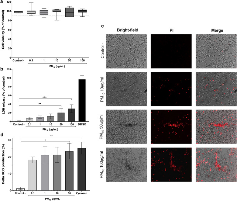
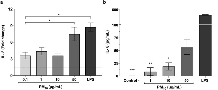
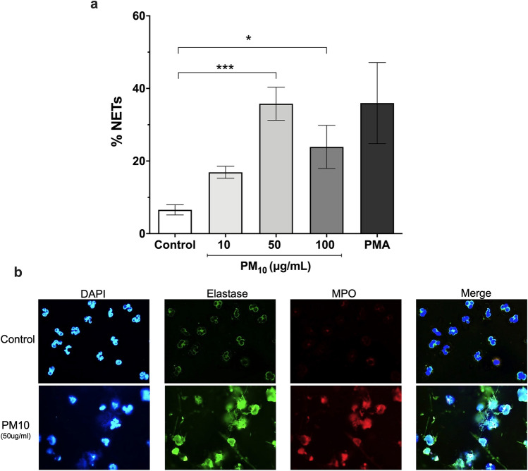
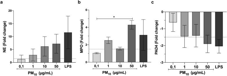
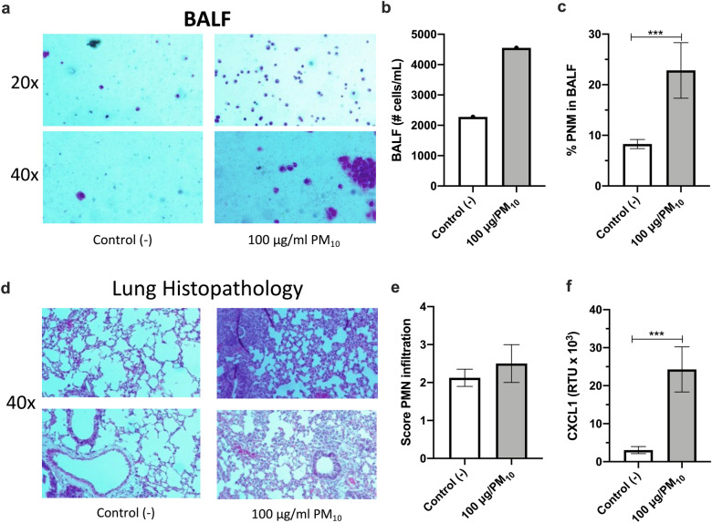
The epidemiological association between exposure to particulate matter (PM10) and various respiratory and cardiovascular problems is well known, but the mechanisms driving these effects remain unclear. Neutrophils play an essential role in immune defense against foreign agents and also participate in the development of inflammatory responses. However, the role of these cells in the PM10 induced inflammatory response is not yet fully established. Thus, this study aims to evaluate the effect of PM10 on the neutrophil-mediated inflammatory response. For this, neutrophils from healthy adult human donors were in vitro exposed to different concentrations of PM10. The cell viability and cytotoxic activity were evaluated by MTT. LDH, propidium iodide and reactive oxygen species (ROS) were quantified by flow cytometry. Interleukin 8 (IL-8) expression, peptidyl arginine deiminase 4 (PAD4), myeloperoxidase (MPO), and neutrophil elastase (NE) expression were measured by RT-PCR. IL-8 was also quantified by ELISA. Fluorescence microscopy was used to evaluate neutrophil extracellular traps (NETs) release. The in vivo inflammatory responses were assessed in BALB/c mice exposed to PM10 by histopathology and RT-PCR. The analysis shows that PM10 exposure induced a cytotoxic effect on neutrophils, evidenced by necrosis and LDH release at high PM10 concentrations. ROS production, IL-8, MPO, NE expression, and NETs release were increased at all PM10 concentrations assessed. Neutrophil infiltration in bronchoalveolar lavage fluid (BALF), histopathological changes with inflammatory cell infiltration, and CXCL1 expression were observed in PM10-treated mice. The results suggest that lung inflammation in response to PM10 could be mediated by neutrophils activation. In this case, these cells migrate to the lungs and release pro-inflamatory mediators, including ROS, IL-8, and NETs. Thus, contributing to the exacerbation of respiratory pathologies, such as allergies, infectious and obstructive diseases.


Conflict of interest statement

The authors declare no competing interests.

Figures

Figure 1
Figure 1
Cytotoxic effects of PM10 and ROS production in PMNs. (a) Cell viability assay by MTT and (b) LDH release in PMNs exposed to different concentrations of PM10 (0.1, 1, 10, 10, 50 and 100 µg/mL) for 5 h. Negative control (culture medium); positive control (DMSO). (c) Necrotic effects in PMNs exposed to different concentrations of PM10 (10, 50, and 100 µg/mL) for 30 min by PI staining. Photographs of each slide were observed at 200X magnification. (d) Intracellular ROS production in PMNs exposed to different concentrations of PM10 (0.1, 1, 10, and 50 µg/mL) for 30 min, determined by flow cytometry. Negative control (culture medium); positive control (zymosan). ROS levels are presented as the mean fluorescent intensity of DHR of treated cells relative to control. All data are represented as mean ± SEM of 8–14 healthy donors; *p < 0.05; **p < 0.01; ***p < 0.001, by Kruskal–Wallis analysis with Dunn's post hoc test.
Figure 2
Figure 2
PM10 exposure induces IL-8 in PMNs. (a) IL-8 mRNA expression and (b) IL-8 release in PMNs exposed to different concentrations of PM10 (0.1, 1, 10 and 50 µg/mL) for 5 h. Negative control (culture medium); positive control (LPS). The dotted line illustrates the 1.5-fold increase over the untreated negative control. All data are represented by the mean ± SEM of 8 healthy donors. * p < 0.05; **p < 0.01; ***p < 0.001, by Kruskal–Wallis analysis with post hoc Dunn's test. In (b), the asterisk represents differences compared with the LPS stimulated cells.
Figure 3
Figure 3
NETs formation in PMNs exposed to PM10. PMNs were exposed to 10, 50, and 100 μg/mL PM10 for 3 h. Negative control (culture medium); positive control (PMA). (a) NETs release was analyzed microscopically, and MPO, NE, and DNA were quantified with ImageJ software. (b) Representative fluorescence microscopy images of NETs. Images show colocalization of MPO (red), NE (green) with DNA fibers released (blue) from released NETs. All data are represented as mean ± SEM of 9 healthy donors. *p < 0.05; ***p < 0.001 by Kruskal–Wallis analysis with Dunn's post hoc test.
Figure 4
Figure 4
PM10 exposure alters NETS-associated gene expression in PMNs. mRNA expression of (a) NE (b) MPO and (c) PAD4 in PMNs exposed to different concentrations of PM10 (0.1, 1, 10 and 50 µg/mL) for 5 h. Negative control (culture medium); positive control (50 ng/ml LPS). The dotted line illustrates the value of 1.5-fold change compared with the untreated negative control. All data are represented as mean ± SEM of 15–18 healthy donors. *p < 0.05 by Kruskal–Wallis analysis with Dunn's post hoc test.
Figure 5
Figure 5
Intranasal treatment with PM10 increases cellularity and inflammatory infiltrate in the lung. BALB/c mice (n = 9 per group) were treated intranasally with 100 µg PM10 for five days. On day six post-treatment, the animals were euthanized, and bronchoalveolar lavage and lung tissue samples were collected. Wright's staining (a) quantification of total inflammatory cells (b) and quantification of PMNs in BALF (c) Representative images (d) and an inflammatory score of PMNs infiltrate (e) in hematoxylin–eosin stained lung sections, 1 = no infiltrate, 2 = mild infiltrate, 3 = moderate infiltrate, 4 = severe infiltrate. CXCL1 mRNA levels in lung lysate were measured by RT-PCR (f). All data are represented as mean ± SEM. ***p < 0.001 by Wilcoxon test.

References

    1. Organización Mundial de la Salud. Las nuevas Directrices mundiales de la OMS sobre la calidad del aire tienen como objetivo evitar millones de muertes debidas a la contaminación del aire. https://www.who.int/news/item/22-09-2021-new-who-global-air-quality-guid... (2021).
    1. World Health Organization. Ambient (outdoor) air pollution. https://www.who.int/news-room/fact-sheets/detail/ambient-(outdoor)-air-q.... pp 6–8 (2018).
    1. Fasola S, et al. Effects of particulate matter on the incidence of respiratory diseases in the pisan longitudinal study. Int. J. Environ. Res. Public Health. 2020;17:2540. doi: 10.3390/ijerph17072540. - DOI - PMC - PubMed
    1. Liang F, et al. Long-term exposure to fine particulate matter and cardiovascular disease in China. J. Am. Coll. Cardiol. 2020;75:707–717. doi: 10.1016/j.jacc.2019.12.031. - DOI - PubMed
    1. Kelly FJ, Fussell JC. Size, source and chemical composition as determinants of toxicity attributable to ambient particulate matter. Atmos. Environ. 2012;60:504–526. doi: 10.1016/j.atmosenv.2012.06.039. - DOI

Publication types