Review
. 2022 Jun;18(6):352-362.
doi: 10.1038/s41584-022-00783-7. Epub 2022 May 9.

Mechanisms and clinical implications of intervertebral disc calcification

Affiliations
Review

Mechanisms and clinical implications of intervertebral disc calcification

Uruj Zehra et al. Nat Rev Rheumatol. 2022 Jun.

Abstract

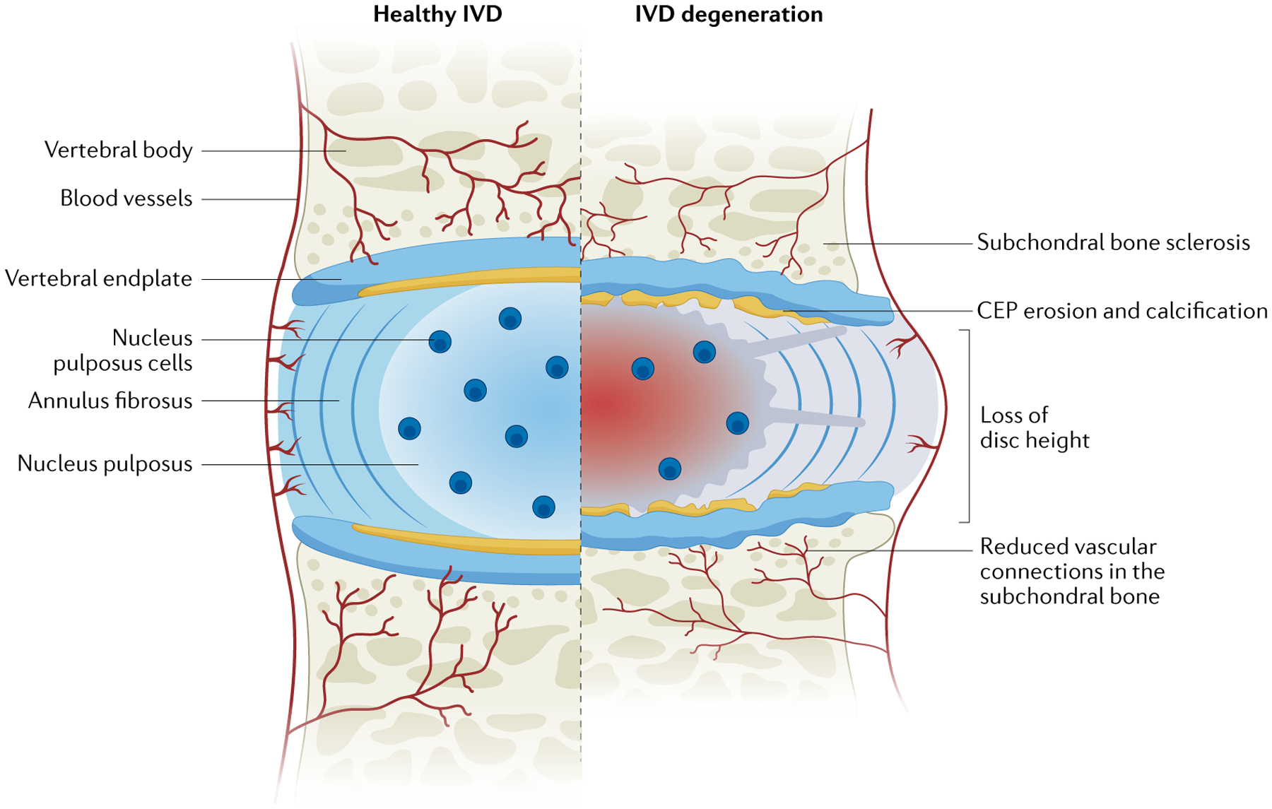
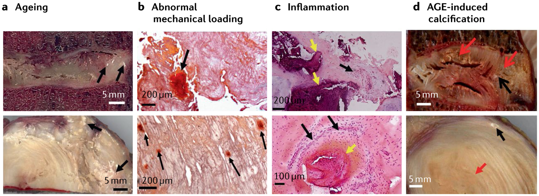
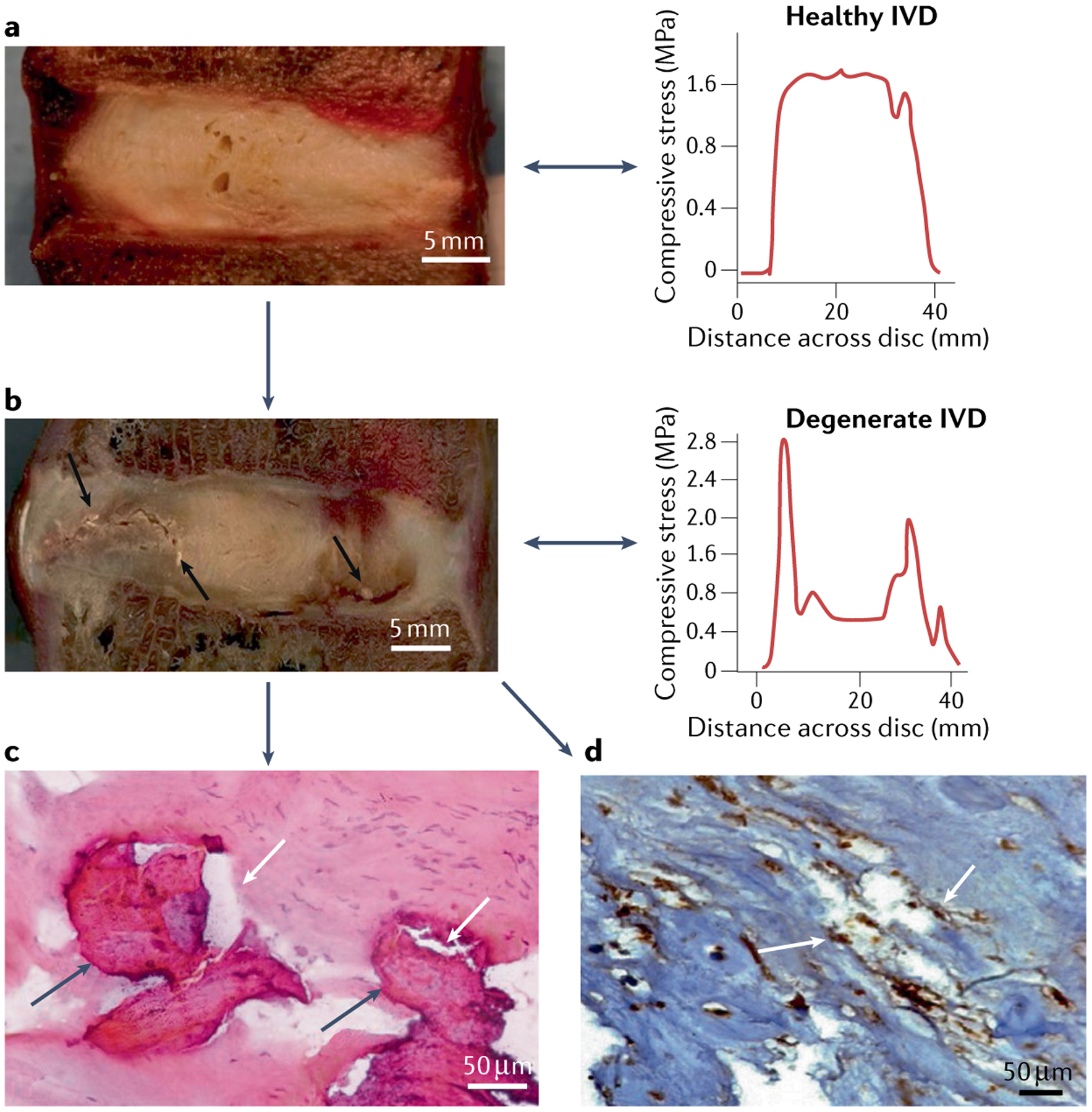
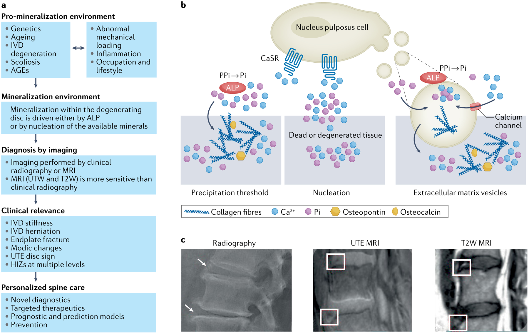
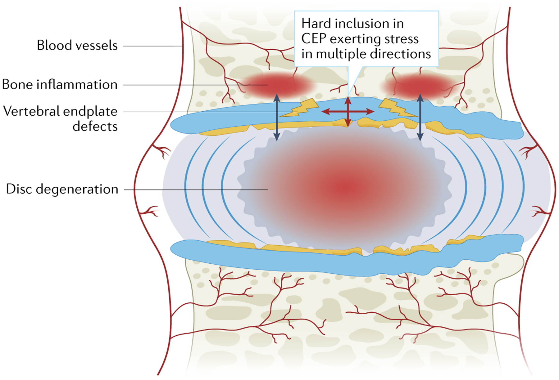
Low back pain is a leading cause of disability worldwide. Intervertebral disc (IVD) degeneration is often associated with low back pain but is sometimes asymptomatic. IVD calcification is an often overlooked disc phenotype that might have considerable clinical impact. IVD calcification is not a rare finding in ageing or in degenerative and scoliotic spinal conditions, but is often ignored and under-reported. IVD calcification may lead to stiffer IVDs and altered segmental biomechanics, more severe IVD degeneration, inflammation and low back pain. Calcification is not restricted to the IVD but is also observed in the degeneration of other cartilaginous tissues, such as joint cartilage, and is involved in the tissue inflammatory process. Furthermore, IVD calcification may also affect the vertebral endplate, leading to Modic changes (non-neoplastic subchondral vertebral bone marrow lesions) and the generation of pain. Such effects in the spine might develop in similar ways to the development of subchondral marrow lesions of the knee, which are associated with osteoarthritis-related pain. We propose that IVD calcification is a phenotypic biomarker of clinically relevant disc degeneration and endplate changes. As IVD calcification has implications for the management and prognosis of degenerative spinal changes and could affect targeted therapeutics and regenerative approaches for the spine, awareness of IVD calcification should be raised in the spine community.


Conflict of interest statement

Competing interests

The authors declare no competing interests.

Figures

Fig. 1 |
Fig. 1 |. Comparison of a healthy and a degenerated intervertebral disc in the spine.
The intervertebral disc (IVD) is a shock-absorbing structure with three main components: the inner nucleus pulposus, the outer annulus fibrosus and the cartilaginous endplates (CEPs), which anchor the disc to the adjacent vertebrae. The healthy nucleus pulposus is a highly hydrated, gelatinous, proteoglycan-rich tissue. The healthy annulus fibrosus encloses the nucleus pulposus and is a highly organized fibrous structure composed of concentric lamellae of tilted collagen fibres with scattered proteoglycans. The degenerated IVD is reduced in height compared with its healthy counterpart owing to extracellular matrix depletion, a fibrous and dehydrated nucleus pulposus, severe structural modifications of annulus fibrosus collagen fibres, extensive damage to the CEP and sclerosis of the subchondral bone. Reprinted from REF., Springer Nature Limited.
Fig. 2 |
Fig. 2 |. Calcification in degenerated discs.
a | Sagittal (top image) and transverse (bottom image) sections of aged intervertebral discs (IVDs) showing several calcified spots (black arrows); scale bar = 5 mm. b | Histological sections of scoliotic discs stained with Alizarin red stain showing that abnormal mechanical loading can induce mineral deposition in the IVDs (black arrows); scale bars = 200 μm. c | Inflammation and calcification are highly associated with each other. H&E stained disc sections of nucleus pulposus (top image) and inner annulus (bottom image) showing the coexistence of inflammation and calcification, calcified deposits (yellow arrows) and adjacent inflammation (black arrows); scale bars upper 200 μm and lower 100 μm. d | Advanced glycation endproduct (AGE)-induced calcification can be seen in the sagittal (top image) and transverse sections (bottom image) of the disc showing the yellowing effect of AGE (red arrows) and calcification (black arrows), suggesting a role of AGEs in calcification; scale bars = 5 mm.
Fig. 3 |
Fig. 3 |. Images showing presence of fissures near calcified spots.
a | Sagittal image of a cadaveric healthy disc with a representative graph of intradiscal stress; scale bar = 5 mm. b | Sagittal cadaveric section of a degenerated intervertebral disc with fissures (black arrows) near calcified spots with representative images of intradiscal stress; scale bar = 5 mm. The representative images of intradiscal stress show the stress profile of discs: the stress profile of the healthy disc shows an even distribution of stress throughout the disc whereas the stress profile of the degenerated disc shows multiple stress peaks. c | Haematoxylin and eosin stained section of a degenerated intervertebral disc showing coexistence of calcification (dark grey arrows) and fissures (white arrows); scale bar = 50 μm. d | Bone morphogenetic protein 2 immuno-stained sections of a degenerated intervertebral disc, showing association of both calcification and fissures (white arrows); scale bar = 50 μm.
Fig. 4 |
Fig. 4 |. Disc calcification: mechanisms, diagnosis and clinical relevance.
a | Overview of disc calcification, diagnosis and clinical relevance. Mineralization within the degenerating disc has received increasing recognition as being important as the spinal phenotypes identified are associated with pain and disability. Disc mineralization deserves further attention in order to improve patient stratification by novel diagnostics to aid targeted therapeutics and even generate prognostic models to enable prevention of disc disease. b | The initiating factors and mechanisms of disc mineralization. Mineralization within the degenerating disc may involve three mechanisms. In one mechanism, calcium (Ca2+) and inorganic monophosphate (Pi) ions accumulate above physiological levels, reaching the threshold of precipitation; in this process alkaline phosphatase (ALP) activity drives accumulation of Pi. In degenerated IVDs there is enhanced expression of calcium-sensing receptor (CaSR), which is involved in Ca2+ sensing and downstream signalling and as such may contribute to the process of intervertebral disc (IVD) calcification. In another mechanism, Ca2+ and Pi present even within physiological levels may precipitate owing to the presence of nucleating agents in dead or degenerated tissue. In a third mechanism, extracellular vesicles secreted by cells are enriched with Ca2+, Pi, and ALP and also contain biological messages (such as microRNAs) involved in bone-formation signalling pathways. Extracellular vesicle biology in the IVD is only beginning to emerge. c | Imaging methods for diagnosing disc mineralization. Mineralization within the intervertebral disc (shown by arrows and boxes) imaged with the aid of multiple imaging modalities, such as plain radiographs, ultra-short time-to-echo (UTE) MRI and T2-weighted MRI (T2W). AGEs, advanced glycation end products; HIZ, high-intensity zone on T2-weighted MRI; UDS, ultra-short time-to-echo disc sign.
Fig. 5 |
Fig. 5 |. A conceptual overview of the initiation of Modic changes and disc degeneration.
Hard inclusions in the cartilaginous endplate (CEP) can initiate cracks in multiple directions that can lead to vertebral endplate defects. A break in the endplate barrier would facilitate the escape of water, free movement of various cytokines and inflammatory cells and constant crosstalk between the disc and vertebral body, predisposing the disc to decompression, degeneration and Modic changes.

Similar articles

Cited by

References

    1. Francisco V et al. A new immunometabolic perspective of intervertebral disc degeneration. Nat. Rev. Rheum 18, 47–60 (2022). - PubMed
    1. Samartzis D et al. A population-based study of juvenile disc degeneration and its association with overweight and obesity, low back pain, and diminished functional status. J. Bone Joint Surg. Am 93, 662–670 (2011). - PubMed
    1. Adams MA & Dolan P Intervertebral disc degeneration: evidence for two distinct phenotypes. J. Anat 221, 497–506 (2012). - PMC - PubMed
    1. Buckwalter JA Aging and degeneration of the human intervertebral disc. Spine 20, 1307–1314 (1995). - PubMed
    1. Feng G, Zhang Z, Dang M, Rambhia KJ & Ma PX Nanofibrous spongy microspheres to deliver rabbit mesenchymal stem cells and anti-miR-199a to regenerate nucleus pulposus and prevent calcification. Biomaterials 256, 120213 (2020). - PMC - PubMed

Publication types

MeSH terms