. 2022 May;29(5):451-462.
doi: 10.1038/s41594-022-00764-0. Epub 2022 May 9.

The HMCES DNA-protein cross-link functions as an intermediate in DNA interstrand cross-link repair

Affiliations

The HMCES DNA-protein cross-link functions as an intermediate in DNA interstrand cross-link repair

Daniel R Semlow et al. Nat Struct Mol Biol. 2022 May.

Abstract

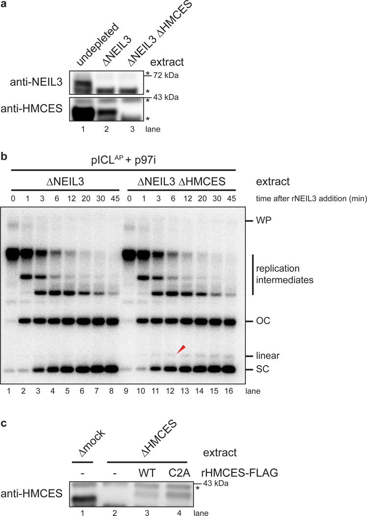
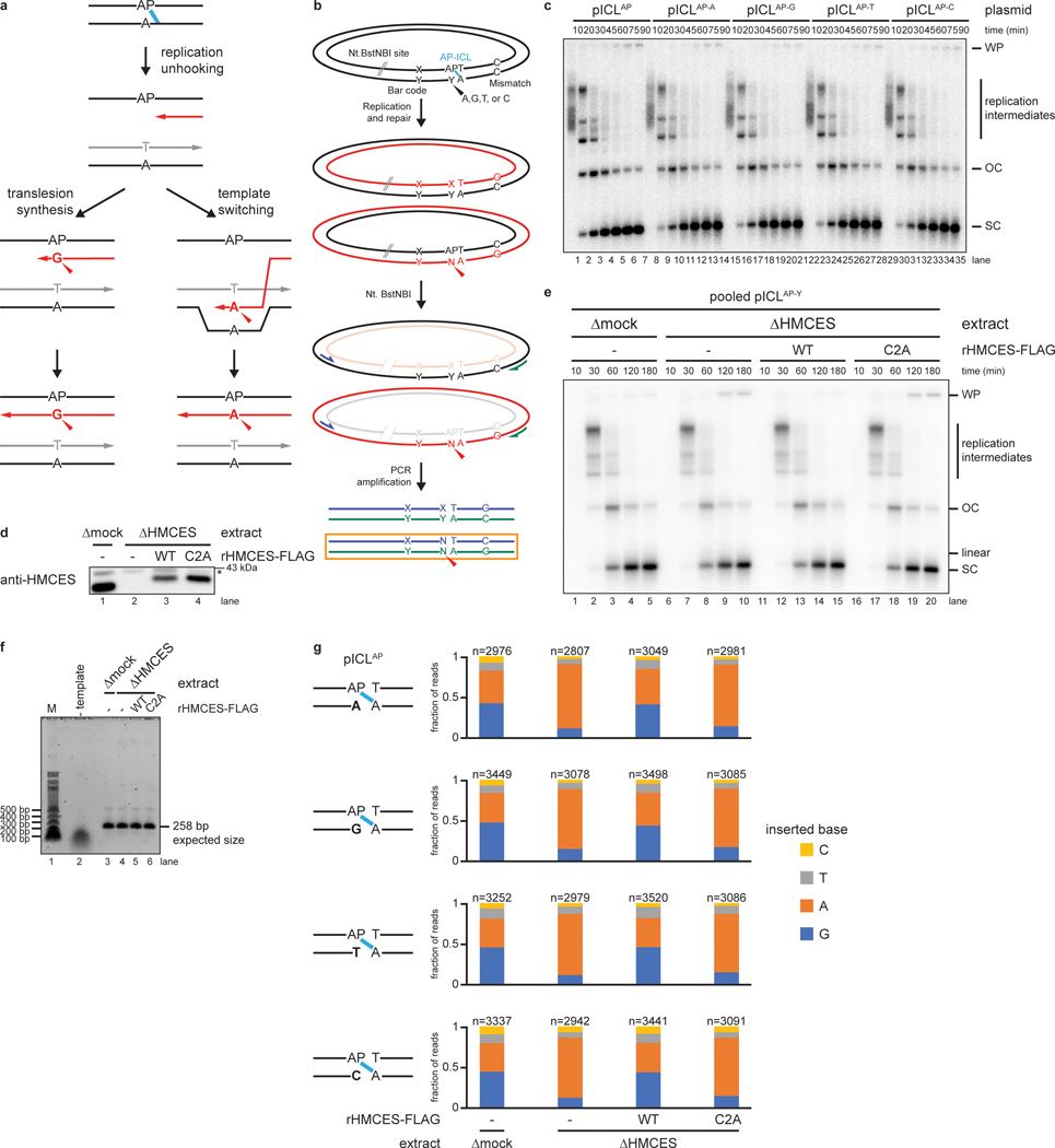
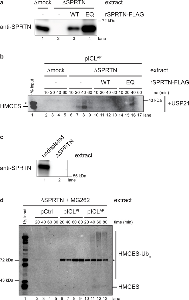
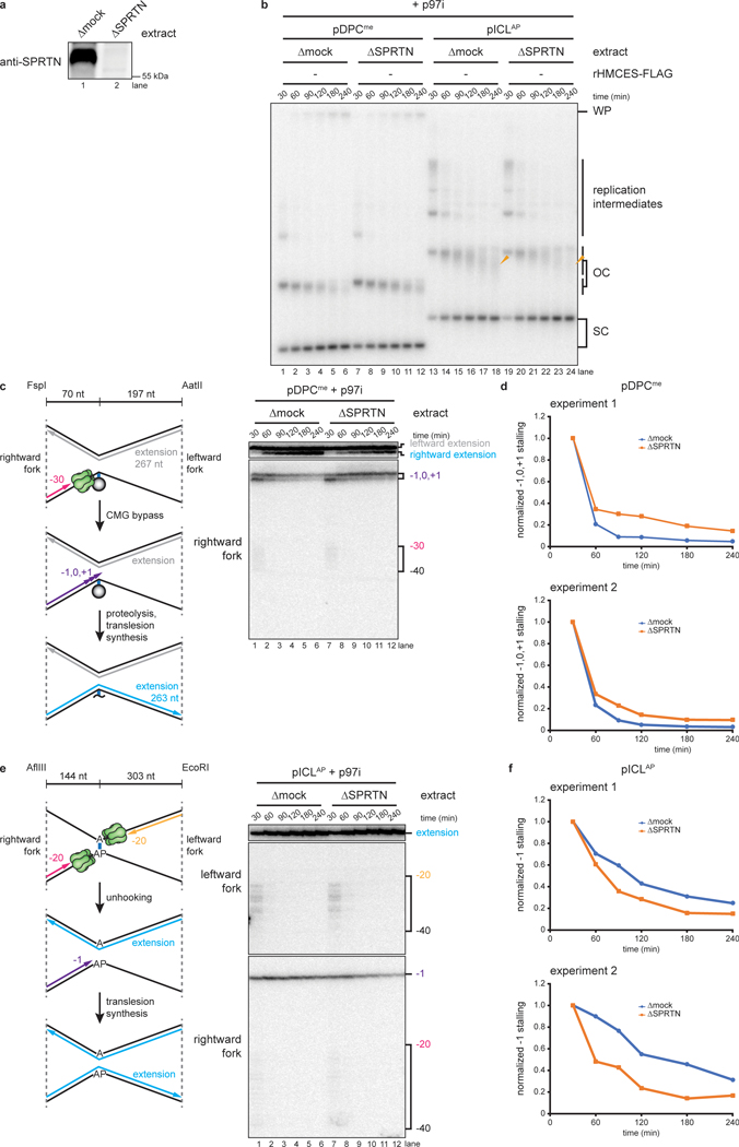
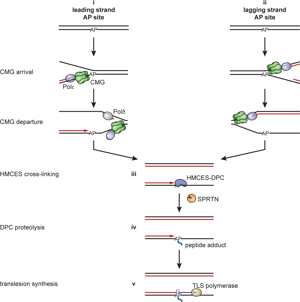
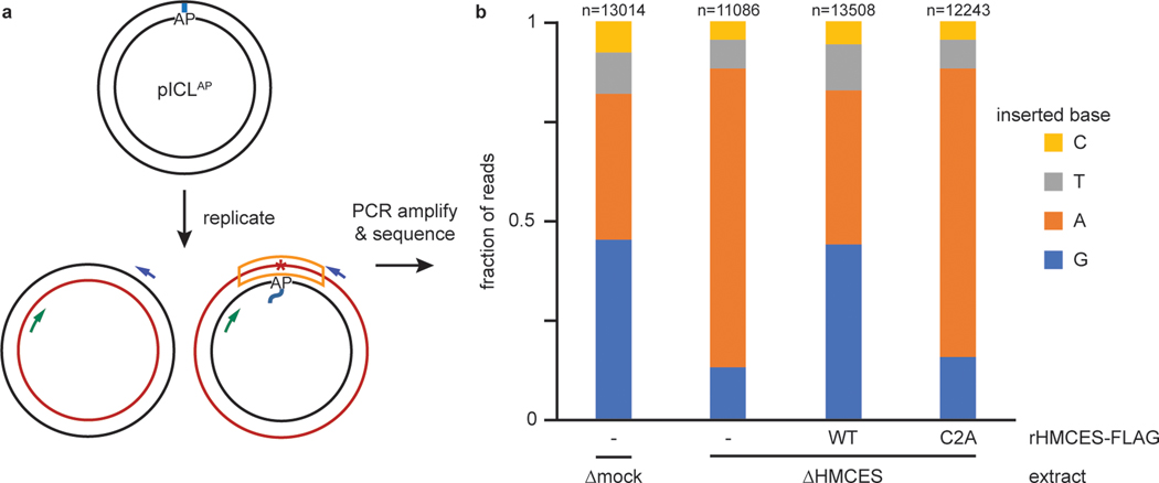
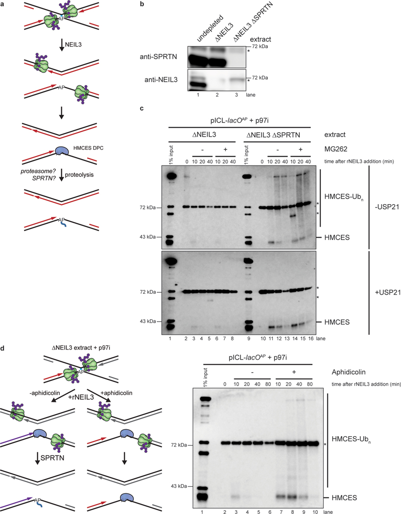
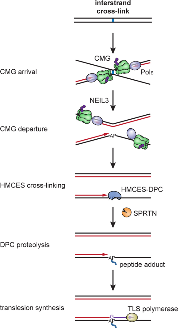
The 5-hydroxymethylcytosine binding, embryonic stem-cell-specific (HMCES) protein forms a covalent DNA-protein cross-link (DPC) with abasic (AP) sites in single-stranded DNA, and the resulting HMCES-DPC is thought to suppress double-strand break formation in S phase. However, the dynamics of HMCES cross-linking and whether any DNA repair pathways normally include an HMCES-DPC intermediate remain unknown. Here, we use Xenopus egg extracts to show that an HMCES-DPC forms on the AP site generated during replication-coupled DNA interstrand cross-link repair. We show that HMCES cross-links form on DNA after the replicative CDC45-MCM2-7-GINS (CMG) helicase has passed over the AP site, and that HMCES is subsequently removed by the SPRTN protease. The HMCES-DPC suppresses double-strand break formation, slows translesion synthesis past the AP site and introduces a bias for insertion of deoxyguanosine opposite the AP site. These data demonstrate that HMCES-DPCs form as intermediates in replication-coupled repair, and they suggest a general model of how HMCES protects AP sites during DNA replication.


Figures

Extended Data Fig. 1
Extended Data Fig. 1. An HMCES-DPC shields AP sites
a, Purified recombinant FLAG-tagged Xenopus laevis HMCES proteins were resolved by SDS-PAGE and visualized by staining with InstantBlue. rHMCESΔPIP harbors W321A and L322A mutations that disrupt a conserved PIP-box that was previously show to mediate interaction with PCNA. Asterisk, contaminating bands. b, A 5’ end radiolabeled 20mer oligonucleotide with a single deoxyuracil (-AP site) or AP site (+AP site) was incubated with rHMCES proteins shown in (a) for 60 min. Samples were then resolved on a denaturing polyacrylamide gel and visualized by autoradiography. Reactions contained 1 nM oligonucleotide and 50 nM rHMCES. c, Schematic of species produced by digestion of pICLAP replication intermediates with HincII and APE1. Digestion with HincII generates a 5.6 kb linear plasmid species while additional AP site cleavage by APE1 is expected to generate 2.3 kb and 3.3 kb species. d, HMCES immunodepletion. The extracts used in the reactions shown in e were blotted for HMCES. Asterisk, non-specific band. e, pICLAP was replicated with [α−32P]dATP in the indicated egg extracts (shown in d). Samples were treated with proteinase K, phenol:chloroform extracted, and digested with HincII or with HincII and APE1. Digested DNAs were resolved on a native agarose gel and visualized by autoradiography. X structures indicate HincII-digested plasmids before ICL unhooking. f, Quantification of APE1 cleavage efficiency for the reactions shown in e. Cleavage efficiency was quantified as the intensity (Int) of 2.3 kb and 3.3 kb fragment bands in each lane divided by the total intensity of linear species bands ([Int2.3kB + Int3.3kB]/[Int2.3kB + Int3.3kB + Int5.6kB]). The efficiency of HMCES-DPC formation in mock-depleted extract was estimated by subtracting the extent of rAPE1 cleavage in mock-depleted extract from the extent of cleavage in HMCES-depleted extract at 45 min (arrowheads), when the absolute signal resulting from rAPE1 cleavage is maximal.
Extended Data Fig. 2
Extended Data Fig. 2. HMCES suppresses DSB formation specifically during NEIL3-dependent ICL repair
a, b, d, f, and h, HMCES and NEIL3 immunodepletions. The extracts used in Fig. 2a (a), Extended Data Fig. 2c (b), Extended Data Fig. 2e (d), Extended Data Fig. 2g (f), and Fig. 2c (h) were blotted for HMCES and NEIL3 as indicated. Asterisks, non-specific bands. c, pICLAP was replicated with [α−32P]dATP in mock- or HMCES-depleted extracts. Replication intermediates were analyzed as in Fig. 2a alongside pCtrl replication products that were linearized by digestion with HincII. e, pICLAP was replicated with [α−32P]dATP in mock- or HMCES-depleted extracts supplemented with rHMCES, as indicated. Replication intermediates were analyzed as in Fig. 2a. g, pCtrl, pICLPt, or pICLAP were replicated with [α−32P]dATP in mock- or HMCES-depleted extract and replication intermediates were analyzed as in Fig. 2a.
Extended Data Fig. 3
Extended Data Fig. 3. HMCES suppresses ATR-dependent CHK1 phosphorylation during NEIL3-dependent ICL repair
a, b, and d, HMCES immunodepletions. The extracts used in Fig. 2d (a), Extended Data Fig. 3c (b), and Extended Data Fig. 3e (d) were blotted for HMCES. Asterisks, non-specific bands. c, pICLAP was replicated in mock- or HMCES-depleted extracts supplemented with ATR inhibitor AZD6738 (ATRi), as indicated. Replication reactions were separated by SDS-PAGE and blotted for phospho-CHK1 and MCM6 (loading control). Accumulation of phosphorylated CHK1 was blocked by AZD6738 treatment, indicating that CHK1 phosphorylation is dependent on ATR. e, pCtrl, pICLPt, or pICLAP was replicated in mock- or HMCES-depleted extracts, as indicated. Replication reactions were analyzed as in c. HMCES depletion increased the accumulation of phosphorylated CHK1 only during replication of pICLAP, indicating a specific role for HMCES in NEIL3-dependent ICL repair.
Extended Data Fig. 4
Extended Data Fig. 4. TLS inhibition does not enhance DSB formation during NEIL3-dependent ICL repair
a, REV1 and HMCES immunodepletion. The extracts used in the replication reactions shown in b were blotted for REV1 and HMCES. Asterisks, non-specific bands. b, pICLAP was replicated with [α−32P]dATP in the indicated extracts (shown in a). Replication intermediates were analyzed as in Fig. 2a.
Extended Data Fig. 5
Extended Data Fig. 5. Synchronization of ICL unhooking by NEIL3-depletion and add back
a, NEIL3 and HMCES immunodepletion. The extracts used in the replication reactions shown in Fig. 3b were blotted for HMCES and NEIL3. Asterisks, non-specific bands. b, pICLAP was replicated in the presence of [α−32P]dATP in the indicated extracts (shown in a) supplemented with p97 inhibitor for 60 min. rNEIL3 was then added to the reactions to allow ICL unhooking. Replication intermediates were analyzed as in Fig. 2a. c, HMCES immunodepletion. The extracts used in Fig. 4 were blotted for HMCES. Asterisk, non-specific band.
Extended Data Fig. 6
Extended Data Fig. 6. Nucleotide identity opposite the AP site does not effect NEIL3-dependent ICL unhooking or TLS
a, Model of two alternative mechanisms of AP site bypass. Translesion synthesis (left branch) uses a specialized TLS polymerase for untemplated insertion of a nucleotide opposite the non-coding AP site. In this pathway, the nucleotide opposite the AP site in the parental plasmid does not influence insertion by the TLS polymerase. Template switching (right branch) uses the newly synthesized DNA of the other sister chromatid as a template for error-free bypass of the AP site. In this case, the inserted nucleotide is expected to co-vary with the nucleotide opposite the AP site in the parental plasmid. b, Plasmid design for next generation sequencing. We prepared four different plasmids, each containing an AP-ICL and a different nucleotide positioned opposite the AP site (Y), as well as a unique barcode (X-Y). An Nt.BstNBI restriction site allows specific cleavage of the AP site-containing strand to prevent its amplification during PCR. A dC-dC mismatch allows reads produced from amplification of the nascent strand that has bypassed the AP site (PCR product in orange box) to be distinguished from reads derived from the corresponding parental strand (upper PCR product). c, The four AP-ICL plasmids, each containing a different nucleotide opposite the AP-site (described in b), were replicated in undepleted egg extract supplemented with [α−32P]dATP. Replication intermediates were analyzed as in Fig. 2a. The base opposite the AP site had no apparent effect on replication or repair efficiency. d, HMCES immunodepletion. The extracts used to generate the sequencing libraries described in Fig. 5 were blotted for HMCES. Asterisk, non-specific band. e, The four AP-ICL plasmids described in b were pooled and replicated with [α−32P]dATP in the same extracts (shown in panel d) and used to generate sequencing libraries described in Fig. 5. Replication intermediates were analyzed as in Fig. 2a. f, Analysis of PCR amplicons used for sequencing. In parallel to the reactions shown in e, pooled pICLAP plasmids were replicated in the indicated extracts (shown in d, but lacking [α−32P]dATP) supplemented with rHMCES, as indicated. DNA was extracted and digested with Nt.BstNBI (to cleave AP site-containing strands). The region of the replicated plasmids surrounding the ICL was then amplified by PCR. PCR amplicons were resolved by native agarose gel electrophoresis and visualized by Sybr Gold staining. g, Analysis of sequencing reads derived from individual pICLAP plasmids. The barcode was used to distinguish sequencing reads derived from the four different AP-ICL containing plasmids described in b. For each extract condition, we obtained >30,000 mapped reads, the vast majority (87.4%−87.8%) of which either perfectly matched the reference sequence or had a single point mutation corresponding to the position opposite the AP site. Of these reads, >11,000 in each condition derived from the nascent DNA stand produced upon bypass of the AP site. The fraction of reads corresponding to insertion of a given nucleotide opposite the AP site are plotted for each plasmid and extract condition. n, number of pooled nascent strand reads obtained for each condition. The result shows that the nucleotide opposite the AP site in the parental plasmid template does not influence the distribution of nascent DNA nucleotides inserted opposite the AP site after unhooking. Next generation sequencing read counts can be found in Supplementary Table 2.
Extended Data Fig. 7
Extended Data Fig. 7. SPRTN protease activity is required for HMCES removal
a and c, SPRTN immunodepletions. The extracts used in the replication reactions shown in Extended Data Fig. 7b (a) and Extended Data Fig. 7d (c) were blotted for SPRTN. b, pICLAP was replicated in mock- or SPRTN-depleted egg extract supplemented with wild-type (WT) or catalytically defective E89Q-mutated (EQ) rSPRTN, as indicated. Chromatin was recovered under stringent conditions, treated with the deubiquitylating enzyme USP21, and associated proteins were separated by SDS-PAGE and blotted for HMCES. Asterisk, non-specific band. d, pCtrl, pICLPt, or pICLAP were replicated in SPRTN-depleted egg extract supplemented with proteasome inhibitor MG262. Chromatin was analyzed as in Fig. 6c. Chromatin-associated HMCES is only observed in the pICLAP replication reaction, implying a specific role for HMCES in NEIL3-dependent ICL repair. Asterisk, non-specific band.
Extended Data Fig. 8
Extended Data Fig. 8. SPRTN-dependent proteolysis of the HMCES-DPC is not required for TLS past the AP site
a, SPRTN immunodepletion. The extracts used in b-f were blotted for SPRTN. b, A plasmid containing a methylated HpaII DPC that is refractory to degradation by the proteasome (pDPCme) or pICLAP were replicated with [α−32P]dATP in mock- or SPRTN-depleted egg extracts supplemented with p97 inhibitor. Replication intermediates were analyzed as in Fig. 4a. c, Left, schematic of nascent strands generated during DPC repair. FspI and AatII cut 70 nucleotides to the left and 197 nucleotides to the right of the ICL, respectively, generating characteristic −30 stall, −1 to +1 stall, and strand extension products. Right, pDPCme was replicated as in b and nascent DNA strands were isolated, digested with FspI and AatII, and resolved by denaturing polyacrylamide gel electrophoresis. As previously reported, SPRTN-depletion delays TLS past the methylated HpaII DPC, as evidenced by the persistence of rightward fork −1, 0, and +1 stall products and a delay in formation of rightward fork extension products. d, The persistence of the rightward fork −1, 0, and +1 stall products in c was quantified by dividing the summed intensity of the −1, 0, and +1 stall product bands in each lane by the intensity of the full-length rightward extension product band. Quantifications were normalized to the accumulation of the −1, 0, and +1 stall products at the 0 min time timepoint. Quantifications from two independent experiments are shown. e, Left, schematic of nascent strands generated during AP-ICL repair, as in Fig. 4b. Right, pICLAP was replicated as in b and nascent DNA strands were analyzed as in Fig. 4b. In contrast to TLS past the methylated HpaII-DPC shown in c, SPRTN-depletion accelerated TLS past the HMCES-DPC, as evidenced by the faster disappearance of rightward leading strand −1 stall products. This result indicates that TLS past the AP site does not require SPRTN-dependent proteolysis of the HMCES-DPC formed during NEIL3-dependent ICL repair. f, The persistence of the rightward fork −1 stall product in e was quantified as in d. Quantifications from two independent experiments are shown.
Extended Data Fig. 9
Extended Data Fig. 9. A proposed general model for AP site protection by HMCES during DNA replication
(i) AP sites encountered in the leading strand template due to spontaneous depurination/depyrimidination or incomplete BER are bypassed by CMG. This leads to uncoupling of DNA unwinding by CMG from leading strand DNA synthesis by Pol ε and HMCES-DPC formation at a ssDNA/dsDNA junction. (ii) AP sites in the lagging strand template are bypassed without CMG uncoupling, and HMCES cross-links to the AP site fully embedded in ssDNA. In both scenarios, Pol δ then extends the lagging strand up to the HMCES-DPC, where upon synthesis stalls (iii). In both cases, the HMCES-DPC abuts a ssDNA/dsDNA junction, leading to proteolysis by SPRTN (iv). The HMCES-DPC and the peptide adduct generated after proteolysis stabilize the AP site until the lesion is bypassed by TLS (v) or an alternative, error-free mechanism (not depicted).
Fig. 1:
Fig. 1:. HMCES cross-links to AP sites during NEIL3-dependent ICL repair.
a, Model of replication-coupled ICL repair pathways. b, Schematic of repair products. AseI and XhoI digestion allows resolution of the top (87 nt) and bottom (85 nt) strands. AP-ICL unhooking by NEIL3 generates an AP-site that cross-links to HMCES and generates a discrete adduct in the top strands after proteinase K digestion. c, Detection of AP-site adducts by strand-specific Southern blotting. pCtrl or pICLAP was replicated in egg extract. The p97i was added to prevent activation of the FA pathway. At the indicated times, DNA was isolated, treated with proteinase K, and digested with AseI and XhoI. The repair products were separated on a denaturing polyacrylamide sequencing gel and visualized by Southern blotting with the indicated strand-specific probes. Size markers were generated by replicating pCtrl and pICLAP in extracts supplemented with [α−32P]dATP, which generates radiolabeled nascent strands that were similarly processed and resolved on the same sequencing gel. d, HMCES immunodepletion. The extracts used in the replication reactions shown in e were blotted for HMCES. Asterisk, non-specific band. e, pICLAP was replicated using the extracts in d and supplemented with p97i and rHMCES, as indicated. Repair products were visualized by body labeling or strand-specific Southern blotting as in c.
Fig. 2:
Fig. 2:. HMCES suppresses DSB formation during NEIL3-dependent ICL repair.
a, pICLAP was replicated with [α−32P]dATP in mock- or HMCES-depleted extracts supplemented with rHMCESWT or rHMCESC2A, as indicated. Replication intermediates were separated on a native agarose gel and visualized by autoradiography. SC, supercoiled; OC, open circular; WP, well products. Red arrowheads indicate linear species; blue arrowheads indicate well-products. b, Experimental strategy to synchronize AP-ICL unhooking by NEIL3-depletion and add back. p97i is added to replication reactions to prevent CMG unloading and accumulate replication forks that have converged at the ICL. rNEIL3 is added to stalled forks to activate unhooking. c, pICLAP was replicated with [α−32P]dATP as described in (b) using the NEIL3- or NEIL3- and HMCES-depleted extracts shown in Extended Data Fig. 2a. Replication intermediates were analyzed as in a. d, pICLAP was replicated in mock- or HMCES-depleted egg extracts (shown in Extended Data Fig. 2g) supplemented with rHMCES, as indicated. Replication reactions were separated by SDS-PAGE and blotted for phospho-CHK1 and MCM6 (loading control).
Fig. 3:
Fig. 3:. HMCES-DPC formation does not impede CMG translocation.
a, Models for timing of HMCES-DPC formation. Left branch, HMCES-DPC formation precedes CMG bypass of the AP site, which is predicted to delay rightward leading strand approach to the AP site. Right branch, CMG bypass of AP site precedes HMCES-DPC formation, resulting in no delay in rightward leading strand approach. b, Top, Schematic of nascent strands generated during ICL repair. AflIII cuts 147 nucleotides to the left and 540 nucleotides to the right of the ICL, respectively, generating characteristic −20 to −40 stall, −1 stall, and strand extension products. Bottom, pICLAP was replicated with [α−32P]dATP and p97i in the indicated extracts (shown in Extended Data Fig. 4a). After 60 minutes, rNEIL3 was added and nascent DNA strands were isolated at the indicated times, digested with AflIII, and resolved by denaturing polyacrylamide gel electrophoresis. Top, middle, and bottom panels show sections of the same gel to visualize extension, leftward leading strands, and rightward leading strands, respectively. Contrast was adjusted to visualize rightward fork stall products. c, The persistence of −20 to −40 stall products in b was quantified by dividing the summed intensities of the −20 to −40 stall product bands in each lane by the intensity of the rightward fork −1 stall product band. Quantifications were normalized to the accumulation of −20 to −40 stall products at the 0 min time time point relative to NEIL3 addition. Quantifications from two independent experiments are shown.
Fig. 4:
Fig. 4:. HMCES-DPC formation impedes translesion synthesis.
a, pICLAP was replicated with [α−32P]dATP and p97i in mock- or HMCES-depleted extracts (shown in Extended Data Fig. 5c) supplemented with rHMCES, as indicated. Replication intermediates were analyzed as in Fig. 2a. Orange arrowheads indicate degraded open circular plasmids. b, Nascent DNA strands from the pICLAP replication reactions shown in a were digested with AflIII and EcoRI (which cut 143 and 307 nt to the left and right of the ICL, respectively) and analyzed as in Fig. 3b. c, The persistence of the rightward fork −1 stall product in b was quantified by dividing the intensity of the −1 stall product band in each lane by the intensity of the full length extension product band. Quantifications were normalized to the accumulation of the −1 stall product at the 30 min time timepoint. Quantifications from three independent experiments are shown.
Fig. 5:
Fig. 5:. The HMCES-DPC promotes dG insertion during translesion synthesis.
a, Diagram depicting the strand that is sequenced after NEIL3-dependent ICL repair. Blue and green arrows indicate PCR primers; red asterisk indicates potential point mutation; the sequenced strand is boxed in orange. See Extended Data Fig. 6b for details of experimental strategy and plasmid design. b, The four pICLAP plasmids described in Extended Data Fig. 6b were pooled and replicated using the indicated extracts (shown in Extended Data Fig. 6d), and sequencing libraries were prepared as described in Extended Data Fig. 6b. The fraction of reads corresponding to insertion of a given nucleotide opposite the AP site is plotted for each extract condition. n, number of pooled nascent strand reads obtained for each condition (see Extended Data Fig. 6g for deconvolution of pooled reads). Next generation sequencing read counts can be found in Supplementary Tables 2–5.
Fig. 6:
Fig. 6:. SPRTN degrades HMCES-DPCs formed during ICL repair.
a, Potential mechanisms of HMCES-DPC proteolysis during ICL repair. b, NEIL3 and SPRTN immunodepletion. The extracts used in the replication reactions shown in c were blotted for NEIL3 and SPRTN. Asterisks, non-specific bands. c, pICLAP was replicated in the indicated egg extracts supplemented with p97i for 60 min. Reactions were then treated with the proteasome inhibitor MG262 (or DMSO control) and rNEIL3 to promote ICL unhooking. Chromatin was recovered under stringent conditions, DNA was digested, and released proteins were separated by SDS-PAGE and blotted for HMCES. Top panel, chromatin was directly analyzed. Bottom panel, recovered chromatin was treated with the deubiquitylating enzyme USP21 before SDS-PAGE. Asterisks, non-specific bands. d, Left, experimental strategy to explore nascent strand approach in HMCES proteolysis. pICLAP was replicated in NEIL3-depleted egg extract supplemented with p97i for 60 min. Reactions were then treated with the polymerase inhibitor aphidicolin (or DMSO control) and rNEIL3 to allow for ICL unhooking. Right, chromatin was recovered under the indicated conditions and analyzed as in c. Asterisk, non-specific band.
Fig. 7:
Fig. 7:. Model for AP site protection by HMCES during NEIL3-dependent ICL repair.
Fork convergence at an ICL activates NEIL3-dependent unhooking and introduces an AP site in the leading strand template. The AP site is bypassed by CMG but stalls DNA polymerase, resulting in uncoupling of DNA unwinding from leading strand synthesis by Polε. This exposes the AP site at a ssDNA/dsDNA junction, where it is cross-linked by HMCES. The ssDNA/dsDNA junction abutting the HMCES-DPC activates proteolysis by SPRTN, but the resulting peptide adduct continues to stabilize the AP site until the lesion is bypassed by TLS. HMCES-DPC formation introduces a bias for dG insertion opposite the AP site.

References

    1. Krokan HE & Bjørås M. Base excision repair. Cold Spring Harb Perspect Biol 5, a012583 (2013). - PMC - PubMed
    1. Lindahl T. Instability and decay of the primary structure of DNA. Nature 362, 709–15 (1993). - PubMed
    1. Mohni KN et al. HMCES Maintains Genome Integrity by Shielding Abasic Sites in Single-Strand DNA. Cell 176, 144–153.e13 (2019). - PMC - PubMed
    1. Thompson PS, Amidon KM, Mohni KN, Cortez D. & Eichman BF Protection of abasic sites during DNA replication by a stable thiazolidine protein-DNA cross-link. Nat Struct Mol Biol 26, 613–618 (2019). - PMC - PubMed
    1. Aravind L, Anand S. & Iyer LM Novel autoproteolytic and DNA-damage sensing components in the bacterial SOS response and oxidized methylcytosine-induced eukaryotic DNA demethylation systems. Biol Direct 8, 20 (2013). - PMC - PubMed

Publication types